
Epiceraticelus Crosby & Bishop, 1931
|
publication ID |
https://doi.org/ 10.11646/zootaxa.4646.1.11 |
|
publication LSID |
lsid:zoobank.org:pub:EBF7F6A3-D6C8-4422-A1A6-6612BAA9ACD7 |
|
DOI |
https://doi.org/10.5281/zenodo.5922994 |
|
persistent identifier |
https://treatment.plazi.org/id/03B587E7-735E-1870-FF23-FC75FD9BFE7F |
|
treatment provided by |
Plazi |
|
scientific name |
Epiceraticelus Crosby & Bishop, 1931 |
| status |
|
Epiceraticelus Crosby & Bishop, 1931 View in CoL
Epiceraticelus Crosby & Bishop, 1931: 380 View in CoL .
Type species. Epiceraticelus fluvialis Crosby and Bishop 1931 , by monotypy.
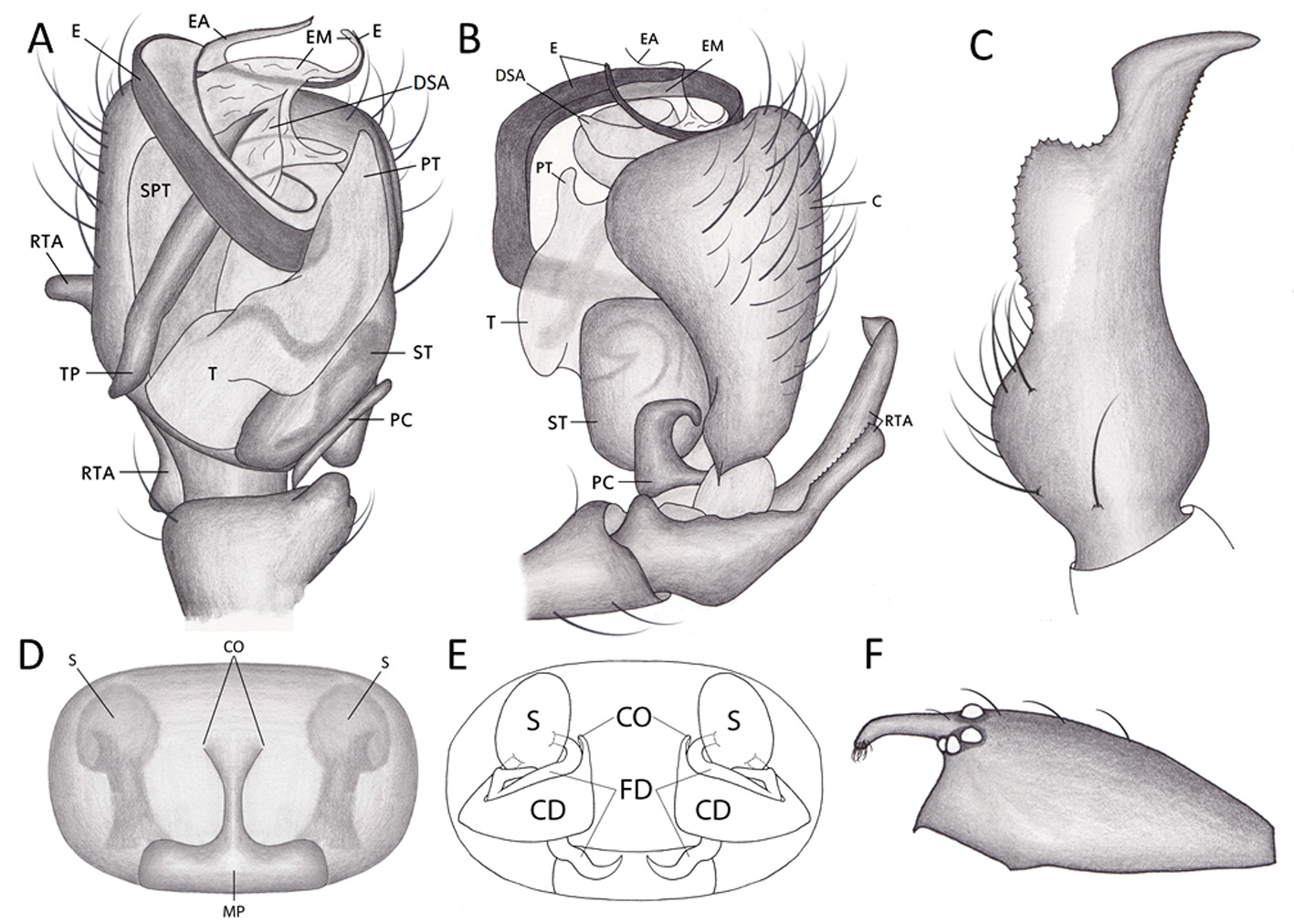
Diagnosis. Males may be separated from similar North American genera (e.g. Ceraticelus , Ceratinella , Grammonota , Scylaceus , and Walckenaeria ) by possessing a large palpal tibial apopysis that is serrated on the medial margin ( Figs. 2A View FIGURE 2 , 3C View FIGURE 3 , 4C View FIGURE 4 , 6C View FIGURE 6 ). Females may be separated from other erigonines by having an epigynum split in half by two parallel copulatory grooves with copulatory openings positioned anteriorly and a widened rectangular median plate posteriorly ( Figs. 2D View FIGURE 2 , 3E View FIGURE 3 , 4E View FIGURE 4 , 6D View FIGURE 6 ). Males of Epiceraticelus are easily distinguished from Scylaceus pallidus by the long sinuous embolic division and by the fine serrations medially on the retrolateral tibial apophysis. Additionally, the narrow “pistol-shaped” tibial apophysis of S. pallidus is distinctive ( Paquin and Dupérré 2003, Figs. 1230, 1231). Epiceraticelus and Scylaceus pallidus females are similar somatically and have a similar posteriorly-placed median plate, but in Epiceraticelus , it is triangular in shape rather than broadly rectangular ( Figs. 2D View FIGURE 2 , 3E View FIGURE 3 , 4E View FIGURE 4 , 6D View FIGURE 6 ). Moreover, the epigynum in S. pallidus has spermathecae that are more posteriolateral in position ( Figs. 2C View FIGURE 2 , 3F View FIGURE 3 , 4D View FIGURE 4 , 6E View FIGURE 6 and Paquin & Dupérré 2003, Fig. 1232).
Description. Length: 1.1–1.3. Cephalothorax: eyes eight, AME smallest, ALE and PLE equal, PME approximately two-thirds the size of PLE, posterior eye row slightly procurved, anterior eye row slightly recurved; eyes ringed with black. Chelicerae with six or seven promarginal teeth, five retromarginal denticles. Carapace oval, two-thirds as wide as long with one to two medial setae; a pair of small setae between PME and AME of each side; anterior portion of carapace three times as high as section near pedicel, clypeal modifications variable, dusky orange to tan, sternum nearly as long as wide, pointed posteriorly between coxae of leg IV. Abdomen: oval, covered with simple setae, concolorous gray, venter gray with dark gray patches around spinnerets and near epigynum; epiandrous fusules absent; colulus twice as long as wide; anterior spinnerets longer than posterior, cone-shaped. Tracheal system desmitracheate (sensu Millidge 1984). Legs: light orange, unmarked; relative length 4, 1, 2, 3; patella–tibia I three-fourths as long as carapace; metatarsi I–III each with a single trichobothrium; paired tarsal claws with eight teeth, median claw with three teeth. Trichobothrium on metatarsus IV lacking. Trichobothrium on metatarsus I at 0.4–0.5. Tibial spines 1111, often 0000. Palp of male complex ( Figs. 2B View FIGURE 2 , 3 View FIGURE 3 A–B, 4B, 6A–B), with a well-developed retrolateral tibial apophysis which is finely serrated on medial margin; embolic division with elongate tail-piece oriented with long axis parallel to long axis of palp; tegulum with distal protegulum; distal suprategular apophysis present; long sinuous embolus present ( Figs. 3 View FIGURE 3 A–B, 6A–B). Epigynum ( Figs. 3 View FIGURE 3 EF, 6D–E) with dual sinuous median epigynal grooves widening into a posterior median plate (narrower and separated in E. fluvialis , while wider and fused in E. mandyae n. sp.), spermathecae ventrally visible, separated by more than 1.5 times their diameter; internally, spermathecae anterior of copulatory and fertilization ducts, laterally directed and partially covering spermathecae posteriorly.
Distribution. New York south to Georgia, west to Texas ( Fig. 1 View FIGURE 1 ).
Natural History. May be found in the leaf litter of damp forests in winter and early spring. Forests commonly consist of various species of pine and oak.


No known copyright restrictions apply. See Agosti, D., Egloff, W., 2009. Taxonomic information exchange and copyright: the Plazi approach. BMC Research Notes 2009, 2:53 for further explanation.
|
Kingdom |
|
|
Phylum |
|
|
Class |
|
|
Order |
|
|
Family |
Epiceraticelus Crosby & Bishop, 1931
| Draney, Michael L., Milne, Marc A., Ulyshen, Michael & Madriz, Gabrielle 2019 |
Epiceraticelus
| Crosby, C. R. & Bishop, S. C. 1931: 380 |