
Paraphasma Redtenbacher, 1906
|
publication ID |
https://doi.org/10.11646/zootaxa.5122.1.1 |
|
publication LSID |
lsid:zoobank.org:pub:EC13A69D-D6FA-4926-AC59-648A5626C9B9 |
|
DOI |
https://doi.org/10.5281/zenodo.10718233 |
|
persistent identifier |
https://treatment.plazi.org/id/03B587AA-FFD3-FFC1-FF2A-FC2DFBB0F1C8 |
|
treatment provided by |
Plazi |
|
scientific name |
Paraphasma Redtenbacher, 1906 |
| status |
|
Paraphasma Redtenbacher, 1906 View in CoL
Paraphasma Redtenbacher, 1906: 114–117 View in CoL [type species: Paraphasma marginale Redtenbacher, 1906 View in CoL by subsequent designation]; Handlirsch, 1930: 760; Bradley & Galil, 1977: 203; Vanschuytbroeck & Cools, 1981: 24; Sellick, 1997: 121, fig. 128; Zompro, 2000: 95 [type species designation]; Bragg, 2001: 640; Zompro, 2002b: 6 [repetition of type species designation]; Zompro, 2004: 158–159, 317, fig. 92a,b; Otte & Brock, 2005: 251; Conle et al., 2011: 32; Jourdan et al., 2014: 493; Conle et al., 2020: 126; Chiquetto-Machado & Cancello, 2021: 1–41 View Cited Treatment [phylogenetic analysis], figs 1, 25, 26.
Oestrophora Redtenbacher, 1906: 124 View in CoL [type species: Oestrophora triangulifera Redtenbacher, 1906 View in CoL by monotypy]; Bragg, 2001: 638; Zompro, 2004: 102; Otte & Brock, 2005: 231; Conle et al., 2020: 127; Chiquetto-Machado & Cancello, 2021: 23, 25 [= Paraphasma Redtenbacher, 1906 View in CoL ].
Mantis, Fabricius, 1775: 274 View in CoL [in part]; Fabricius, 1787: 227 [in part]; Fabricius, 1793: 12 [in part].
Necroscia, Westwood, 1859: 128 [in part].
Olcyphides, Kirby, 1904a: 410 View in CoL [in part]; Rehn, 1907: 165; Otte & Brock, 2005: 231 [in part].
Phasma, Fabricius, 1798: 186 View in CoL [in part]; Latreille, 1807: 87 [in part]; Serville, 1831: 57 [in part]; Gray, 1835: 22 [in part]; Burmeister, 1838: 583 [in part]; Serville, 1839: 266 [in part]; De Haan, 1842: 123 [in part]; Westwood, 1859: 117 [in part].
Prexaspes, Kirby, 1904a: 413 View in CoL [in part]; Brock, 1998b: 34; Otte & Brock, 2005: 282 [in part].
Stratocles, Redtenbacher, 1906: 102 View in CoL [in part].
Trigonophasma, Kirby, 1904a: 372 [in part].
Species included: Paraphasma conspersum Redtenbacher, 1906 View in CoL ; Paraphasma indistinctum Chiquetto-Machado View in CoL sp. nov.; Paraphasma laterale ( Fabricius, 1775) View in CoL ; Paraphasma marginale Redtenbacher, 1906 View in CoL ; Paraphasma minus Redtenbacher, 1906 View in CoL ; Paraphasma sooretama Chiquetto-Machado View in CoL sp. nov.; Paraphasma spinicauda View in CoL ChiquettoMachado sp. nov.; Paraphasma trianguliferum ( Redtenbacher, 1906) View in CoL ; Paraphasma umbretta ( Lichtenstein, 1796) View in CoL ; Paraphasma fasciatum Gray, 1835 View in CoL [nomen dubium].
Diagnosis. Representatives of Paraphasma are winged stick insects with moderate sexual dimorphism and body length varying from 42 to 75 mm. The phallic organ contains the clearest characters to distinguish this genus from other Pseudophasmatidae , with the following features being diagnostic of Paraphasma : presence of a ‘locking’ sclerite on the inner face of the ventral lobe, bearing protuberances that fit into folds on the surface of the dorsal lobe; base of the phallic organ with three apodemes, one or two of which forming distinct spatulate expansions.
Additionally, the following combination of external characters can be used for the differentiation of Paraphasma from other genera: Body mostly brown or black, but most species exhibiting a characteristic color pattern with a pair of yellowish lateral stripes extending along head, prothorax, mesothorax and costal region of tegmina and hindwings. Head sub-rectangular or subquadrate in dorsal view, with almost flat vertex and well-developed ocelli; antennae distinctly longer than the body. Openings of the prothoracic pair of defensive glands very conspicuous; profemur distinctly curved and compressed basally, in females often with anterodorsal carina distinctly raised medially; mesoand metafemur lacking distinct carinae. Tegmina short, not reaching median region of metanotum and at most 3x longer than wide, anal region with conspicuous reticulate venation; hindwing reaching at least abdominal tergite VII, anal region basally yellow or orangish (colorless in dried specimens) and with a brownish stripe extending along apical and posterior margins. Males: subgenital plate longer than tergites IX and X combined, with convex posterior region; thorn pads weakly developed, usually bearing less than 20 minute teeth. Females: tergite X tectiform, with posterior margin slightly emarginate; cerci short, straight and cylindrical; praeopercular organ as a longitudinal protuberance with shiny aspect; subgenital plate lanceolate, extending approximately to posterior margin of tergite X and bearing an internal longitudinal carina.
Redescription of male. Small and delicate stick insects; body length 42–62 mm. Color: Body and appendages predominantly black or brown; ventral region of body in lighter shades. Most species with a pair of light, yellowish lateral stripes extending along head, prothorax, mesothorax and costal region of tegmina and hindwings (stripes may become greenish in dried specimens) and with a light brown or yellowish dorsomedian line extending along head, pro- and mesothorax. Head, prothorax, mesothorax, antennae and legs sometimes with light brown or yellowish regions. Hindwing with translucent anal region, with a yellow or orangish basal area (colorless in dried specimens) surrounded by a brownish stripe along apical and posterior margins; stripe widening along apical margin. Live specimens exhibit one of two possible general color patterns: (1) cryptic pattern, with predominance of brownish or orangish pale shades and lateral stripes (if present) pale yellow; or (2) bright pattern, with marked contrast between the mostly shiny black body and bright yellow lateral stripes. Head: Smooth; sub-rectangular or subquadrate in dorsal view, slightly elongate or about as long as wide; vertex slightly convex or flat. Compound eye large and very prominent, covering nearly half of head length, in lateral view slightly elongate or almost round.Ocelli well-developed, distinctly apart from each other; median one more distant from the lateral pair.Antennae filiform, very long, distinctly surpassing end of abdomen, sometimes for more than half the length of the latter; scape dorsoventrally compressed; pedicel cylindrical, slightly shorter than scape; first flagellomere about 3x longer than pedicel. Thorax: Prothorax smooth; as long as or slightly longer than head; distinctly narrower than head; weakly convex dorsally and ventrally; laterally flat. Pronotum sub-rectangular, with slight constriction on anterior third; anterolateral corners with rounded indentations, outlining openings of pair of defensive glands; posterior margin convex; pair of gentle dorsolateral carinae originating posterior to defensive glands and extending until nearly posterior margin. Mesothorax slightly rugose, 1.5–1.8x longer than prothorax; about as wide as prothorax on anterior half and gradually widening on posterior half; weakly convex dorsally and ventrally, laterally flat; sub-rectangular in cross-section on anterior half and approximately cylindrical on posterior half. Mesonotum gently elevated posteriorly, with a weak longitudinal carina extending along each lateral margin; mesepisternum with a more pronounced carina extending along ventral margin. Metathorax and median segment smooth, parallel-sided, as wide as posterior region of mesothorax; both distinctly convex dorsally, weakly convex laterally, ventrally flat; metathorax approximately 3x longer than median segment; both combined 1.5–2x longer than mesothorax. Metepisternum with a longitudinal carina extending along ventral margin. Legs: Fairly long and slender. Profemur as long as or slightly longer than combined length of mesothorax, metathorax and median segment; curved and compressed basally; approximately trapezoidal in cross-section, with carinate edges and distinct ventromedian carina. Mesofemur 0.6x length of profemur; as long as or slightly longer than pro- and mesothorax combined. Metafemur slightly shorter than profemur. Meso- and metafemur sub-rectangular in cross-section, with dorsal and ventral faces slightly convex; edges weakly carinate; ventromedian carina absent. Tibiae slightly shorter than corresponding femur, 1.5–2.5x longer than corresponding tarsus; rectangular or trapezoidal in cross-section; ventromedian carina absent; with conspicuous area apicalis. Pro- and metabasitarsus slightly longer than following three tarsomeres combined; mesobasitarsus approximately as long as following three tarsomeres combined. Wings: Tegmina short, not reaching median region of metanotum; in dorsal view 2–3x longer than wide; posterior margin rounded or weakly angulate; apical margin rounded, truncate or acuminate; shoulder pad prominent, varying from dull protuberance to sharp spine; anal region with conspicuous reticulate venation. Hindwing well-developed, reaching abdominal tergite VII or VIII. Abdomen: 1.6–1.8x longer than head, thorax and median segment combined; dorsally and ventrally smooth, but tergites IX and/or X sometimes slightly carinate longitudinally. Segment II the longest; segments gradually shortening from III to VI; segment VII about two-thirds length of VI. Tergites VIII–X distinctly shorter than preceding ones; tergite IX as long as or slightly longer than VIII, 1.3–1.6x longer than X. Segments II–VI equally wide. Tergite VII as wide as VI anteriorly and widening posteriorly; tergite VIII distinctly wider than preceding ones; tergite IX as wide as or narrower than VIII. Tergite X as wide as or narrower than IX; wider than or as wide as long; strongly convex; posterior margin emarginate, truncate or concave, in caudal view inverted U-shaped. Thorn pads usually weakly developed, each bearing at most 20 minute teeth, often less than 10. Cerci 0.9–1.7x the length of tergite X; approximately straight or distinctly incurved; apex spatulate, rounded or acuminate. Vomer with shape varying among species; apex simple. Epiproct small, hardly visible under tergite X; posterior margin rounded. Sternite VIII distinctly shorter than corresponding tergite; wider than long. Subgenital plate longer than tergites IX and X combined, 2–3x longer than sternite VIII; distinctly divided into anterior and posterior region. Anterior region of subgenital plate with somewhat soft exoskeleton, enabling folding for exposure of phallic organ; in some species originating a pair of slender lateral projections, directed posteriorly. Posterior region of subgenital plate convex; as long as or slightly longer than anterior region; shape of posterior margin strongly varying among species. Cerci, posterior margin of tergite X and subgenital plate more densely pilose than remaining regions of terminalia. Phallic organ: Base short, approximately cylindrical, originating a small and lamellate right basal pouch. Dorsal sclerite well-developed, usually wider than long and covering a substantial portion of the organ; anterior marginal gently concave. Distal process of dorsal sclerite directed to the left and posteriorly, in angle varying from 45º to almost 90º in relation to longitudinal axis of phallic organ; in most species short and wide, sometimes relatively slender. Dorsal and ventral lobes large and robust, distinctly separate from each other on the right but partially or almost completely fused on the left. Dorsal lobe the longest; sometimes undivided, but usually divided into main body on the right and a dorsal smaller pouch on the left. Ventral lobe undivided. ‘Locking’ sclerite of the ventral lobe always present, morphologically unique in each species; bearing one to three protuberances that fit into membranous or slightly sclerotized folds on the inner face of dorsal lobe. Three base apodemes present, of which one or two project into dorsal lobe as a large spatulate expansion; one apodeme very small, situated on right side of base near right basal pouch.
Redescription of female. Small to medium-sized stick insects; longer and stouter than males; body length 52–75 mm. Color: As in male. Head: As in male except for the antennae proportionally shorter (but still distinctly surpassing end of abdomen). Thorax: As in male except for the following proportions: prothorax as long as or slightly shorter than head; mesothorax 1.3–1.8x longer than prothorax; metathorax 3–3.7x longer than median segment; metathorax and median segment combined 1.7–2.5x longer than mesothorax. Legs: As in male, but slightly shorter in relation to body and profemur often with anterodorsal carina strongly raised medially. Wings: As in male but hindwing slightly longer in relation to body, sometimes reaching tergite IX. Abdomen: 1.5–1.8x longer than head, thorax and median segment combined; dorsally and ventrally smooth, but tergite X sometimes gently carinate longitudinally. Segments II and III the longest; gradually shortening from IV to VII. Tergites VIII–X distinctly shorter and slightly narrower than preceding ones; tergite VIII at most two-thirds as long as VII. Tergite IX as long as or slightly shorter than VIII; 1.3–1.4x longer than X. Tergite X as long as or slightly longer than wide; tectiform; posterior margin gently emarginate. Cerci short, straight and cylindrical; 0.6–1.2x length of tergite X; apex blunt. Epiproct hardly visible under tergite X; posterior margin rounded. Sternite VII often with rounded indentation on posterior margin; praeopercular organ as a longitudinal protuberance with shiny aspect, sometimes surpassing posterior margin of sternite VII. Subgenital plate approximately lanceolate, reaching but not surpassing posterior margin of tergite X; apex gently acuminate; inner face longitudinally carinate. Cerci, tergite X and subgenital plate densely pilose.
Description of egg. Capsule approximately cylindrical, with varying degree of elongation, gently narrowing towards polar area and operculum; polar area approximately flat; capsule covered with bristles; length and density of bristles variable among species. Operculum perpendicular in relation to longitudinal axis of capsule; approximately oval, slightly higher than wide; bearing bristles. Micropylar plate oval, somewhat elongate, positioned medially on capsule. Median line short and inconspicuous. Internal micropylar plate open; oval and elongate; internal median line distinct, more than half as long as plate. Egg in varying shades of brown.
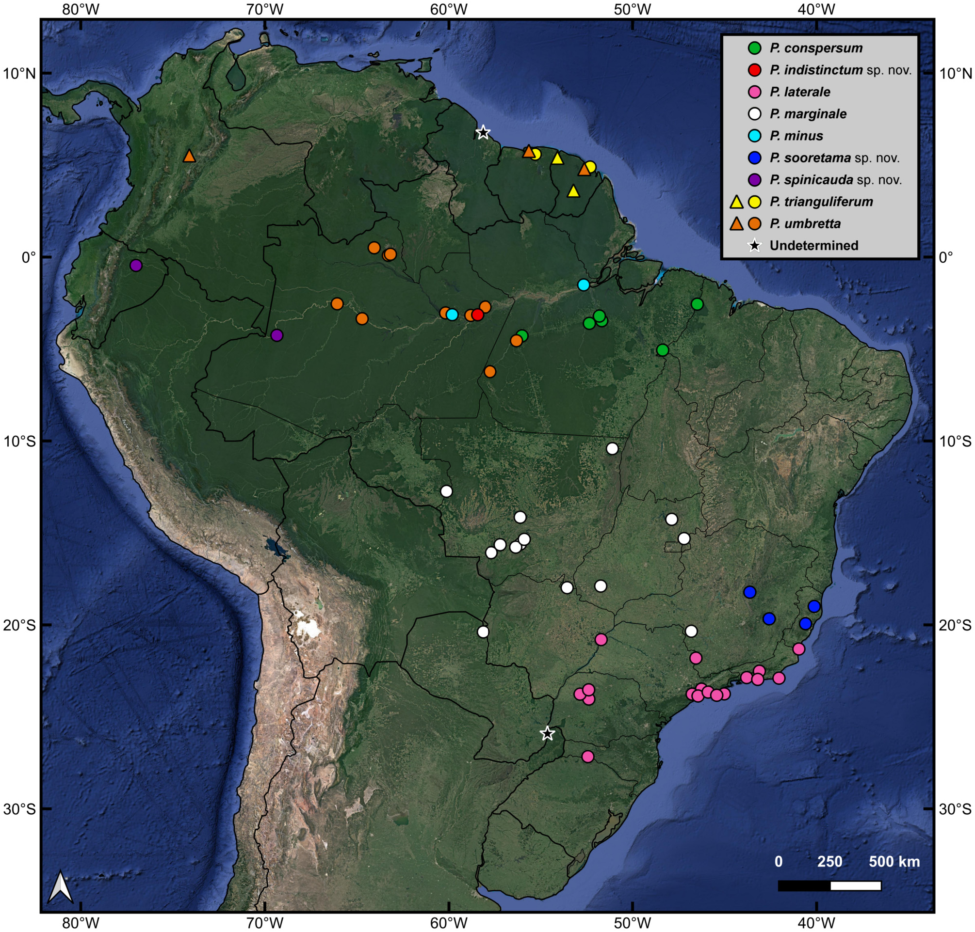
Distribution ( Fig. 1 View FIGURE 1 ). Paraphasma is distributed throughout central and northern South America, with records from most Brazilian states and from French Guiana, Suriname, Guyana, Colombia, Ecuador, Paraguay and northeast Argentina. The genus is also very likely to occur in Venezuela, Peru and Bolivia. The southernmost record of Paraphasma is from the district of Nova Teutônia, Santa Catarina, Brazil (27º09’53”S; 52º25’21”W) (a male of P. laterale in the MZUSP collection), and the northernmost record is from the region of Demerara, Guyana (06º46’N; 58º08’W) [a specimen identified by Rehn (1906) as Olcyphides fasciatus , an old combination of Paraphasma fasciatum (nomen dubium)].
Remarks. The representatives of Paraphasma exhibit little variation in external morphology. The head, thorax, legs, hindwings and female abdomen are very similar among the species. The tegmina show some, albeit small, morphological variation. Structures of the male terminalia such as the tergite X, cerci, vomer and subgenital plate vary considerably among some species, but are almost invariable in P. conspersum , P. indistinctum sp. nov., P. laterale , P. marginale , P. minus and P. sooretama sp. nov. The main characters for species delimitation come from the phallic organ.
According to our phylogenetic analysis of Paraphasma ( Chiquetto-Machado & Cancello 2021) , the genus is supported by two synapomorphies of external morphology (tegmina with spiniform shoulder pads; male subgenital plate with acuminate posterior margin) and three of the phallic organ (dorsal sclerite distinctly wider than long; presence of a ‘locking’ sclerite of the ventral lobe; base apodemes forming spatulate expansions). However, the two external synapomorphies, as well as the one regarding the shape of the dorsal sclerite, were secondarily lost in one or more species of Paraphasma (see Chiquetto-Machado & Cancello 2021: fig. 25).
No known copyright restrictions apply. See Agosti, D., Egloff, W., 2009. Taxonomic information exchange and copyright: the Plazi approach. BMC Research Notes 2009, 2:53 for further explanation.
|
Kingdom |
|
|
Phylum |
|
|
Class |
|
|
Order |
|
|
Family |
Paraphasma Redtenbacher, 1906
| Chiquetto-Machado, Pedro I., Morales, Adriana C. & Cancello, Eliana M. 2022 |
Paraphasma
| Chiquetto-Machado, P. I. & Cancello, E. M. 2021: 1 |
| Conle, O. V. & Hennemann, F. H. & Bellanger, Y. & Lelong, P. & Jourdan, T. & Valero, P. 2020: 126 |
| Jourdan, T. & Lelong, P. & Bellanger, Y. 2014: 493 |
| Conle, O. V. & Hennemann, F. H. & Gutierrez, Y. 2011: 32 |
| Otte, D. & Brock, P. D. 2005: 251 |
| Zompro, O. 2004: 158 |
| Zompro, O. 2002: 6 |
| Bragg, P. E. 2001: 640 |
| Zompro, O. 2000: 95 |
| Sellick, J. T. C. 1997: 121 |
| Vanschuytbroeck, P. & Cools, J. 1981: 24 |
| Bradley, J. C. & Galil, B. S. 1977: 203 |
| Handlirsch, A. 1930: 760 |
| Redtenbacher, J. 1906: 117 |
Oestrophora
| Chiquetto-Machado, P. I. & Cancello, E. M. 2021: 23 |
| Conle, O. V. & Hennemann, F. H. & Bellanger, Y. & Lelong, P. & Jourdan, T. & Valero, P. 2020: 127 |
| Otte, D. & Brock, P. D. 2005: 231 |
| Zompro, O. 2004: 102 |
| Bragg, P. E. 2001: 638 |
| Redtenbacher, J. 1906: 124 |
Stratocles, Redtenbacher, 1906: 102
| Redtenbacher, J. 1906: 102 |
Olcyphides
| Otte, D. & Brock, P. D. 2005: 231 |
| Rehn, J. A. G. 1907: 165 |
| Kirby, W. F. 1904: 410 |
Prexaspes, Kirby, 1904a: 413
| Otte, D. & Brock, P. D. 2005: 282 |
| Brock, P. D. 1998: 34 |
| Kirby, W. F. 1904: 413 |
Trigonophasma
| Kirby, W. F. 1904: 372 |
Necroscia
| Westwood, J. O. 1859: 128 |
Phasma, Fabricius, 1798: 186
| Westwood, J. O. 1859: 117 |
| De Haan, W. 1842: 123 |
| Serville, J. G. A. 1839: 266 |
| Burmeister, H. 1838: 583 |
| Gray, G. R. 1835: 22 |
| Serville, J. G. A. 1831: 57 |
| Latreille, P. A. 1807: 87 |
| Fabricius, J. C. 1798: 186 |
Mantis
| Fabricius, J. C. 1793: 12 |
| Fabricius, J. C. 1787: 227 |
| Fabricius, J. C. 1775: 274 |