
Russula rubiginosus R. Q. Ji, M.L. Xie & J. J. Zhou, 2022
|
publication ID |
https://doi.org/ 10.11646/phytotaxa.575.2.3 |
|
DOI |
https://doi.org/10.5281/zenodo.7415296 |
|
persistent identifier |
https://treatment.plazi.org/id/03B55D1D-E25F-6D51-FA97-FD6DD278FD3A |
|
treatment provided by |
Plazi |
|
scientific name |
Russula rubiginosus R. Q. Ji, M.L. Xie & J. J. Zhou |
| status |
sp. nov. |
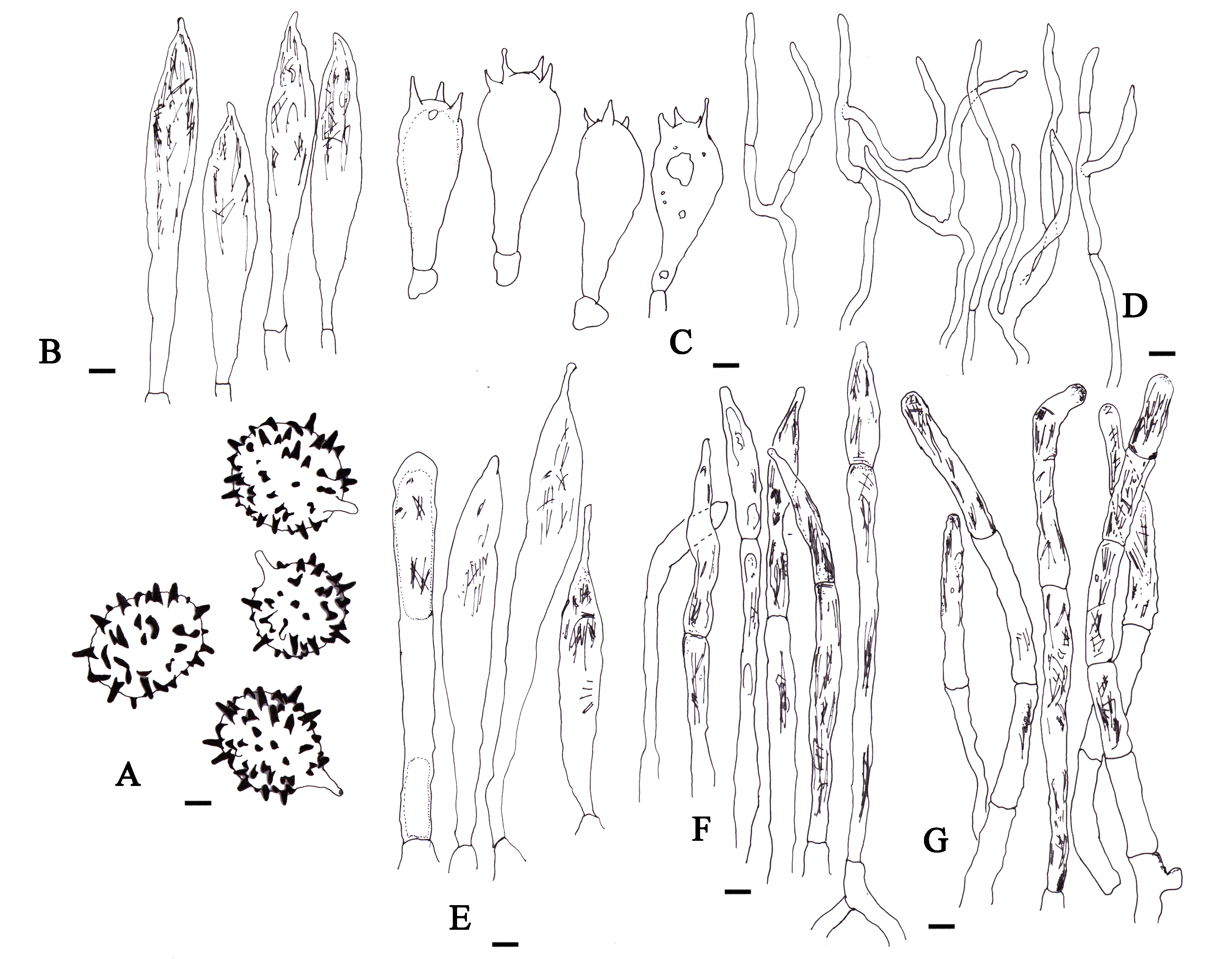
Russula rubiginosus R. Q. Ji, M.L. Xie & J. J. Zhou View in CoL , sp. nov., Figures 3 View FIGURE 3
MycoBank 843715
Diagnosis:— Differs from Russula subrubescens by small-sized basidiospores and large-sized cystidia.
Etymology GoogleMaps :— Refers GoogleMaps to the rusty spots on the cap and stipe of basidiomata.
Holotype:— CHINA. Heilongjiang Province: Wudalianchi City , ASL 300 m, 126°11’E, 48°37’N, 13 August 2018, Peng-Jie Xing & Yang Xu ( HMJAU58933 , GenBank OL828349 View Materials ) GoogleMaps .
Description:— Basidiomata medium-sized. Pileus 40‒72 mm diam., fleshy, firm, first hemispherical, then turned applanate with depressed center after maturation, margin becoming somewhat sulcate with age, glabrous, corinthain pink (xxvii 3ʹʹ d) to livid pink (xxvii 3ʹʹ f) when juvenile, corinthain red (xxvii 3ʹʹ) to corinthain pink (xxvii 3ʹʹd) with age, center orient pink (ii 11 f) to orange pink (ii 9 f) when juvenile, smiona orange (ii 11 b) to orange pink (ii 9 f) with age; mostly more or less covered with small ochraceous tawny (xv 15ʹ i) to mikado brown (xxix 13ʹʹ i) spots. Lamellae adnate, crowded, up to 6 mm wide, more or less furcate towards the stipe, slightly anastomosing, fragile, light ochraceous salmon (xv 13ʹ d) first, primuline yellow (xvi 19ʹ) when mature or dry, often with rust spots at lamellae. Stipe cylindrical to slightly clavate with rounded base, enlarged towards the base, firm, 40‒60 × 8‒13 mm, surface minutely wrinkled, white (liii) when juvenile, becoming baryta yellow (iv 21f) towards the base when old or dry, appearing ochraceous tawny (xv 15ʹ i) to mikado brown (xxix 13ʹʹ i) patches or spots when handled or with age, especially bottom. Context firm, white (liii) when juvenile, naples yellow (xvi 19ʹ d) with age, distinctly yellowing when dry or bruised. Taste acrid. Odor indistinct.
Basidiospores (40/2/2) 7–10 (12–) × 6–9 μm, Q = (1.2–)1.3–1.25(–1.31), Q av. = 1.19 ± 0.05, broadly ellipsoid, ornamentation of moderately large and distant: 5–7 (–10) amyloid spines in the circle of diameter of 3 μm on basidiospore surface, spines 1.0–2.0 μm high, mostly isolated, occasionally fused in short chains or crests (0–2 fusions in the circle), line connections very rare and short (0–1 line connection in the circle), not forming a mesh. Basidia (30–) 40–53.7 × (13–) 15–17 (–20) μm, clavate to subclavate, thin-walled, oil granules present when viewed with KOH, 4‒spored, sometimes 2‒spored; sterigmata 6‒8 × 2‒3 µm. Subhymenium pseudoparenchymatic. Lamellar trama mainly composed of large sphaerocytes. Pleurocystidia 66–92 × 9–13 μm, fusiform, clavate to subclavate, often with a subacute tip, thin-walled, sometimes with a small appendage and dense crystal inclusions. Cheilocystidia 60–90 × 8–13 µm, mostly similar to pleurocystidia. Pileipellis is composed of suprapellis and subpellis; suprapellis strongly gelatinized, 140–250 μm thick, composed of dense, ascending or erect hyphal ends and protruding and near surface repent pileocystidia, terminal cells 15–45 × 2–3 μm, cylindrical, often constricted towards the apex; subpellis less gelatinized, 40–100 μm thick, hyphae 2–6 µm wide, dense, strongly intricate, often branched. Pileocystidia numerous and often large sized, narrowly to broadly clavate or fusiform, mainly two or three celled, occasionally one-celled, rarely with more cells, thin-walled, obtuse to subacute, usually inflated near apical part, with terminal cells measuring 30–50 × 6–8 µm, contents in Congo red heteromorphous, usually granulose, but sometimes partly crystalline or banded. Stipitipellis cutis composed of thin-walled, septate, cylindrical, hyaline hyphae 2–4 μm wide, caulocystidia present, dispersed, rare, mainly two or three celled, occasionally one celled, rarely with more cells, thin-walled, obtuse to subacute, usually inflated near apical part, with terminal cells measuring 33–65 × 5–7 μm. Clamp connections absent in all tissues.
Habitat and distribution:—Ectomycorrhizal fungi symbiotic with Q. mongolica . Solitary in July to August under Q. mongolica forests. Known from Northeast China.
Additional specimens examined:— CHINA. Heilongjiang Province: Wudalianchi City , ASL 300 m, 126°11’E, 48°37’N, 13 August 2018, Peng-Jie Xing & Yang Xu ( HMJAU58934 View Materials , GenBank OL828348 View Materials ) GoogleMaps . Inner Mongolia Autonomous Region: Zhalantun County, Xiushui Scenic Area , ASL 370 m, 27 July 2013, Sai-Fei Li, Dong Zhao & Guo-Jie Li ( HMAS267826 View Materials , GenBank KX441111 View Materials ); ibid., 28 July 2013, Sai-Fei Li, Dong Zhao & Guo-Jie Li ( HMAS267741 View Materials , GenBank KX441095 View Materials ) GoogleMaps .
No known copyright restrictions apply. See Agosti, D., Egloff, W., 2009. Taxonomic information exchange and copyright: the Plazi approach. BMC Research Notes 2009, 2:53 for further explanation.
|
Kingdom |
|
|
Phylum |
|
|
Class |
|
|
Order |
|
|
Family |
|
|
Genus |