
Stylogomphus thongphaphumensis, Chainthong & Sartori & Boonsoong, 2020
|
publication ID |
https://doi.org/ 10.11646/zootaxa.4763.2.6 |
|
publication LSID |
urn:lsid:zoobank.org:pub:6649FF3C-25C1-4A26-9362-D5945684708C |
|
DOI |
https://doi.org/10.5281/zenodo.3806442 |
|
persistent identifier |
https://treatment.plazi.org/id/03B5265B-FFC7-3F6A-FF5F-DBC36D49F82D |
|
treatment provided by |
Carolina |
|
scientific name |
Stylogomphus thongphaphumensis |
| status |
sp. nov. |
Stylogomphus thongphaphumensis View in CoL sp. nov.
( Figs 1–14 View FIGURES 1–2 View FIGURES 3–7 View FIGURES 8–9 View FIGURES 10–12 View FIGURES 13–14 , 17–27 View FIGURES 15–17 View FIGURES 18 View FIGURES 19–24 View FIGURES 25–39 , 44, 46, 48 View FIGURES 43–48 )
Material examined. THAILAND: Holotype: 1♂, Huai Khayeng, Thong Pha Phum district , Kanchanaburi Prov- ince, 14°36′20″N 98°34′38″E, 206 m a.s.l., larva collected on 14.XII.2014, adult emerged on 30.IV.2015, D. Chain- thong leg. GoogleMaps
Paratypes: Nine final stadia larvae, same locality and collection date as holotype; adults emerged on: 4 ♂♂ 24.XII.2014, 8.III.2015, 18.III.2015, 29.III.2015; 5 ♀♀ 20.XII.2014, 24.XII.2014, 18.II.2015, 26.III.2015.
Etymology. The specific epithet “thongphaphumensis” refers to the district of Kanchanaburi Province that contains the type locality of the new species.
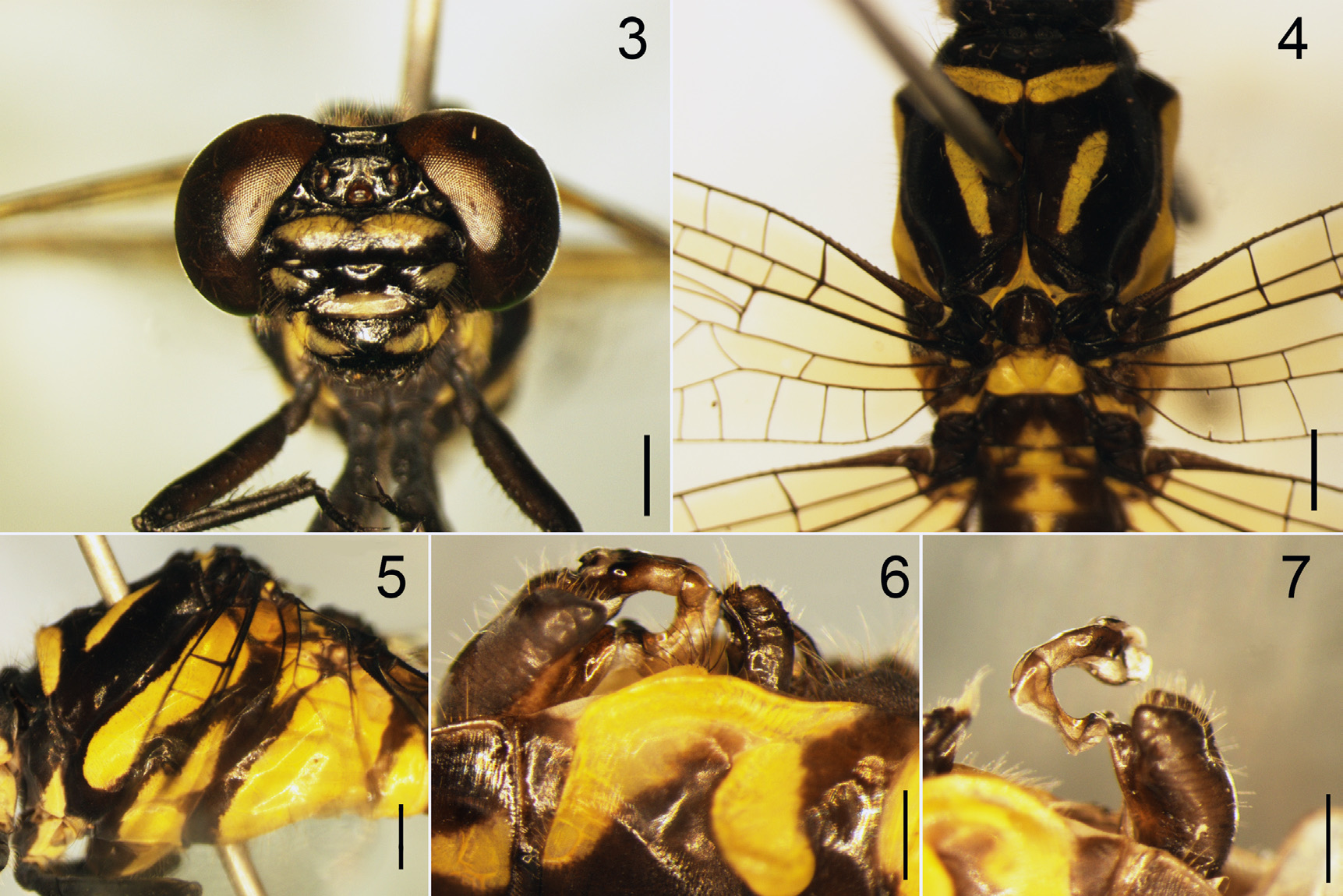
Description of the holotype. General appearance and color pattern as shown in Figures 1 View FIGURES 1–2 , 3–12 View FIGURES 3–7 View FIGURES 8–9 View FIGURES 10–12 .
Head ( Fig. 3 View FIGURES 3–7 ). Black with yellow markings as follows: median lobe of labium black and lateral lobes yellow; mandibles yellow, labrum with a pair of transverse ellipsoid yellow spots; genae black; anteclypeus yellow; postclypeus black, with single large yellow spots laterally; postfrons with a broad yellow band, antefrons black; occiput and vertex black.
Thorax. Colour pattern as shown in Figures 4–5 View FIGURES 3–7 . Prothorax black with yellow laterally. Pterothorax black with yellow markings, as shown in Figure 5 View FIGURES 3–7 ; mesothoracic collar yellow, except on middle; antehumeral stripe narrow, not connected to mesothoracic collar, almost reaching margin of antealar triangle; mesepimeron yellow with black margins all round; metepisternum black with two yellow spots; metepimeron mostly yellow. Legs black, coxae mostly yellow and inner surface black; femora, tibiae and tarsi black.
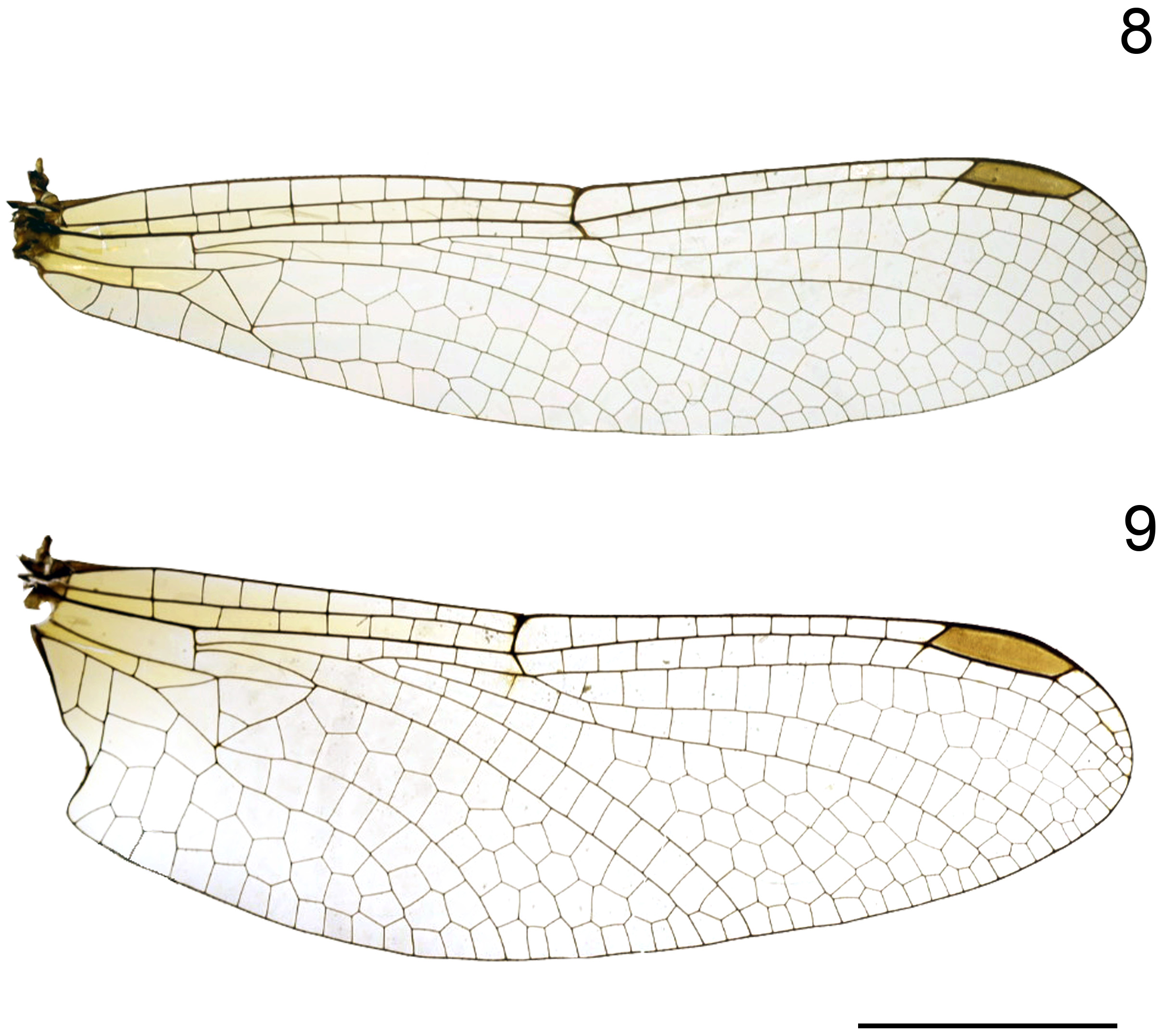
Wings. Hyaline, venation dark brown ( Figs 8–9 View FIGURES 8–9 ); 11 (left) and 10 (right) Ax on Fw, 7 (left) and 8 (right) on Hw, 9 (left) and 8 (right) Px on Fw, 9 (left) and 9 (right) on Hw. Pterostigmata very dark brown, surmounting 3.5 cells in both Fw and Hw; anal triangle 3-celled.
Abdomen. Abdomen predominantly black, with bright yellow markings, as follows ( Fig. 1 View FIGURES 1–2 ): S1 mostly yellow laterally, S2 yellow around auricle, another narrow yellow mid-dorsal stripe on S1and S2; S3 with basal mid-dorsal tiny triangular yellow spot, S4–7 with small dorso-lateral yellow markings at base; S8–10 black.
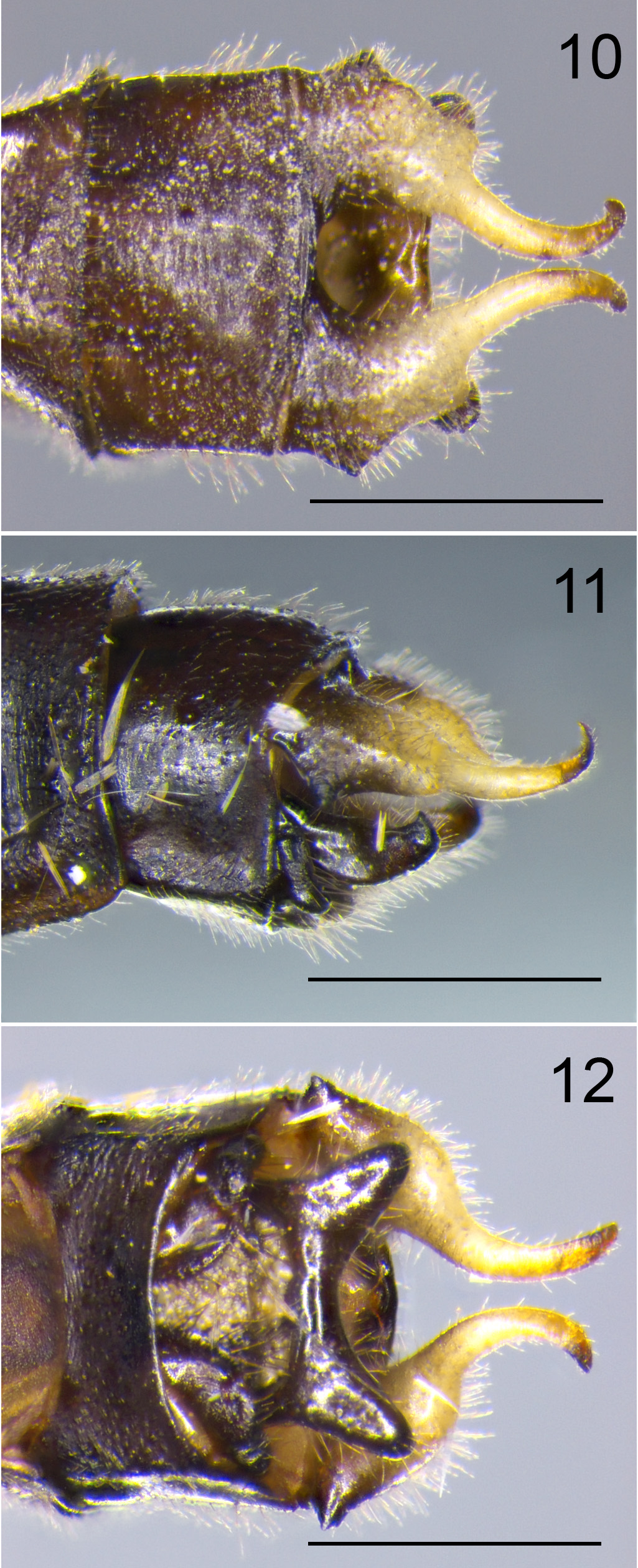
Anal appendages ( Figs 10–12 View FIGURES 10–12 ). Cerci creamy white, approximately 1.5x length of S10, with ventrolateral black tooth about one-fifth from base, short and turned outward; in dorsal view ( Fig. 10 View FIGURES 10–12 ) constricted in the middle, apical half slender. Epiproct black, wider than long, approximately 0.7x length of S10 and 0.4x of cerci, lateral lobes diverging with blunt tips; the median cleft U-shaped and shallow in ventral view, covered with minute setae, widening toward apex ( Figs 11–12 View FIGURES 10–12 ).
Accessory genitalia. Anterior and posterior hamulus dark brown, anterior hamulus in lateral view slender and smoothly curved. Posterior hamulus broad, anterior margin with small denticles, inner-curved spine on upper side. Vesica spermalis ( Figs 6–7 View FIGURES 3–7 ), first segment bifid, prepuce of third segment bulging in lateral view, apex of lobe hammer-shaped.
Measurements (mm): Fw length 23, Hw length 21, abdomen with appendages 28.5.
Variation in paratype males. Minor variations apparent among the four paratype males, when compared with the holotype. The paratype specimens differ in the number of Ax and Px: 8–11 (left) or 11 (right) Ax on Fw, 8 (left) or 8 (right) on Hw, 8–10 (left) or 9–10 (right) Px on Fw, 6 (left) or 6–8 (right) on Hw. Measurements (mm) are: ♂ forewing 23–24, hindwing 21–23; abdomen including appendages 27–29.
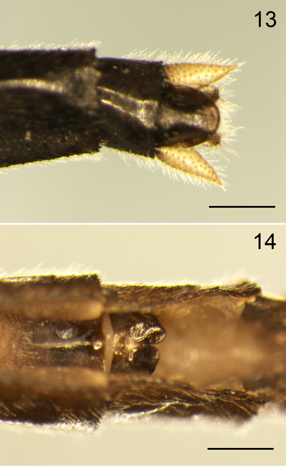
Paratype females. Body colouration similar to male ( Fig. 2 View FIGURES 1–2 ). Yellow areas little broader than those of male. Occiput black without a protuberance; anteclypeus yellow; postclypeus black with pair of large triangular yellow spots; postfrons with a band narrowly interrupted in middle. Ax 10–11 (left), 9–11 (right) on Fw, 8–9 (right), 8–10 (left) on Hw; Px 8–9 (right), 7–9 (left) on Fw, 9–10 (right), 7– 8 (left) on Hw; abdomen almost cylindrical. Yellow markings similar to those of male: S1 and S2 mostly yellow laterally, S3 with dumbbell-shaped yellow markings in lateral view. S4–7 with a half ring yellow markings; S8–10 black; cerci pale, about same length as S10, with gradually tapering, pointed tip ( Fig. 13 View FIGURES 13–14 ); valvula vulvae length 0.4 mm., dark brown ( Fig. 14 View FIGURES 13–14 ), one-third length of S9 sternum, bilobed, with a V-shaped shallow cleft between lobes, the apices blunt and not turned outward. Measurements (mm): ♀ Fw 23–25, Hw 22–23; abdomen including appendages 28–30.
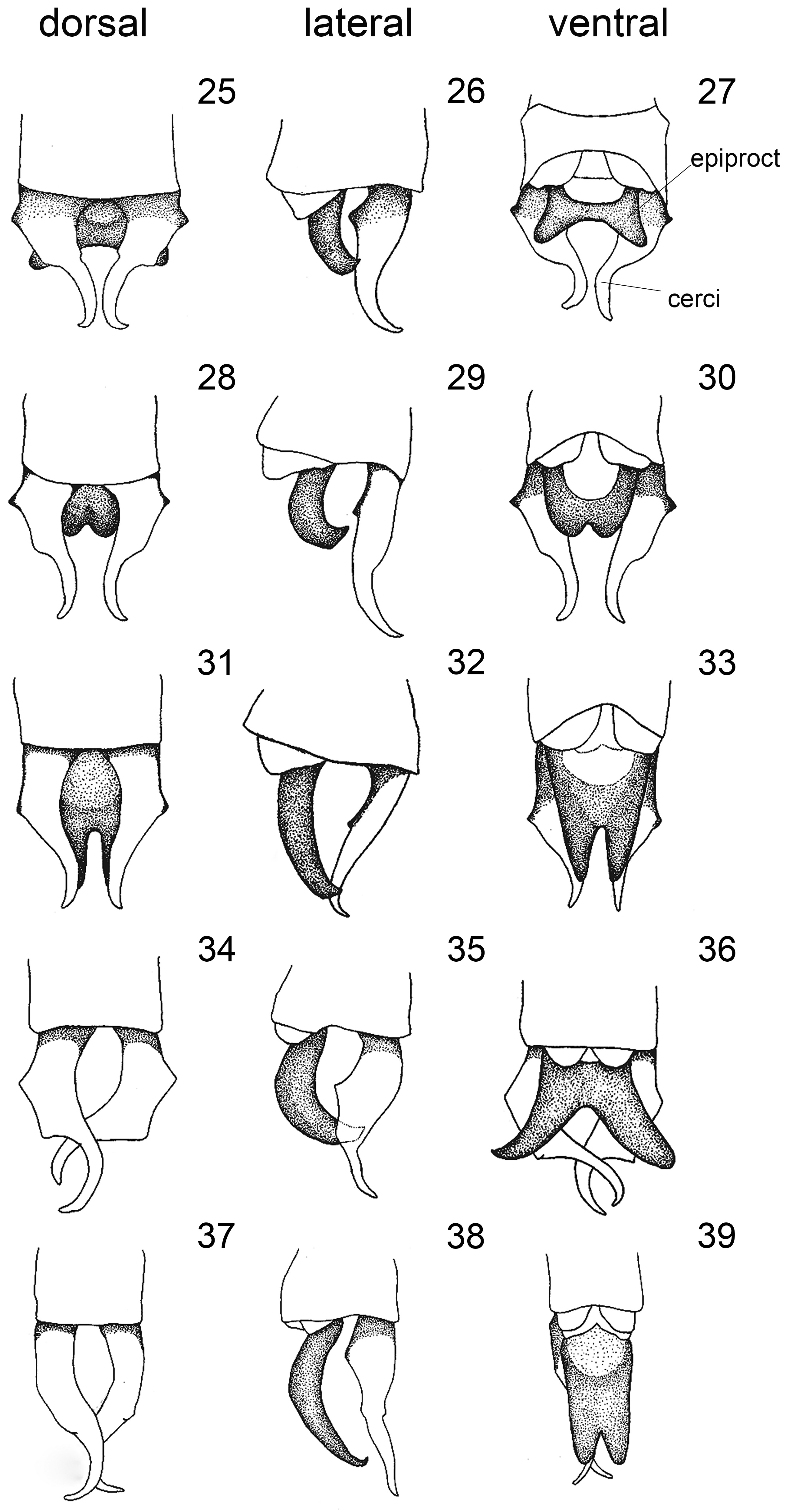
Differential diagnosis: When compared with known Stylogomphus species in Asia, the members of this genus can be divided into two groups by the shape of male anal appendages. The first group has long epiproct as in S. changi Asahina, 1968 ; S. inglisi Fraser, 1922 ; S. ryukyuanus Asahina, 1966 ; S. malayanus ; S. lawrenceae Yang & Davies, 1996 ; and S. delicatus Kompier, 2017 . The second group has short epiproct as in S. shirozui Asahina, 1966 ; S. suzukii Oguma, 1926 ; S. chunliuae Chao, 1954 ; S. tantulus Chao, 1954 ; S. annamensis, Kompier, 2017 ; and S. thongphaphumensis . The comparison between the new species and other known Stylogomphus species indicates close morphological similarity between S. shirozui and S. tantulus in the shape of cerci and epiproct, but epiproct of S. shirozui has pointed tips, and the pterothorax bears Y-shaped markings. The epiproct of S. tantulus is long, approximately half the length of the cerci, lateral lobes diverging more than S. thongphaphumensis ( Chao 1990; Yang & Davies 1996; Sasamoto 2001, 2004; Kompier 2017; Zhang 2019). Within Southeast Asian species ( Figs 25–39 View FIGURES 25–39 ), S. thongphaphumensis is somewhat more similar to S. annamensis from Vietnam than it is to other species ( Figs 28–30 View FIGURES 25–39 ), closely similar in the details of the shape of cerci, strong lateral basal tooth and strong constriction just past half their length, but S. thongphaphumensis is readily distinguishable from other species by the shape of the epiproct. The epiproct of S. thongphaphumensis is wider than long, with lateral lobes diverging with blunt tips, the median cleft U-shaped and shallow ( Figs 25–27 View FIGURES 25–39 ); a short epiproct, shallowly incised into two rounded lobes in S. annamensis ( Figs 28–30 View FIGURES 25–39 ).; the epiproct of S. delicatus is only a little shorter than the cerci, deeply incised to about 3/5 length, resulting in two pointed halves, the incision parallel-sided and the outer edges convergent ( Figs 31–33 View FIGURES 25–39 ); deeply incised to about 2/3 of length and diverged apically in S. lawrenceae ( Figs 34–36 View FIGURES 25–39 ); and relatively straight and not diverged apically in S. malayanus ( Figs 37–39 View FIGURES 25–39 ).
In addition, females of S. lawrenceae , S. annamensis and S. malayanus are known to date, whereas females of S. delicatus are still unknown (Yang & Davies1996; Sasamoto 2001, 2004; Zhang 2019, p. 465). Although the male of S. thongphaphumensis is closer to S. annamensis than to any other Southeast Asian species, the females are closest to S. malayanus , which has the occiput without a Y-shaped horn-like protuberance. In contrast, the occiput of both S. lawrenceae and S. annamensis has a Y-shaped horn-like protuberance. The females of S. thongphaphumensis can be separated from other Southeast Asian species by the shape of the valvula vulvae, one-third the length of the S9 sternum, with a V-shaped shallow cleft between the lobes and the apices blunt and not turned outward.
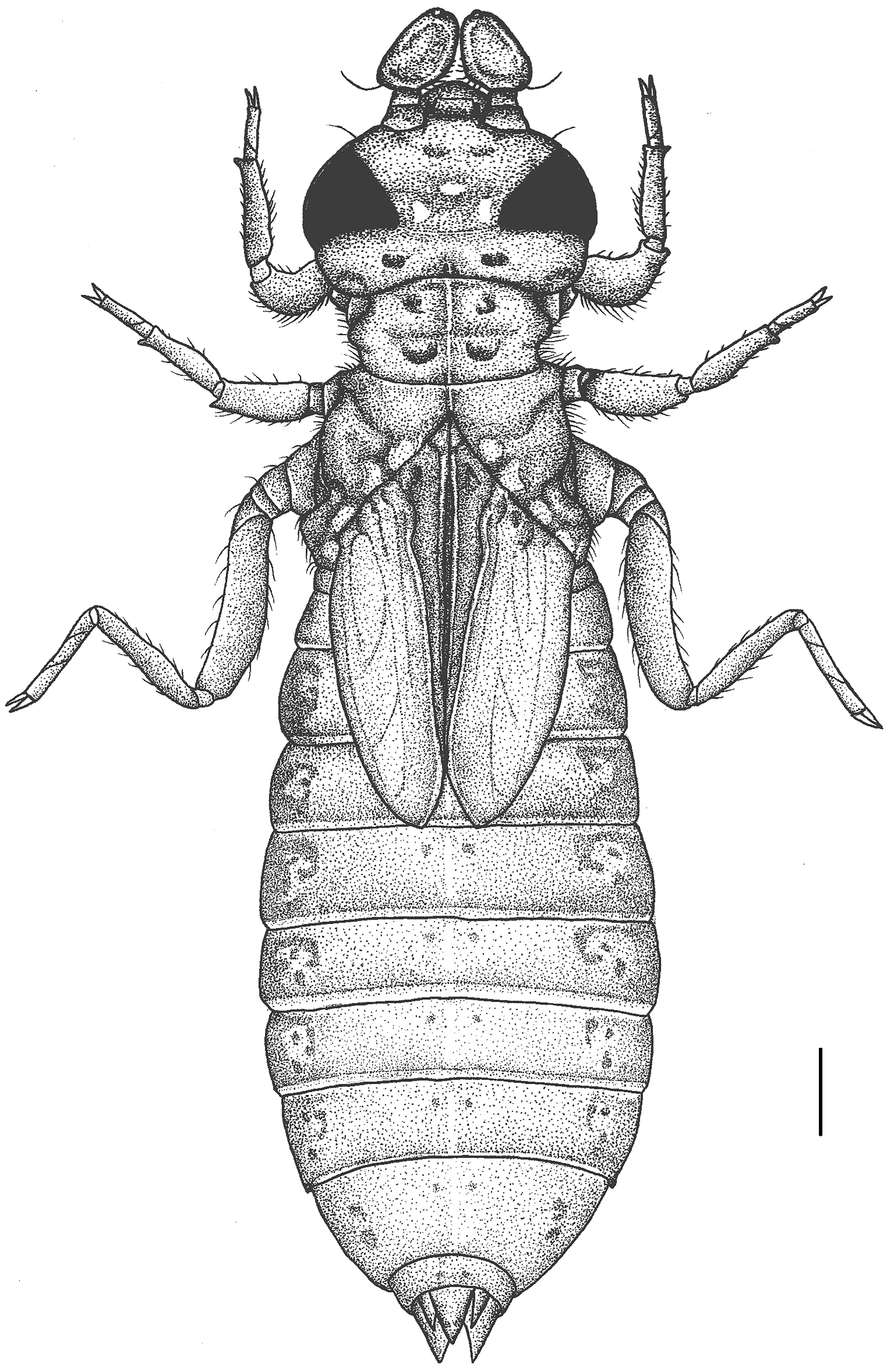
Description of the last stadium larva. General appearance and details of structures as shown in Figs 17–24 View FIGURES 15–17 View FIGURES 18 View FIGURES 19–24 , 44, 46, 48 View FIGURES 43–48 .
Colouration. Larvae uniformly dark brown and black; only the end of femora, tibiae and tarsi with alternating yellow.
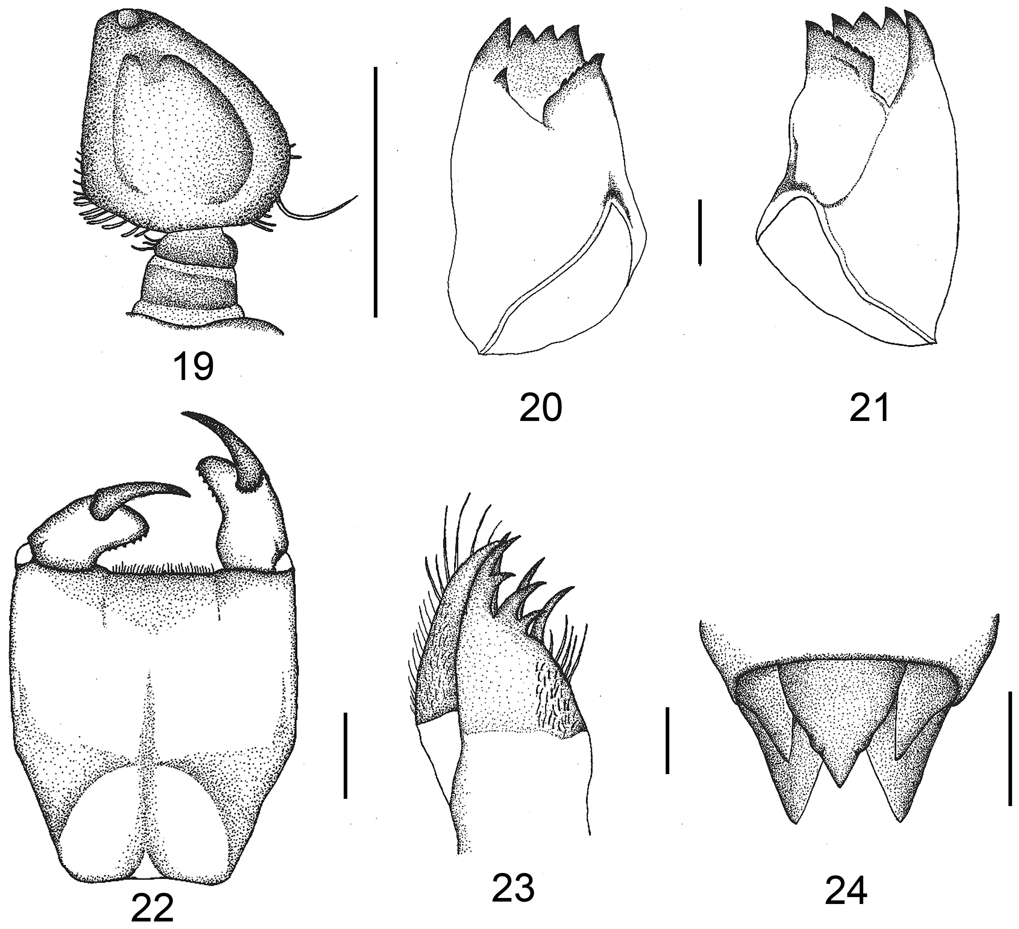
Head. Head wider than long, frontal part stouter, posterior lobe of the head shorter than the eye length, eyes large and broadest across, with three large ocelli. Antennae four-segmented, first two segments small and rather circular, third segment right-angled and triangular, inner side almost linear with many short flat setae, outer side obtusely curved with a long seta, fourth segment tiny ( Figs 19 View FIGURES 19–24 , 44 View FIGURES 43–48 ). Mandibles as in Figs 20–21 View FIGURES 19–24 and mandibular formula: L 1234 0 a(m1–5 or m1–6)b / R 1234 y a(m1–3 or m1–4)b with a>b in both mandibles ( Figs 20–21 View FIGURES 19–24 ). Maxillae: galeolacinia with seven moderately incurved teeth, three dorsal teeth nearly equal in length, four ventral teeth of different sizes, apical one largest; stipes and palp setose ( Fig. 23 View FIGURES 19–24 ).
Labium. Flat and not protruding when at rest ( Figs 22 View FIGURES 19–24 , 48 View FIGURES 43–48 ). Prementum-postmentum articulation reaching posterior margin of procoxae. Prementum subrectangular, longer than wide, sides convex, convergent basally, with small teeth and minute setae at lateral margins; apical margin convex, with ventral row of 4–5 short, subquadrate, reddish brown teeth and dorsal rows of whitish piliform setae on apical border; labial palp with uniformly inflexed inner edge, yellowish brown, apical lobe reddish, rounded, its internal margin with row of 5–8 equidistant teeth. Movable hook reddish brown, sharp and moderately incurved.
Thorax. Prothorax rectangular, dorsal portion raised at sides forming two crescent-shaped ridges. Wing sheaths parallel, reaching almost the distal margin of S4. Femur and tibia of forelegs almost of equal length. Middle tibia slightly longer than femur. Hind femur longer than tibia. When pressed against the abdomen, the hind leg femur reaches the base of S3. Tarsal formula 2–2–3, tarsi yellowish. Minute short rows of setae scattered along femur and tibia of all six legs.
Abdomen. Ovoid, dorsum granular, dark brown, with irregular dark colour markings, with a pair of black middorsal spots on S2–10 ( Figs 18 View FIGURES 18 , 46 View FIGURES 43–48 ). Very tiny lateral spines at S8 and S9, lacking dorsal spines. Anal appendage small ( Fig. 24 View FIGURES 19–24 ); cercus about 2/3 of epiproct, paraprocts slightly longer. Epiproct triangular with tubercles, conical.
Measurements (mm, n=4): Total body length 15.76–16.37; length of abdomen 9.06–9.67; abdominal maximum width 4.07–4.76; head maximum width 3.38–3.76; length of hind femur 1.94–2.17; antennae third segment length 0.90–1.02; antennae fourth segment length 0.06–0.08; epiproct length 0.49–0.66; cerci length 0.38–0.49; paraprocts length 0.56–0.78.
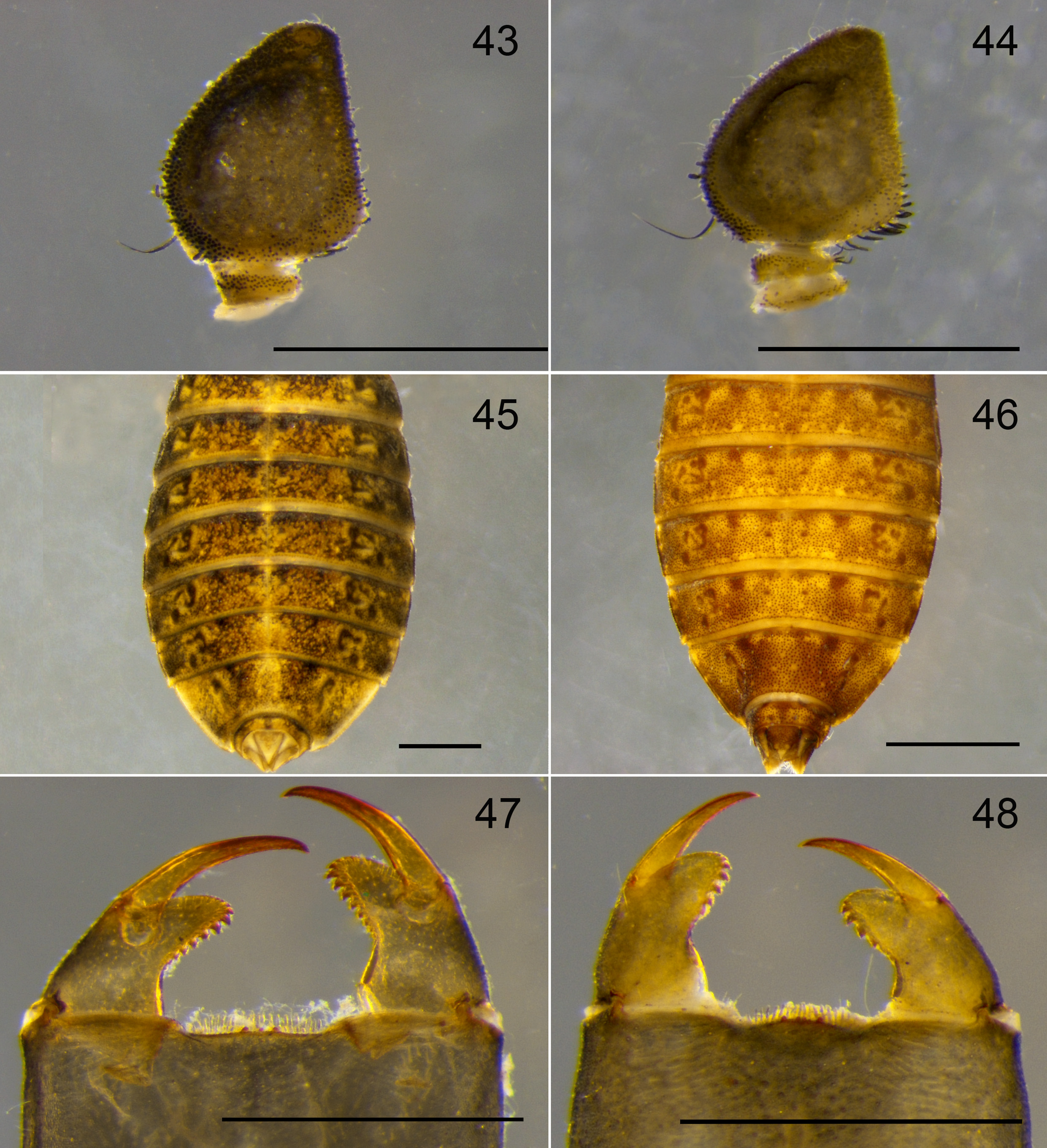
Diagnosis of larvae: Among the Southeast Asian species, the larvae of S. lawrenceae and S. malayanus are described (Yang & Davies 1996, Sasamoto 2001), whereas those of S. annamensis and S. delicatus are still unknown. The larva of S. thongphaphumensis differs from known species by the presence of palpal teeth and seta on the third antennae. The third antennal segment in S. thongphaphumensis is right-angled, triangular; the inner side has many short, flat setae, the outer side has a long seta; there are 5–8 palpal teeth, and the abdomen has a pair of black middorsal spots on S2–10. ( Figs 44, 46, 48 View FIGURES 43–48 ). The larva of S. malayanus has a small number of short, flat setae on the inner side and a long setae on the outer side of the third antennal segment, there are 7–10 palpal teeth, and the abdomen lacks the black mid-dorsal spot ( Figs 43, 45, 47 View FIGURES 43–48 ). Conversely, the larva of S. lawrenceae has the third antennal segment with dense flat seta on the inner side and without long setae on the outer side, and there are 7 palpal teeth.
Habitat and Ecology. The larvae of S. thongphaphumensis occur in headwater streams, where they are found in the sandy gravel substrate ( Figs 15–16 View FIGURES 15–17 ). The stream bed was mostly composed of boulders (5%), cobbles (30%), gravel (30%), sand (30%) and leaf litter (5%). During the odonate larval sampling, environmental and physicochemical parameters were measured. The streams ranged from 5.52–6.90 m in width and 15–16.67 cm in depth, the water velocity was about 0.33–0.63 m /sec, air temperature ranged from 22.50–30.00°C, the water temperature ranged from 21.74–27.29°C, pH ranged from 6.22–7.95, dissolved oxygen (DO) ranged from 6.2–7.12 mg /L, total dissolved solids (TDS) ranged from 159.67–232.67 mg /L, and conductivity from 252.67–372.67 µS/cm.


No known copyright restrictions apply. See Agosti, D., Egloff, W., 2009. Taxonomic information exchange and copyright: the Plazi approach. BMC Research Notes 2009, 2:53 for further explanation.
|
Kingdom |
|
|
Phylum |
|
|
Class |
|
|
Order |
|
|
Family |
|
|
Genus |