
Tipula (Vestiplex) levisoni Starkevich & Podenas, 2022
|
publication ID |
https://doi.org/ 10.11646/zootaxa.5115.1.9 |
|
publication LSID |
lsid:zoobank.org:pub:5FF52BDB-1928-4C4A-ACA2-47B5B869A611 |
|
DOI |
https://doi.org/10.5281/zenodo.6347071 |
|
persistent identifier |
https://treatment.plazi.org/id/7D95A140-6F3B-4577-8862-F64D35DBDD8F |
|
taxon LSID |
lsid:zoobank.org:act:7D95A140-6F3B-4577-8862-F64D35DBDD8F |
|
treatment provided by |
Plazi |
|
scientific name |
Tipula (Vestiplex) levisoni Starkevich & Podenas |
| status |
sp. nov. |
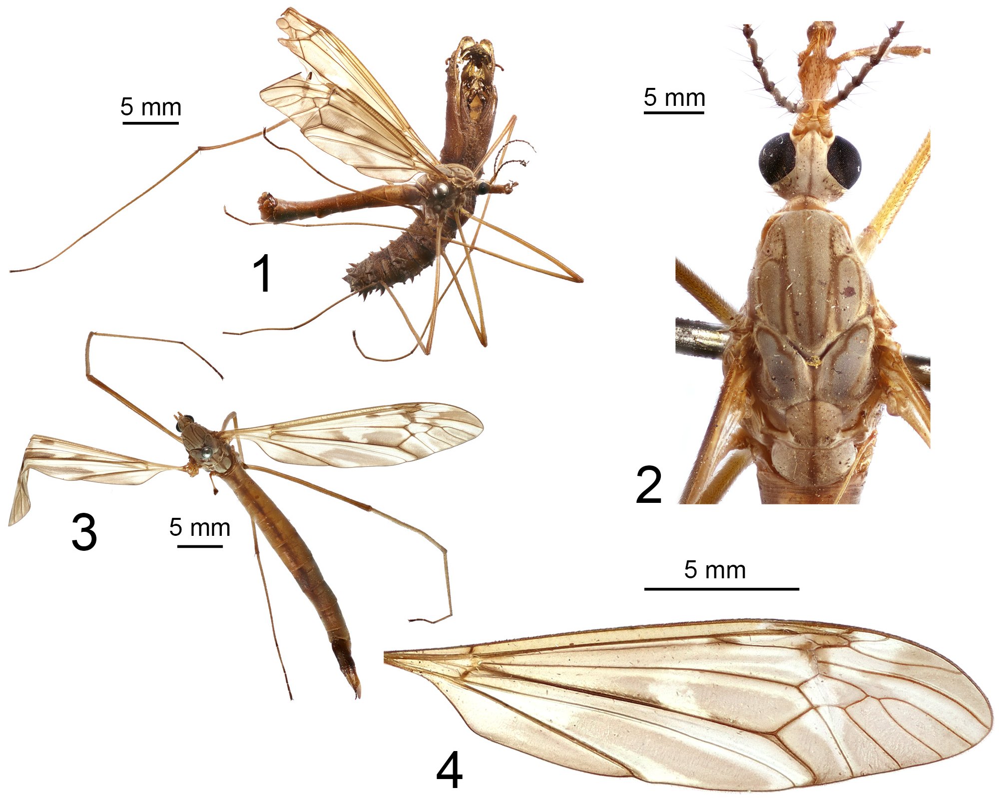
Tipula (Vestiplex) levisoni Starkevich & Podenas sp. nov.
( Figs 1–20 View FIGURES 1–4 View FIGURES 5–14 View FIGURES 15–20 )
Type material. HOLOTYPE ♂, labelled: INDIA, Kashmir , Gulmarg, 29.vi.1931, Fletcher coll., B.M.1932.13 [larval exuvium on same pin, male genitalia slide No. PS 0375m] ( BMNH) . PARATYPES: INDIA, same data as holotype, 26.vii.1931 [genitalia slide No. PS 0376f] (1 ♀, BMNH) ; same data as holotype, 9.vii.1931 (1 ♀, BMNH) . NEPAL, Baitadi , Tinkar Khola, 13,000 ft. [3962 m], 5.vii.1953. J.B. Tyson; W. Nepal Expedition, B.M.1953-592 (1 ♂, BMNH) .
Diagnosis. Tipula (V.) levisoni sp. nov. can be recognized by its brownish-yellow body, elongated antenna which reaches the base of the abdomen and the epandrium forming a large, crescent in outline, saucer-shaped plate with deep posterior U-shaped notch bordered by denticles on either side. The gonocoxite is unarmed with a rectangular ventromesal part. Females are large with an elongated abdomen and a body length longer than 30 mm. Cercus slender with upturned tip and ventral margin distally with small, distinct serrations. The hypovalva comprises a pale, elongated, slender filament. The median incision between the hypovalvae is deeper than the posterior margin of sternite 8, with the lateral incision deep and narrow.
Description. Male. Body length 18.9–21.0 mm, wing length 20.6–21.2 mm (N=2). General body colour brownish yellow ( Figs 1, 2 View FIGURES 1–4 ).
Head. Vertex and occiput grey with narrow brown median line ( Fig. 2 View FIGURES 1–4 ). Rostrum brownish yellow, dorsally thinly dusted with grey pruinescence. Nasus distinct. Antenna 13-segmented, when bent backward almost reaching base of abdomen. Scape and pedicel yellow, first flagellar segment yellow, brown at tip, remaining flagellomeres brown. Each flagellomere, except first, with basal enlargement with small incision and narrowing apex; below narrowing, with additional narrowing at apex, making each segment slightly sinuous. Apical flagellomere small. Longest verticils approximately same length as their corresponding segments. Palpus with basal segment pale, second segment pale with brown surface, last segments dark brown.
Thorax. Pronotum yellowish brown with brown median line. Prescutum and presutural scutum with four greyish brown stripes narrowly bordered with brown ( Fig. 2 View FIGURES 1–4 ). Medial pair of stripes fused in basal half, distally narrowly separated. Interspaces yellowish grey, anteriorly with small brown dots. Postsutural scutum with lobes each having greenish-brown nearly oval spot bordered with brown. Scutellum yellowish brown, mediotergite brown, thinly dusted, both sclerites with brown medial line. Pleura yellowish brown, thinly dusted with grey pruinescence. Coxae yellow, thinly dusted, trochanter yellow, femora and tibiae yellow, narrowly darkened at tip; first tarsal segment yellow, darkened at tip, remaining tarsal segments dark brown. Tarsal claw without tooth. Wing patterned with brown ( Fig. 4 View FIGURES 1–4 ). Halter pale with brown knob.
Abdomen. Brownish yellow, terminal segments brown. Tergites with broad brown median and lateral stripes.
Hypopygium ( Figs 5–14 View FIGURES 5–14 ). Brown, broader than abdomen. Epandrium forming large, concave, sclerotised saucer-shaped plate, crescent-shaped in outline ( Figs 5–7 View FIGURES 5–14 ). Main body of plate brown with blackened rim. Posterior margin of plate broadly emarginated medially with deep U-shaped notch, laterally bordered by black denticles on either side; rest of margin toothed. Lateral angle of plate broadly obtuse. Anterior and lateral portions of plate raised into narrow sclerotised border, posterolaterally terminating in large, black and obtuse teeth. Gonocoxite unarmed, ventromesal part rectangular in shape ( Fig. 8 View FIGURES 5–14 ). Outer gonostylus flattened, leaf-shaped ( Fig. 9 View FIGURES 5–14 ). Inner gonostylus, large, curved, claw-shaped with single black, large mid-dorsal tooth. Beak extended into blackened, narrowed rostrum with indistinctly split tip ( Fig. 10 View FIGURES 5–14 ). Aedeagal guide inshape of narrow, elongated tube, base with dorsal flange narrow and elongated, membranous at base, ventral flange flattened; distal margin bent inward forming two microscopic hooks ( Fig. 11 View FIGURES 5–14 ). Gonocoxal fragment large with lateral and medial sclerites well-developed ( Fig. 12 View FIGURES 5–14 ). Medial sclerites fused, anterior apodeme distinct, flattened and narrowed in middle; posterior part broadly flattened, expanded at base, distal parts flattened and arched. Lateral sclerites U-shaped. Sperm pump with small and flattened central vesicle ( Fig. 13 View FIGURES 5–14 ). Compressor apodeme with broad median emargination. Posterior immovable apodeme longer than compressor apodeme, large and extended laterally into broad plate, medially with darker ridge. Anterior immovable apodeme nearly triangular, narrowed at lateral margin. Aedeagus with distal part ventrally membranous ( Fig. 14 View FIGURES 5–14 ).
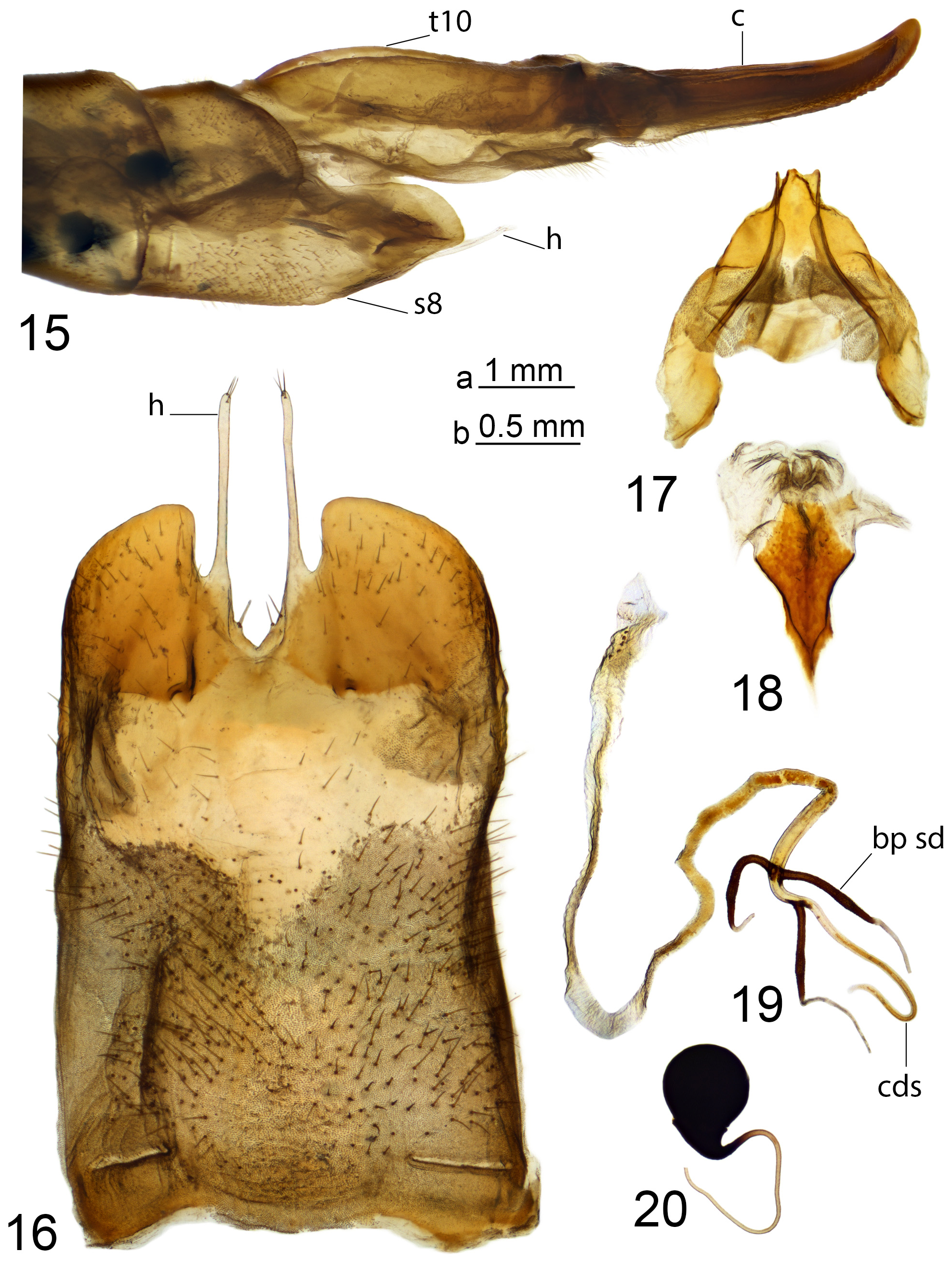
Female. Body length 33.8–38.2 mm, wing length 25.9–27.4 mm (N=2). Generally similar to male, but abdomen more elongate ( Fig. 3 View FIGURES 1–4 ).
Female terminalia ( Figs 15–20 View FIGURES 15–20 ). Tergite 10 shiny brown. Cercus brownish yellow, about as long as tergite 10, slender with up-turned tip ( Fig. 15 View FIGURES 15–20 ). Ventral margin distally with small, distinct serrations. Hypovalva pale, elongated, filamentous, with short trichia at tip ( Fig. 15 View FIGURES 15–20 ). Median incision between hypovalvae deeper than posterior margin of sternite 8. Lateral incision deep and narrow, about three times as deep as maximum width. Posterolateral angle of sternite 8 rounded. Sternite 9 with posterior part shovel-shaped, margin with lateral incisions, medially forming groove; anterior parts flattened ( Fig. 17 View FIGURES 15–20 ). Furca flattened, base narrow ( Fig. 18 View FIGURES 15–20 ). Bursa copulatrix with spermathecal ducts sclerotised at the base, in shape of curved, slender, dark brown sticks ( Fig. 19 View FIGURES 15–20 ). Cul-de-sac of bursa copulatrix curved. Spermatheca pear-shaped ( Fig. 20 View FIGURES 15–20 ).
Etymology. The new species is named after Levison James Wood (1982–), a British army officer, explorer and writer, best known for his remarkable expeditions in the Himalayas, Central America, the Arabian Peninsula and walking the entire length of the River Nile.
Distribution. India (Jammu and Kashmir), Nepal (Sudurpashchim Province).
Remarks. Tipula (V.) levisoni sp. nov. is assigned here as a member of T. (V.) leucoprocta species group proposed by Savchenko (1960, 1964) and as discussed by Starkevich et al. (2020). Males of the leucoprocta species group are distinguished by an unarmed gonocoxite, the epandrium having a large sclerotised plate with an extensive saucer with a raised border, posterolaterally terminating in teeth and a remarkably large gonocoxal fragment. In females, the bursa copulatrix and basal part of the spermathecal ducts are sclerotised. Two members of the leucoprocta species group are known from the Western Himalayas, T. (V.) tanycera ( Pakistan) and T. (V.) mitchelli ( India) .
Tipula (V.) levisoni sp. nov. is closest to T. (V.) tanycera based on body colouration and the shape of the hypopygium. Both species are characterized by a crescent-shaped epandrium, inner gonostylus with middorsal tooth, slender aedeagal guide and the shape of the gonocoxal fragment. Females are characterized by sternite 8 with deep lateral incisions.
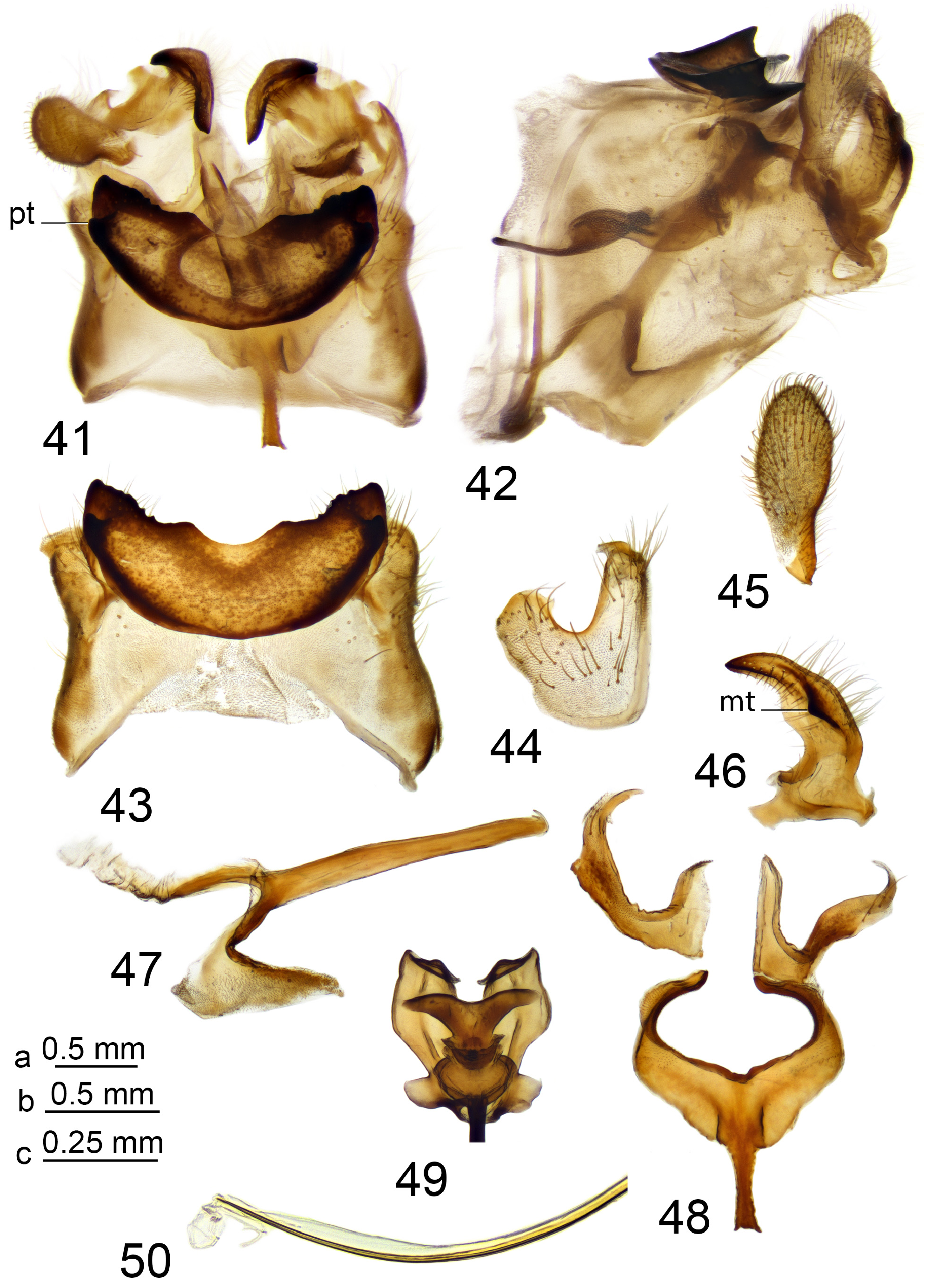
Males can be separated by their antennae which reaches the base of the abdomen in T. (V.) levisoni sp. nov. while in T. (V.) tanycera the antenna is longer,almost reaching the middle of the abdomen. They can also be separated by the following details of the epandrium: T. (V.) levisoni sp. nov. has the posterior margin with a deep median notch and a large posterolateral tooth ( Fig. 7 View FIGURES 5–14 ), while in T. (V.) tanycera the median notch is shallower and has a distinctly smaller posterolateral tooth ( Fig. 43 View FIGURES 41–50 ). The outer gonostylus of T. (V.) levisoni sp. nov. is flattened, leaf-shaped ( Fig. 9 View FIGURES 5–14 ), while in T. (V.) tanycera it is nearly oval ( Fig. 45 View FIGURES 41–50 ). The inner gonostylus bears a middorsal tooth with a broadly expanded base in T. (V.) levisoni sp. nov. ( Fig. 10 View FIGURES 5–14 ). Tipula (V.) tanycera has a middorsal tooth with a distinctly less expanded base ( Fig. 46 View FIGURES 41–50 ).
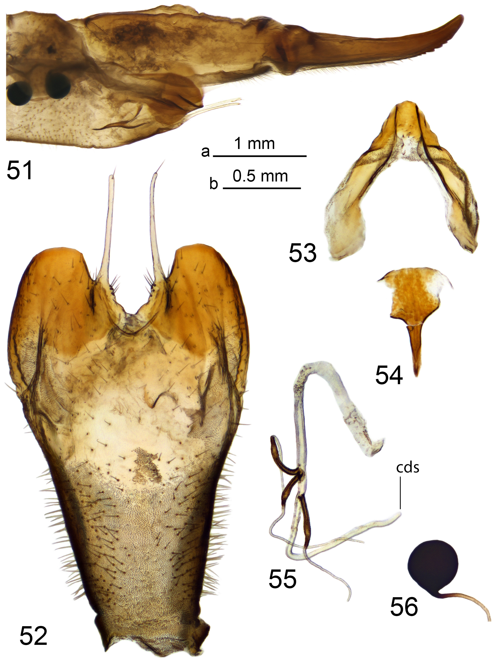
Females can be separated by their body size which exceeds 30 mm in T. (V.) levisoni sp. nov. and is less than 20 mm in T. (V.) tanycera . The cercus has small serrations and a nearly rounded tip in T. (V.) levisoni sp. nov. ( Fig. 15 View FIGURES 15–20 ), while in T. (V.) tanycera the serrations are distinct on the distal end and the cercus tip is pointed ( Fig. 51 View FIGURES 51–56 ).
Another regional species related to T. (V.) levisoni sp. nov. and T. (V.) tanycera is T. (V.) mitchelli , which can be separated by a rectangular epandrium and the inner gonostylus lacking a middorsal tooth.

No known copyright restrictions apply. See Agosti, D., Egloff, W., 2009. Taxonomic information exchange and copyright: the Plazi approach. BMC Research Notes 2009, 2:53 for further explanation.