
Caliroa
|
publication ID |
https://doi.org/ 10.11646/zootaxa.4768.3.1 |
|
publication LSID |
lsid:zoobank.org:pub:C8036F69-F881-4727-96E7-C78AA6C7F920 |
|
DOI |
https://doi.org/10.5281/zenodo.3794885 |
|
persistent identifier |
https://treatment.plazi.org/id/03B387A9-FFD1-FF84-1DC6-24EC37C4FE3C |
|
treatment provided by |
Plazi |
|
scientific name |
Caliroa |
| status |
|
Key to Japanese species of Caliroa View in CoL View at ENA
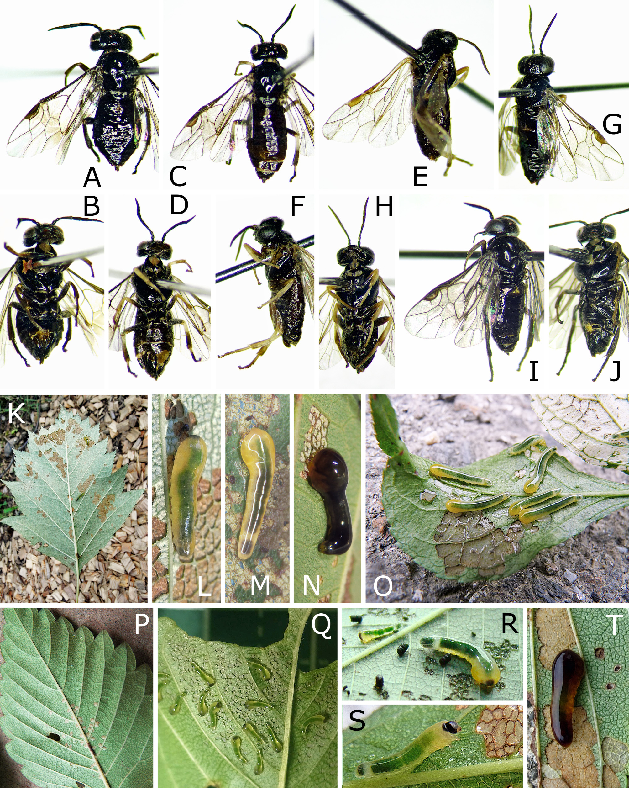
Adults
1 Hind leg dark brown to black, without pale areas, at most narrowly brown on apex of femur ( Fig. 1A, B, I, J View FIGURE 1 )............ 2
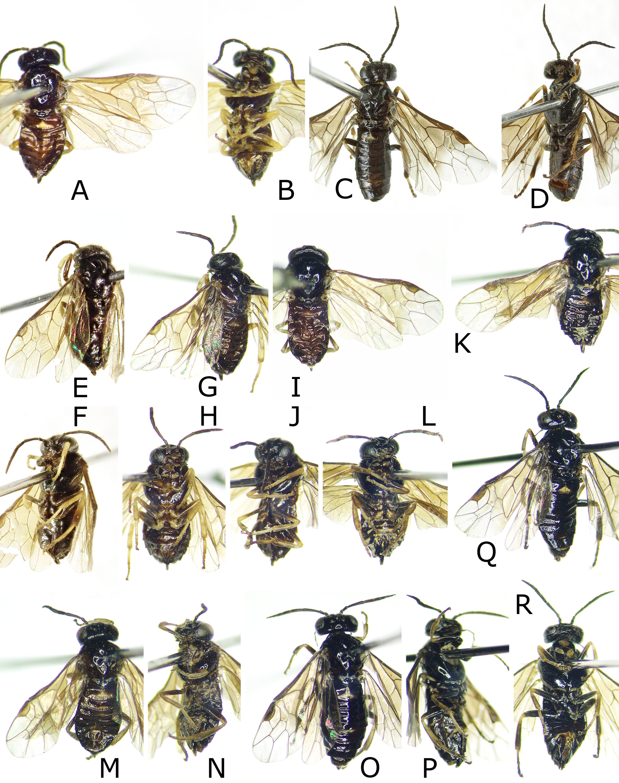
- Hind leg with white, yellow or yellow brown areas ( Figs 1 View FIGURE 1 C–H, 2A–R).......................................... 3
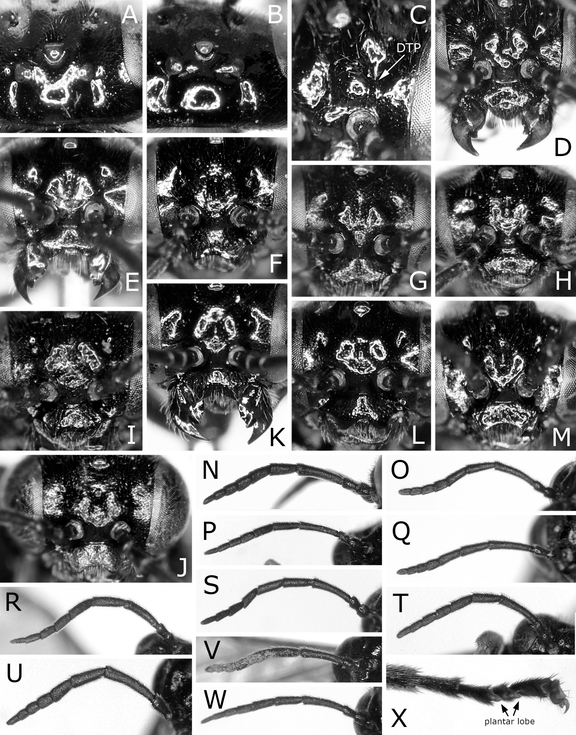
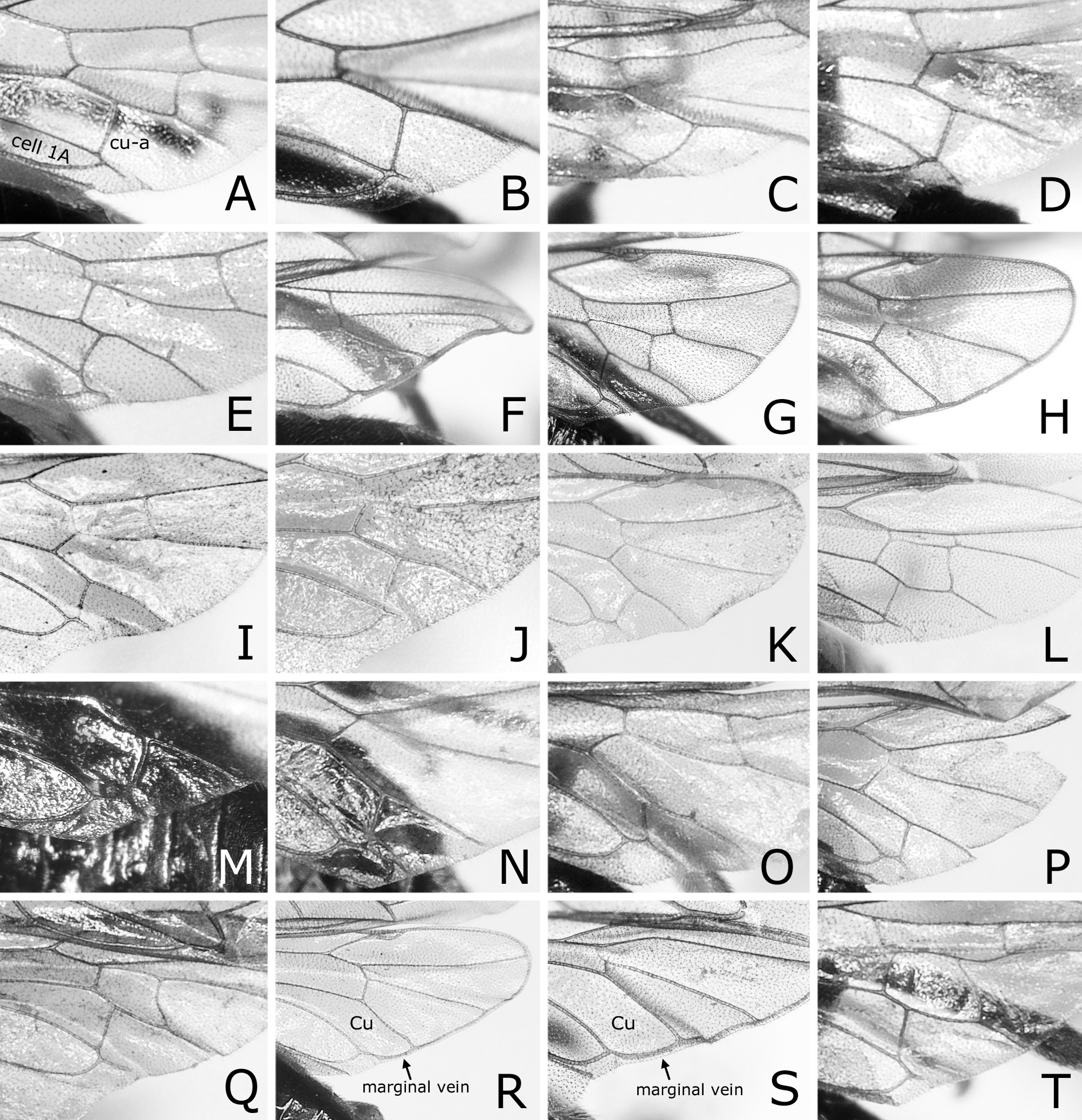
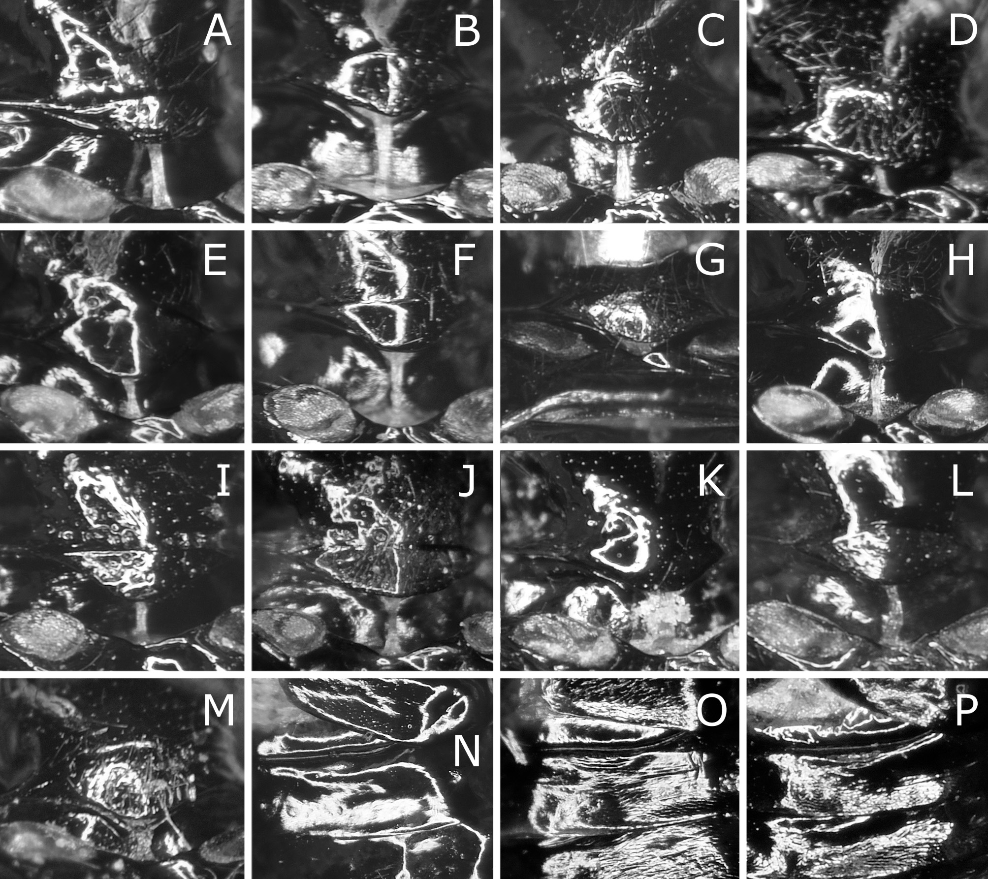
2(1) Postocellar area 1.3–1.5 × as wide as length behind lateral ocellus, with anterior groove laterally ( Fig. 3B View FIGURE 3 ). Forewing with joint of vein Rs and crossvein 2r-rs usually located near or at joint of vein Rs and crossvein 3r-m ( Fig. 1A View FIGURE 1 ). Male hind wing without marginal vein ( Fig. 4C View FIGURE 4 )...................................................................... C. cerasi , ♀ ♂
- Postocellar area 2.1–2.2 × as wide as length behind lateral ocellus, without anterior groove as in Fig. 3A View FIGURE 3 . Forewing with joint of vein Rs and crossvein 2r-rs located far basal to joint of vein Rs and crossvein 3r-m ( Fig. 1I View FIGURE 1 ). Male hind wing with marginal vein ( Fig. 4H View FIGURE 4 ).............................................................................. C. ibukii , ♀ ♂
3(1) Wings almost colorless, sometimes slightly blackish on cells C and Sc and below stigma in forewing ( Fig. 1C, E, G View FIGURE 1 )...... 4
- Wings distinctly brownish or blackish on basal two thirds ( Fig. 2 View FIGURE 2 )............................................... 6
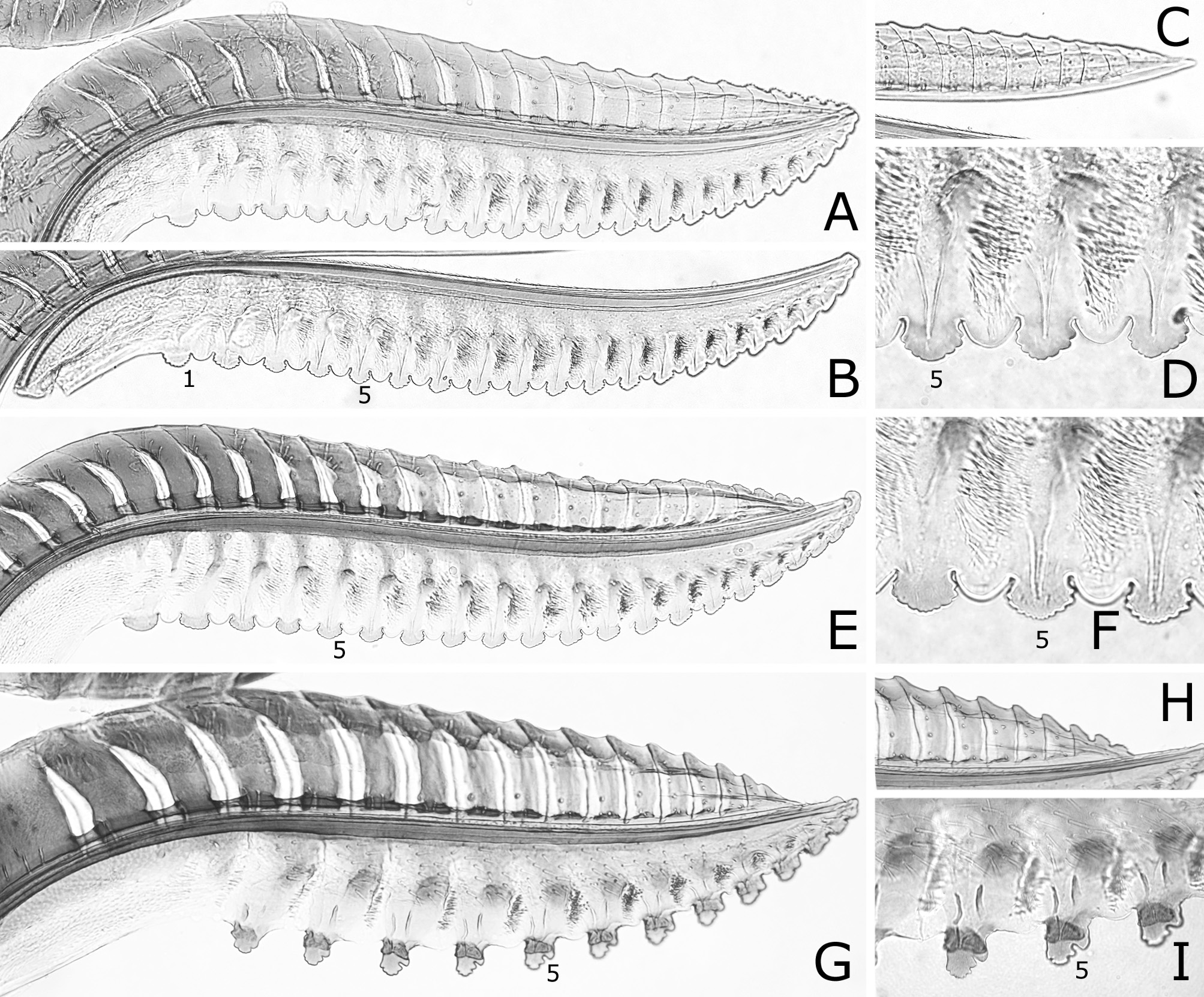
4(3) Forewing slightly blackish on cells C and Sc and usually blackish below stigma ( Fig. 1C View FIGURE 1 ). Lancet with middle serrulae nearly triangular in outline, with acute teeth ( Fig. 6I View FIGURE 6 ; fig. 23 in Lacourt, 2002)........................................... 5
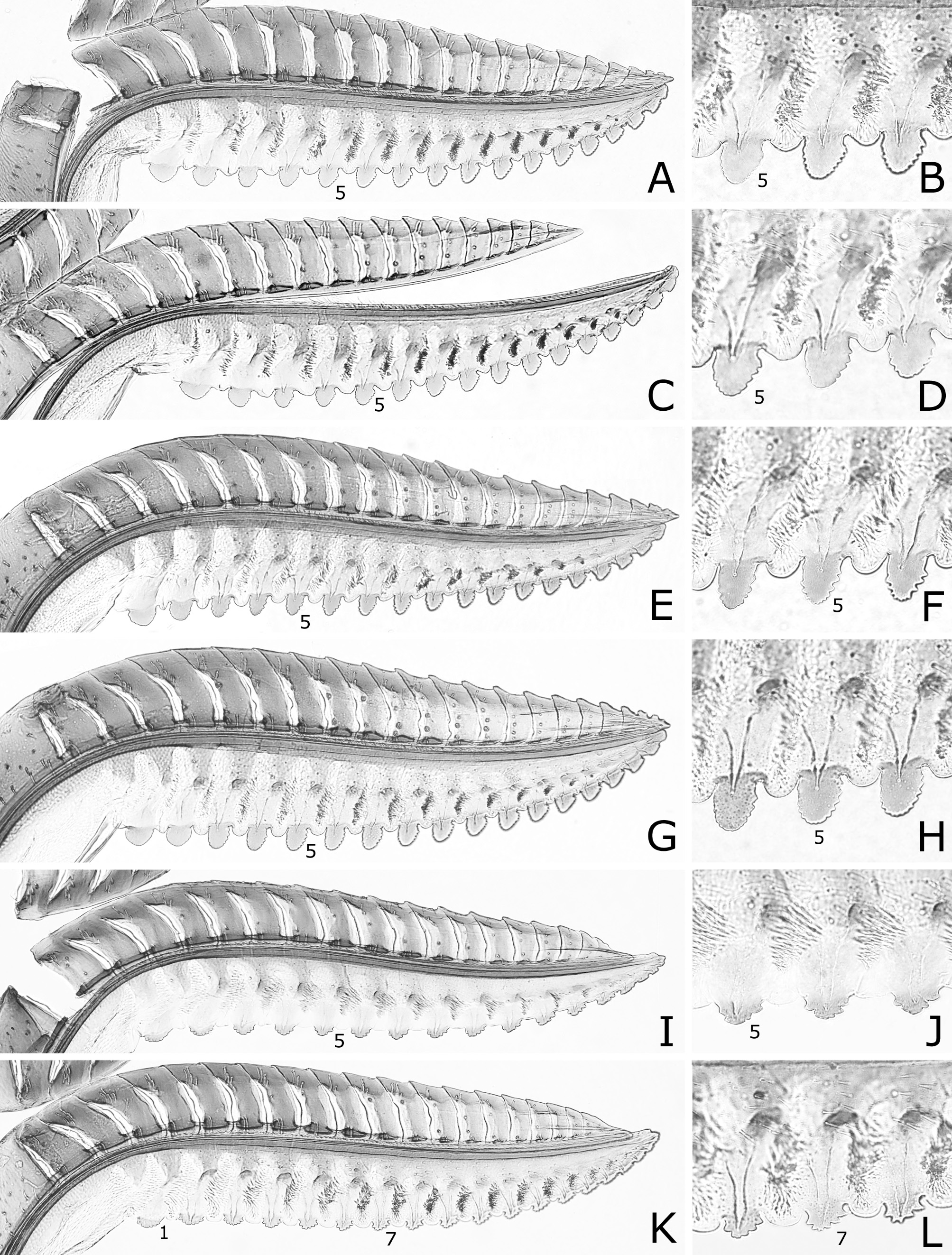
- Forewing almost colorless all over ( Fig. 1E, G View FIGURE 1 ). Lancet with middle serrulae nearly wide oval in outline, with rounded teeth ( Fig. 7D, F View FIGURE 7 ). [Hind wing with joint of vein 1A and crossvein cu-a located basal to apex of cell 1A in female ( Fig. 4E View FIGURE 4 ), with marginal vein in male ( Fig. 4F View FIGURE 4 ).]............................................................... C. oishii , ♀ ♂
5(4) In female, hind wing with joint of vein 1A and crossvein cu-a located apical to apex of cell 1A ( Fig. 4D View FIGURE 4 ); lancet with ctenidia (= groups of setae) of middle annuli extending to or near ventral margin of lancet ( Fig. 6I View FIGURE 6 ).... C. bibaiensis , ♀ [♂ unknown]
- In female, hind wing with joint of vein 1A and crossvein cu-a located basal to apex of cell 1A (fig. 5 in Lacourt, 2002; photos in Taeger et al., 2018); lancet with ctenidia of middle annuli widely separated from ventral margin of lancet (fig. 23 in Lacourt, 2002). [In male, hind wing with marginal vein (fig. 2 in Lacourt, 2002).].............................. C. varipes , ♀ ♂
6(3) In female and male, hind wing with joint of vein 1A and crossvein cu-a located apical to apex of cell 1A and without marginal vein ( Fig. 4I, K View FIGURE 4 , M–P)................................................................................. 7
- In female, hind wing with joint of vein 1A and crossvein cu-a located at or basal to apex of cell 1A and without marginal vein ( Fig. 4J, L, Q, T View FIGURE 4 ). In male, hind wing as in female, or without crossvein cu-a and with marginal vein ( Fig. 4R, S View FIGURE 4 )........ 10
7(6) Mesoscutellar appendage mostly setose ( Fig. 5I, J View FIGURE 5 ) or setose on wide medial area (Fig. K). Apical four flagellomeres combined 0.9–1.4 × as long as first flagellomere ( Fig. 3 View FIGURE 3 T–V)........................................................... 8
- Mesoscutellar appendage only with several setae posterolaterally ( Fig. 5E View FIGURE 5 ) or entirely glabrous. Apical four flagellomeres combined 1.6–2.1 × as long as first flagellomere ( Fig. 3R View FIGURE 3 )...................................... C. vaccini , ♀ ♂ part
8(7) Malar space without setae. Lancet with deep serrulae ( Fig. 9A, B View FIGURE 9 , 10 View FIGURE 10 A–H)........................................ 9
- Malar space with row of setae (in male often partly glabrous). Lancet with very shallow serrulae ( Fig. 9 View FIGURE 9 C–F). [Clypeus with depth of emargination 0.1–0.2 × median length of clypeus ( Fig. 3J View FIGURE 3 ). Apical four flagellomeres combined 0.9–1.0 × as long as first flagellomere ( Fig. 3U View FIGURE 3 ). Mesoscutellar appendage mostly setose ( Fig. 5J View FIGURE 5 )]....................... C. staphyleae , ♀ ♂
9(8) Clypeus with depth of emargination 0.2 × median length of clypeus ( Fig. 3I View FIGURE 3 ). Apical four flagellomeres combined 1.4 × as long as first flagellomere ( Fig. 3T View FIGURE 3 ). Mesoscutellar appendage mostly setose ( Fig. 5I View FIGURE 5 ). Lancet with 14 serrulae ( Fig. 9A, B View FIGURE 9 ).................................................................................... C. aizankei , ♀ [♂ unknown]
- Clypeus with depth of emargination 0.3–0.5 × median length of clypeus ( Fig. 3K, L View FIGURE 3 ). Apical four flagellomeres combined 0.9–1.2 × as long as first flagellomere ( Fig. 3V View FIGURE 3 ). Mesoscutellar appendage setose medially ( Fig. 5K View FIGURE 5 ). Lancet with 17–18 serrulae ( Fig. 10 View FIGURE 10 A–H)............................................................. C. zelkovae , ♀ few specimens
10(6) Apical four flagellomeres combined 1.1–1.2 × as long as first flagellomere (fig. 2H in Hara, 2011). Mesoscutellar appendage entirely glabrous ( Fig. 5H View FIGURE 5 ). Male hind wing without marginal vein (fig. 2M in Hara, 2011).................. C. nara , ♀ ♂
- Combination of character states not as above.............................................................. 11
11(10) Apical four flagellomeres combined 1.5–2.1 × as long as first flagellomere ( Fig. 3R, S View FIGURE 3 ). Mesoscutellar appendage entirely glabrous or narrowly setose posterolaterally ( Fig. 5E, F View FIGURE 5 ). Male hind wing without marginal vein...................... 12
- Apical four flagellomeres combined 0.9–1.3 × as long as first flagellomere ( Fig. 3V, W View FIGURE 3 ). Mesoscutellar appendage mostly setose ( Fig. 5L, M View FIGURE 5 ) or serose on wide medial area ( Fig. 5K View FIGURE 5 ). Male hind wing with marginal vein ( Fig. 4R, S View FIGURE 4 )........... 13
12(11) Hind leg predominantly yellow to yellow brown, darkened on base of coxa and apices of tibia and tarsus and sometimes on base of femur (except for trochantellus) ( Fig. 2B View FIGURE 2 ). Clypeus with depth of ventral emargination 0.2 × median length of clypeus ( Fig. 3H View FIGURE 3 )................................................................................. C. vaccini , ♀ ♂ part
- Hind leg predominantly black, yellow or white on bases of tibia and tarsus ( Fig. 2D View FIGURE 2 ). Clypeus with depth of ventral emargination 0.3–0.5 × median length of clypeus (fig. 2E in Hara, 2011)...................................... C. ouensis , ♀ ♂
13(11) Hind tibia black, yellow on basal third ( Fig. 2R View FIGURE 2 ). Lancet with ctenidia (= groups of setae) of middle annuli ventrally extending to level of base of serrula ( Fig. 10K, L View FIGURE 10 ); middle serrulae shallower than wide, with three or four posterior teeth; areas between serrulae distinctly convex. [Mesoscutellum with only inconspicuous minute punctures ( Fig. 5M View FIGURE 5 ). Mesoscutellar appendage mostly setose ( Fig. 5M View FIGURE 5 ).]............................................................. C. nire , ♀ [♂ unknown]
- Hind tibia brown yellow to brown, at most narrowly darkened apically or dorsoapically ( Fig. 2L, N, P View FIGURE 2 ). Lancet not as above ( Fig. 10 View FIGURE 10 A–J)........................................................................................ 14
14(13) Mesoscutellum usually posterolaterally with one or some relatively large punctures along posterior margin ( Fig. 5K View FIGURE 5 ). Clypeus with depth of emargination 0.3–0.5 × median length of clypeus. Lancet with middle serrulae about as deep as wide, with five or more posterior teeth ( Fig. 10 View FIGURE 10 A–H); areas between serrulae distinctly convex. Male hind wing with section of marginal vein in cell Cu (section arrowed in Fig. 4R View FIGURE 4 ) separated from wing margin in posterior half.................... C. zelkovae , ♀ ♂
- Mesoscutellum with only inconspicuous minute punctures ( Fig. 5L View FIGURE 5 ). Clypeus with depth of ventral emargination 0.2–0.3 × median length of clypeus. Lancet with middle serrulae shallower than wide, with three or four posterior teeth ( Fig. 10I, J View FIGURE 10 ; fig. 3 in Hara & Shinohara, 2013); areas between serrulae slightly convex. Male hind wing with section of marginal vein in cell Cu (section arrowed in Fig. 4S View FIGURE 4 ) separated from wing margin in posterior fourth....................... C. matsumotonis , ♀ ♂


No known copyright restrictions apply. See Agosti, D., Egloff, W., 2009. Taxonomic information exchange and copyright: the Plazi approach. BMC Research Notes 2009, 2:53 for further explanation.