
Grammacephalus Haupt, 1929
|
publication ID |
https://doi.org/10.11646/zootaxa.4688.1.3 |
|
publication LSID |
lsid:zoobank.org:pub:6A91548F-0C20-4711-950A-11ED5B007BA1 |
|
DOI |
https://doi.org/10.5281/zenodo.3691801 |
|
persistent identifier |
https://treatment.plazi.org/id/03B287C3-FF8B-FF8F-FF65-C478FA79F0B5 |
|
treatment provided by |
Plazi |
|
scientific name |
Grammacephalus Haupt |
| status |
|
Key to species of Grammacephalus Haupt View in CoL
The following species were described based on female specimens and are therefore omitted from the key: G. minabicus and G. niveimarginatus .
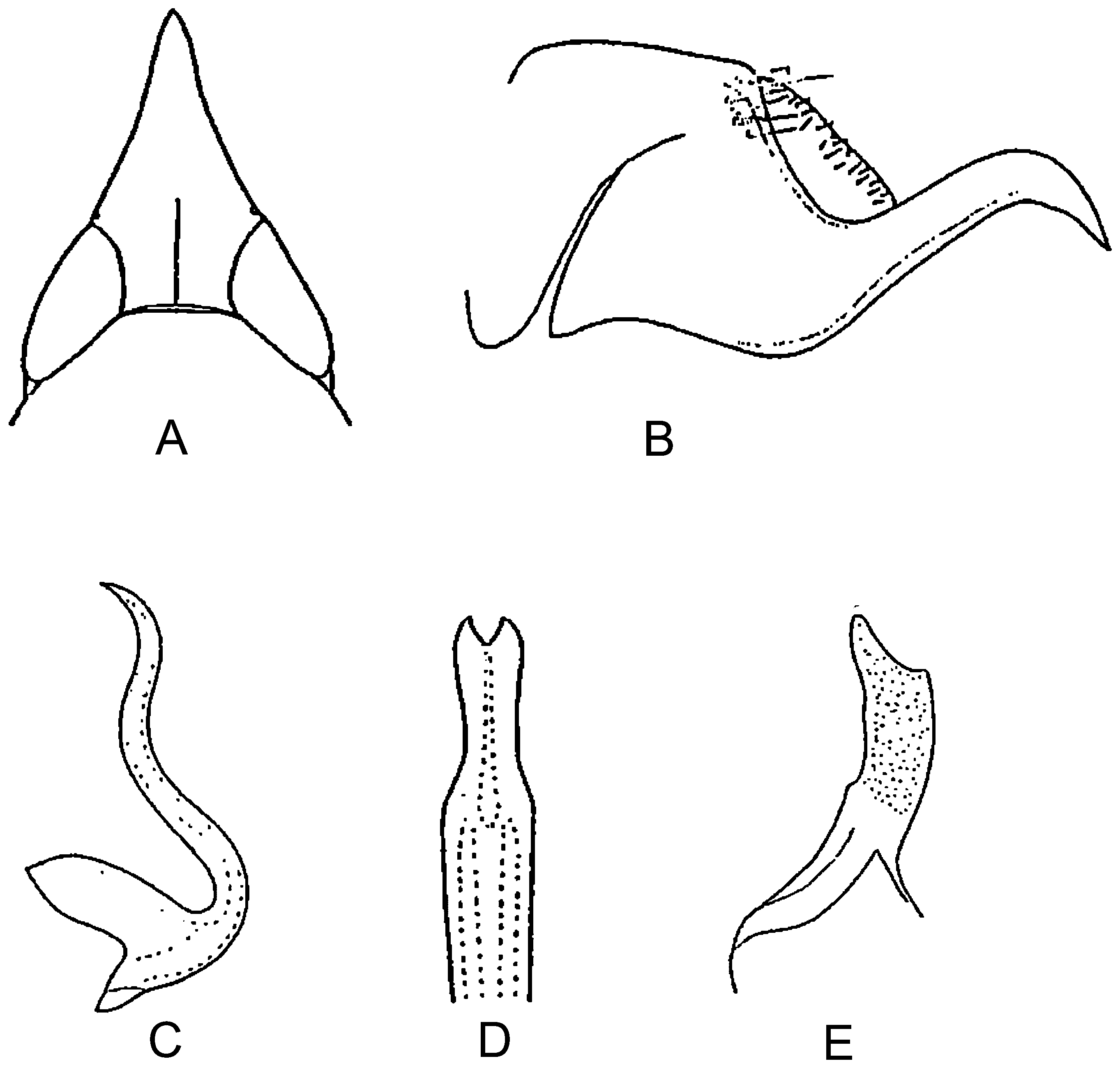
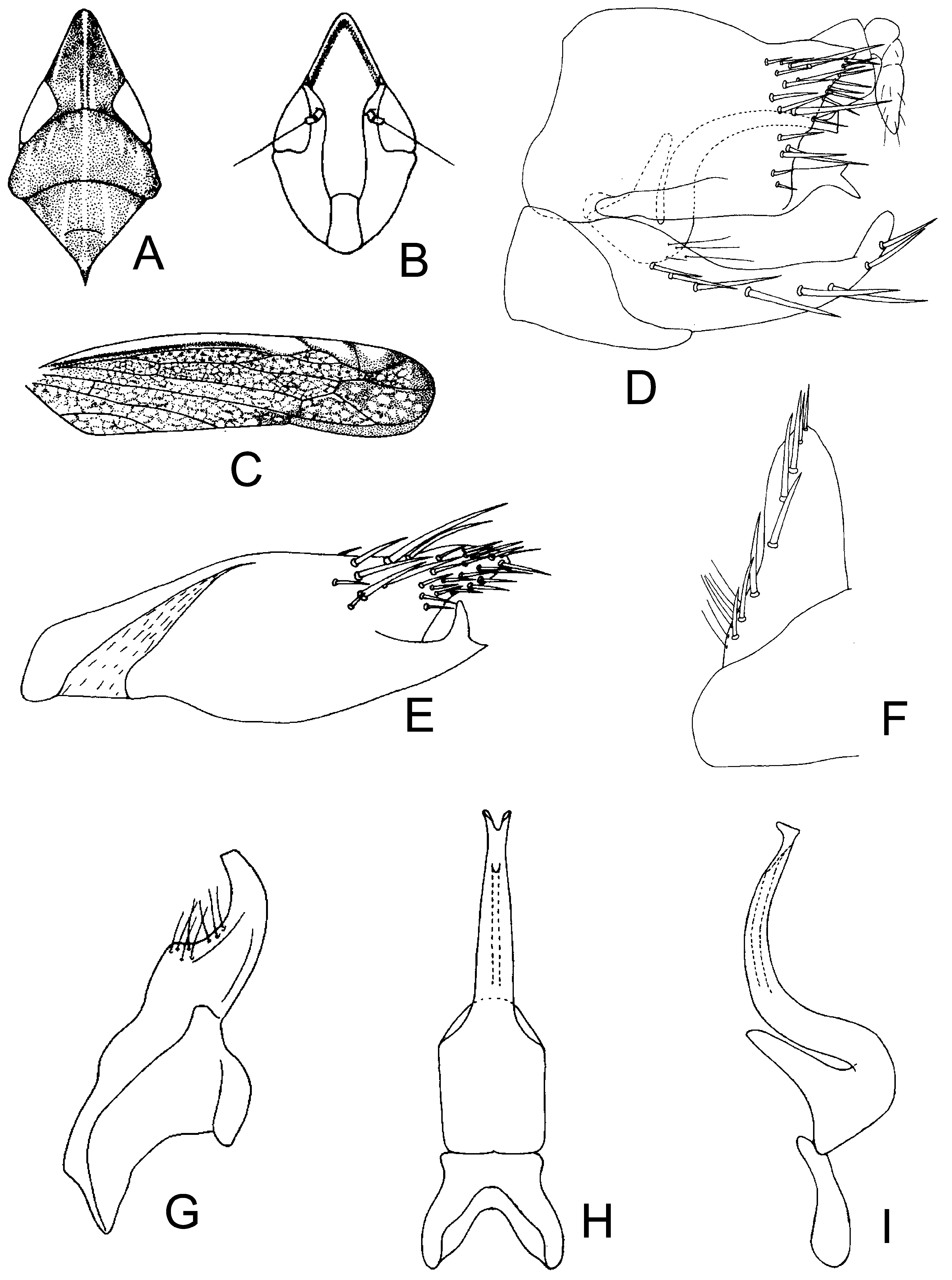
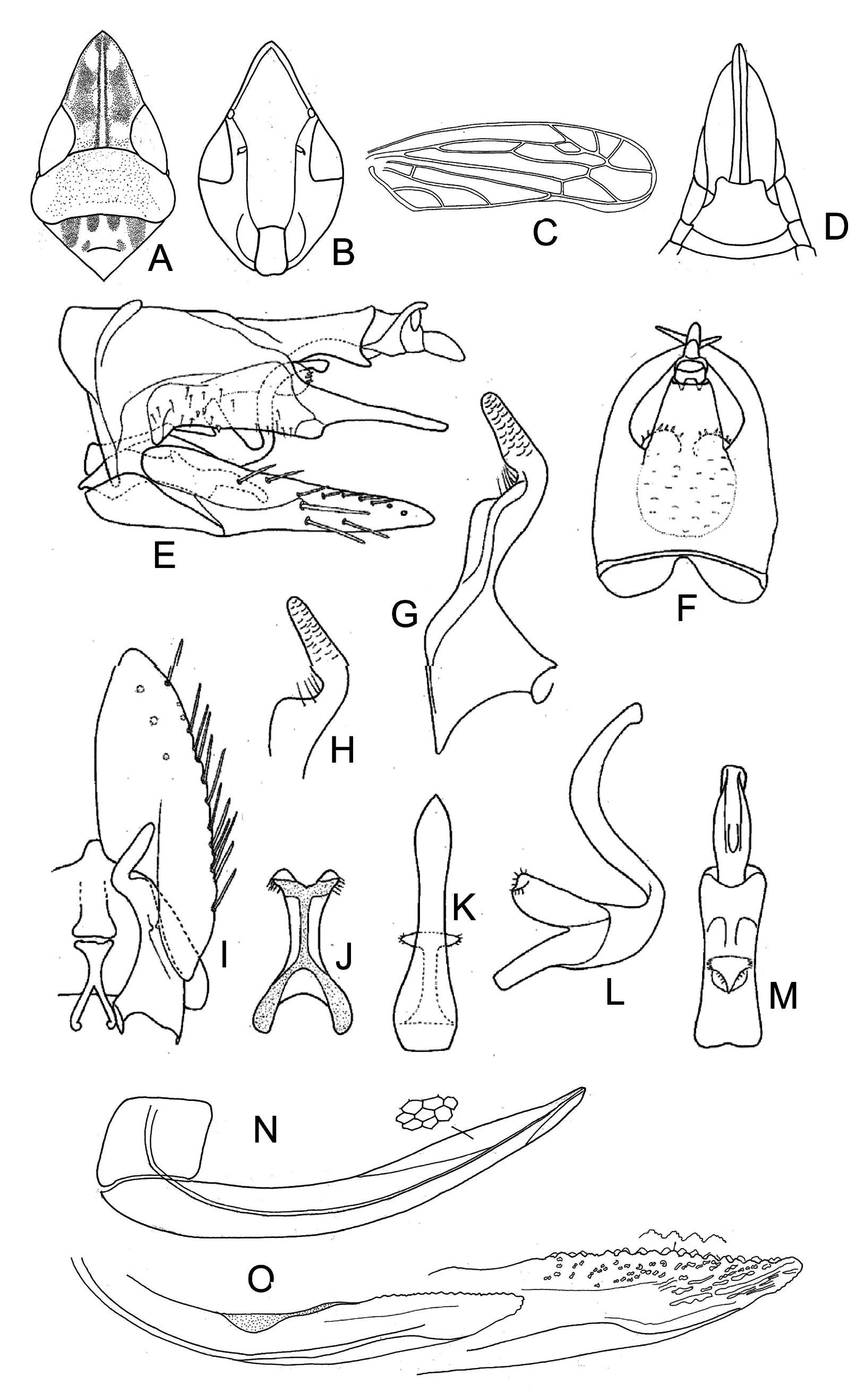
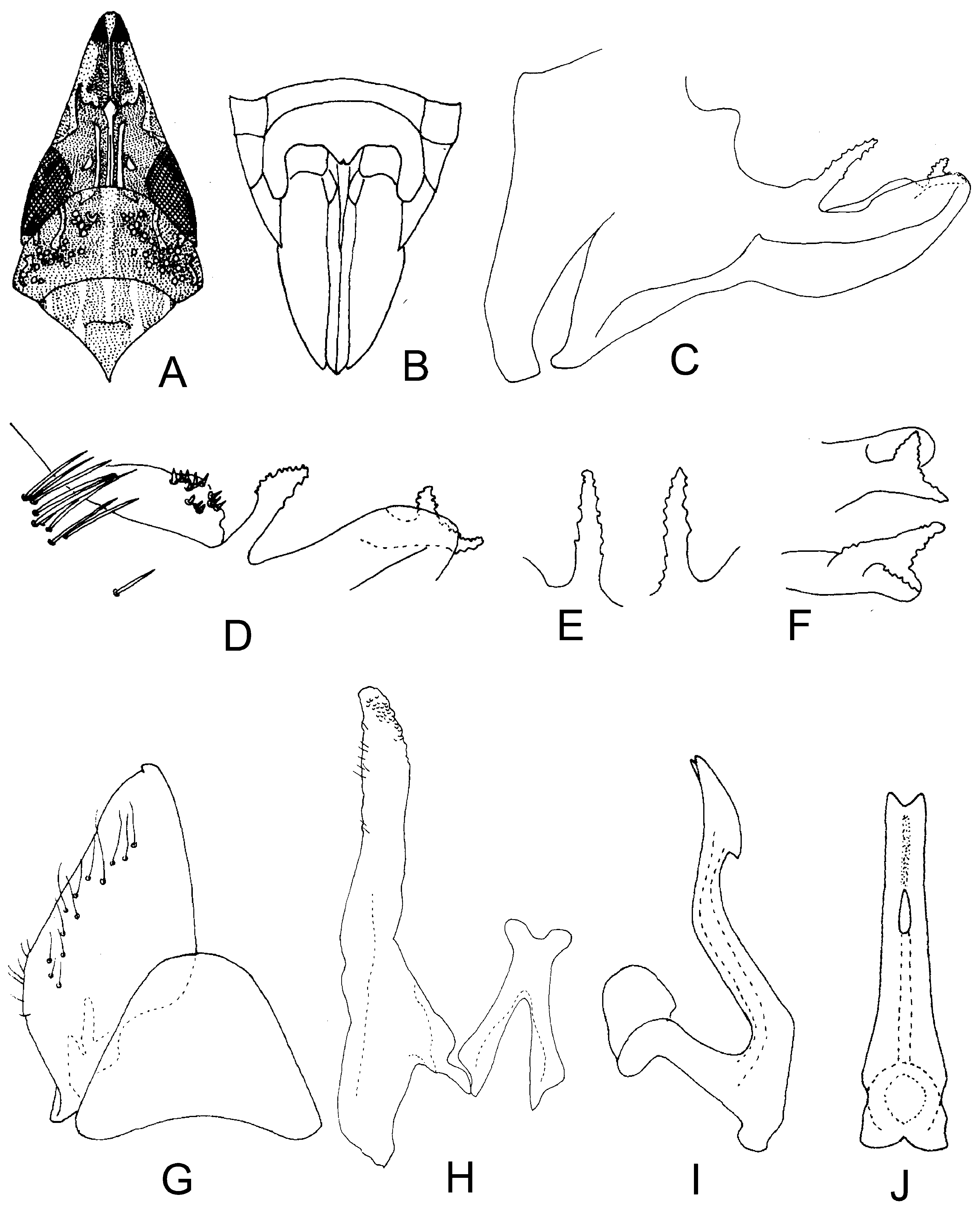
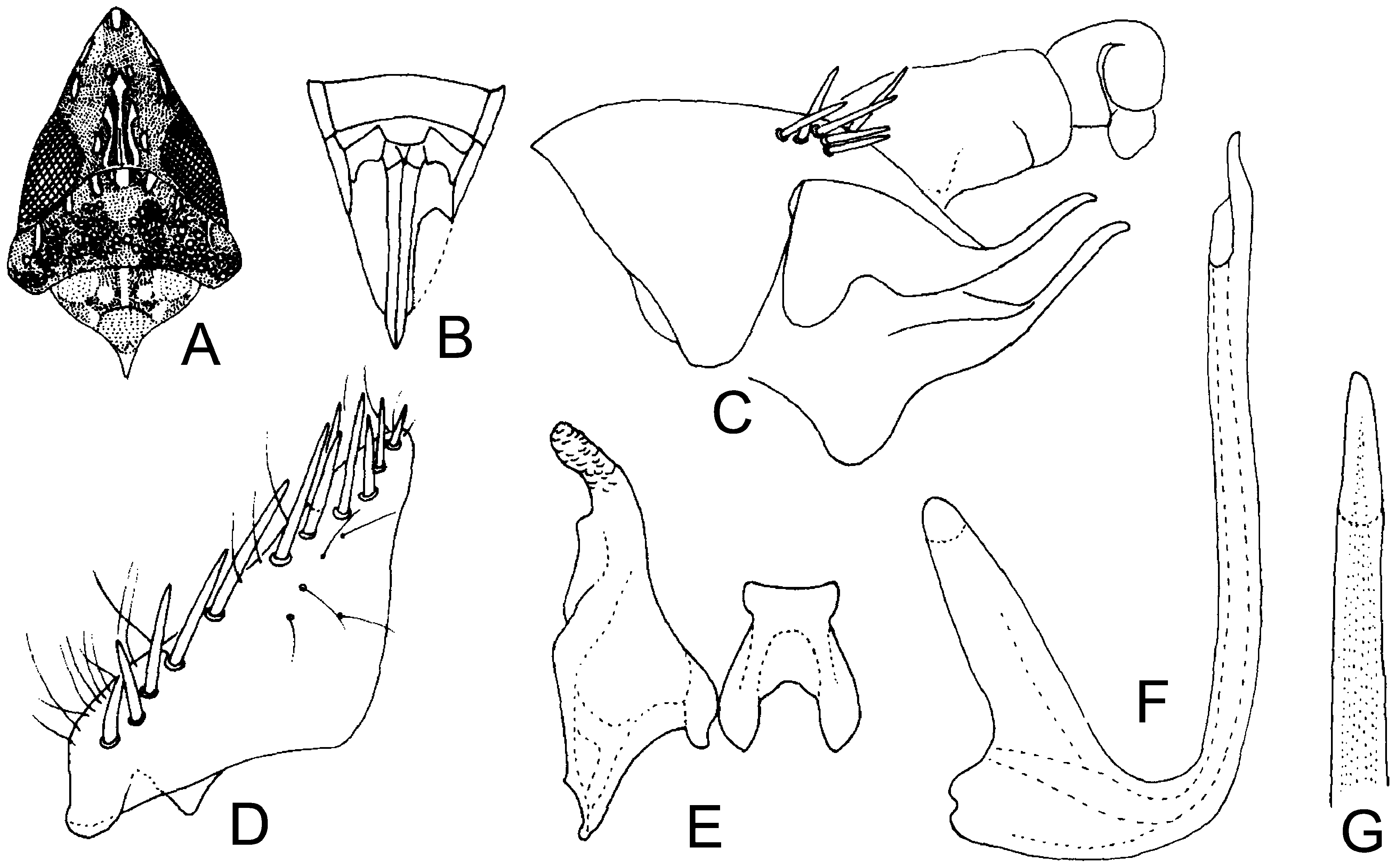
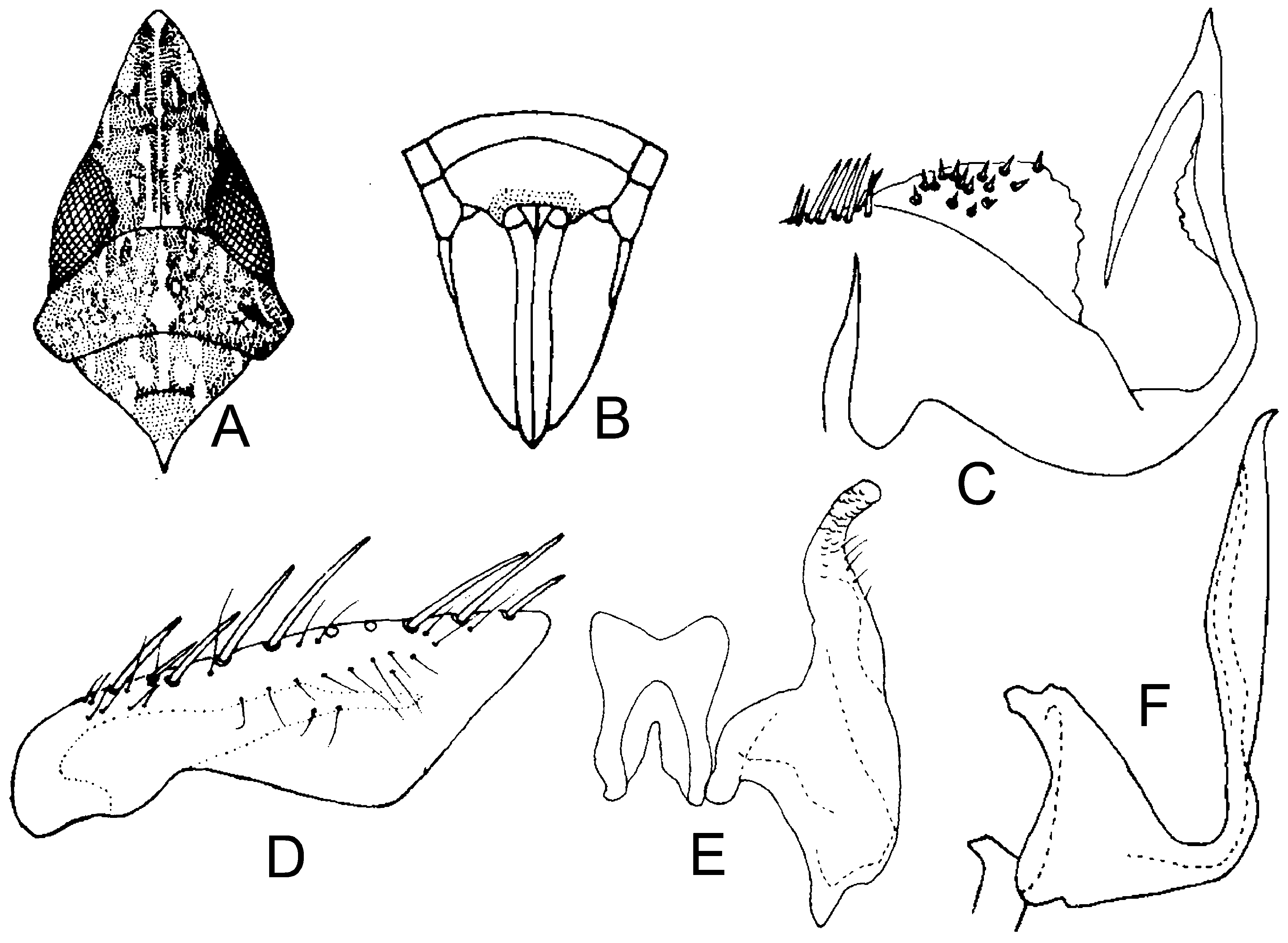
1. Male pygofer process absent ( Fig. 3B View FIGURE 3 )................................................ Grammacephalus genoicus View in CoL
- Male pygofer process present ( Figs 1B View FIGURE 1 , 2D View FIGURE 2 , 4B View FIGURE 4 , 5E View FIGURE 5 , 6 View FIGURE 6 C–F, 8B, 9C, 10C, 12A)..................................... 2
2. Male pygofer process serrate ( Figs 6 View FIGURE 6 C–F, 10C).............................................................. 3
- Male pygofer process not serrate ( Figs 1B View FIGURE 1 , 2D View FIGURE 2 , 4B View FIGURE 4 , 5E View FIGURE 5 , 8B View FIGURE 8 , 9C View FIGURE 9 , 12A View FIGURE 12 )............................................ 4
3. Apex of subgenital plate notched, dorsally upturned ( Fig. 6G View FIGURE 6 ); aedeagal shaft with an apical keel in lateral view ( Fig. 6I View FIGURE 6 )..................................................................................... Grammacephalus kempi View in CoL
- Apex of subgenital plate entire flat ( Fig. 10D View FIGURE 10 ); aedeagal shaft with median expansion laterally, without apical keel in lateral view ( Fig. 10F View FIGURE 10 ).................................................................... Grammacephalus raunoi View in CoL
4. Male pygofer process with bifurcated apex ( Figs 2D View FIGURE 2 , 12A, B View FIGURE 12 ).................................................. 5
- Male pygofer process without apical bifurcation ( Figs 1B View FIGURE 1 , 4B View FIGURE 4 , 5E View FIGURE 5 , 8B View FIGURE 8 , 9C View FIGURE 9 )....................................... 6
5. Subgenital plate with lateral margins slightly incurved at midlength ( Fig. 2F View FIGURE 2 ); aedeagal shaft strongly reflexed basally with pair of short dorsal processes at apex ( Figs 2H, I View FIGURE 2 )........................................... Grammacephalus furcatus View in CoL
- Subgenital plate with lateral margins not incurved at midlength ( Fig. 12D View FIGURE 12 ); aedeagal shaft moderately reflexed basally without appendages ( Figs 12 View FIGURE 12 E–G)................................................... Grammacephalus punjabensis View in CoL sp.n.
6. Aedeagal shaft with subapical curve in lateral view ( Fig. 4C View FIGURE 4 )................................. Grammacephalus pugio View in CoL
- Aedeagal shaft without subapical curve in lateral view ( Figs 1C View FIGURE 1 , 4E View FIGURE 4 , 5L View FIGURE 5 , 8G View FIGURE 8 , 9F View FIGURE 9 )................................... 7
7. Aedeagal shaft not reflexed in lateral view ( Figs 4E View FIGURE 4 , 9F View FIGURE 9 )...................................................... 8
- Aedeagal shaft somewhat reflexed in lateral view ( Figs 1C View FIGURE 1 , 5L View FIGURE 5 , 8G View FIGURE 8 )............................................. 9
8. Male pygofer process pointed with long subapical spur on dorsal surface ( Fig. 4D View FIGURE 4 ); aedeagal shaft long and narrowly bladeshaped in lateral view ( Fig. 4E View FIGURE 4 )...................................................... Grammacephalus harpago View in CoL
- Male pygofer process without subapical spur dorsally ( Fig. 9C View FIGURE 9 ); aedagal shaft tubular and tapered towards apex ( Fig. 9F, G View FIGURE 9 )................................................................................ Grammacephalus rahmani View in CoL
9. Aedeagal shaft strongly reflexed basally, broadened and rounded apically ( Figs 8G, H View FIGURE 8 ).......... Grammacephalus pallidus View in CoL
- Aedeagal shaft not strongly reflexed basally (1C, 5L)........................................................ 10
10. Male pygofer process with slight inward curve near apex ( Fig. 5E View FIGURE 5 ); style apophysis digitate (Fig. H); aedeagal shaft apex acutely tapered in posterior view ( Figs 5K View FIGURE 5 ).............................................. Grammacephalus indicus View in CoL
- Male pygofer process with slight ventral curve near apex ( Fig. 1B View FIGURE 1 ); style apophysis smooth ( Fig. 1E View FIGURE 1 ); aedeagal shaft bifid apically in posterior view ( Fig. 1D View FIGURE 1 ).................................................... Grammacephalus acuticeps View in CoL


No known copyright restrictions apply. See Agosti, D., Egloff, W., 2009. Taxonomic information exchange and copyright: the Plazi approach. BMC Research Notes 2009, 2:53 for further explanation.
|
Kingdom |
|
|
Phylum |
|
|
Class |
|
|
Order |
|
|
Family |
|
|
SubFamily |
Deltocephalinae |
|
Tribe |
Scaphoideini |
|
Genus |