
Helicopsyche (Feropsyche) camuriensis, Johanson, Kjell Arne & Holzenthal, Ralph W., 2004
|
publication ID |
https://doi.org/ 10.5281/zenodo.169460 |
|
publication LSID |
lsid:zoobank.org:pub:5403C332-6597-48D3-BD4D-7AD6D4CDC2CA |
|
DOI |
https://doi.org/10.5281/zenodo.6271556 |
|
persistent identifier |
https://treatment.plazi.org/id/03B1879D-FFBF-DE40-FE98-F96C7B63F983 |
|
treatment provided by |
Plazi |
|
scientific name |
Helicopsyche (Feropsyche) camuriensis |
| status |
sp. nov. |
Helicopsyche (Feropsyche) camuriensis , new species
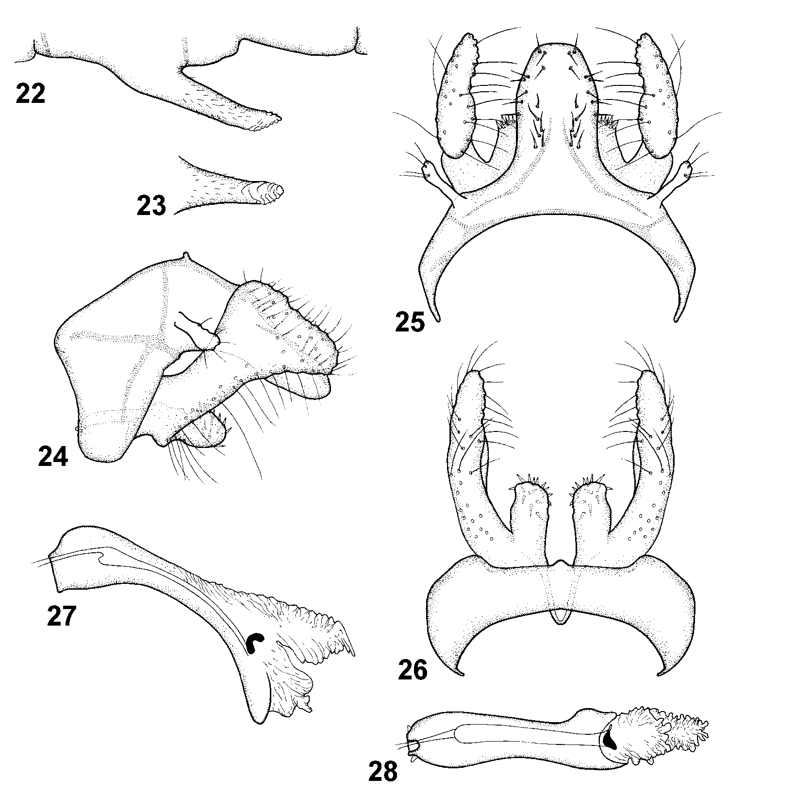
Fig. 22–28 View FIGURES 22 – 28 , 87 View FIGURE 87
Helicopsyche caligata Flint has a clubshaped gonocoxite similar to that of H. camuriensis , but the new species is unique in having a larger anterior lobe on segment IX, the presence of large basimesal lobes on the gonocoxite, and the numerous setae along the dorsal margin of tergum X.
Male. Head: Antennal scape about 2/3 as long as eye diameter. Maxillary palp segments equally long, each segment as long as eye diameter. Cephalic warts round, small, strongly convex, about 1/3 as long as eye diameter, with pale grey and golden brown setae. Forewing pale golden brown, length 3.8–4.1 mm. Sternum VI process ( Fig. 22, 23 View FIGURES 22 – 28 ) about 1/3 its segment length, covered by microtrichiae; nearly straight and slightly tapering in lateral view ( Fig. 22 View FIGURES 22 – 28 ), oriented posteroventrally; narrowing along its length in ventral view ( Fig. 23 View FIGURES 22 – 28 ); apex bearing ventral lamellae ( Fig. 23 View FIGURES 22 – 28 ).
Male genitalia ( Fig. 24–28 View FIGURES 22 – 28 ). Segment IX, in lateral view ( Fig. 24 View FIGURES 22 – 28 ), with anterior lobe hyperboloid, oriented anteriad, present midlaterally; anterodorsal margin nearly straight; anteroventral margin shallowly concave; in dorsal view ( Fig. 25 View FIGURES 22 – 28 ), with inner margin widely hyperboloid; in ventral view ( Fig. 26 View FIGURES 22 – 28 ), with minute posterior process; lateral apodeme present as anteriorly oriented, nearly straight line ( Fig. 24 View FIGURES 22 – 28 ), fading before reaching anterior margin; submarginal line absent; tergal transverse apodeme present; sternal transverse apodeme present. Segment X, in lateral view ( Fig. 24 View FIGURES 22 – 28 ), oriented posteroventrad, nearly straight along its length; dorsally undulate; slightly tapering along its length, apex widely rounded; in dorsal view ( Fig. 25 View FIGURES 22 – 28 ), slightly narrowing apically, apex without notch; with about 14 pairs of about equally long megasetae in group starting at proximal half. Superior appendage ( Fig. 24 View FIGURES 22 – 28 ) tubular, apex slightly clubshaped, oriented posterolaterally. Primary branch of gonocoxite, in lateral view ( Fig. 24 View FIGURES 22 – 28 ), with proximal half about equally broad, strongly widening to clubshaped distal half, with undulate dorsal margin; apex weakly produced, oriented posteriorly; central part of primary branch slightly narrower than height of central part of Tergum X ( Fig. 24 View FIGURES 22 – 28 ); anterodorsal margin nearly straight, smooth; posteroventral margin nearly straight at proximal half; basimesal lobe large, protruding primary branch, apically widely rounded in lateral view ( Fig. 24 View FIGURES 22 – 28 ), tubular in ventral view ( Fig. 26 View FIGURES 22 – 28 ), with nearly straight, subparallel median margins; with about 15 short megasetae on dorsal margin; basal plate, in lateral view ( Fig. 24 View FIGURES 22 – 28 ), narrow, slightly curving ventrally toward anterior apex; in ventral view ( Fig. 26 View FIGURES 22 – 28 ) triangular, apex rounded. Phallus, in lateral view ( Fig. 27 View FIGURES 22 – 28 ), with anterior half nearly straight, gently curving ventrad at midlength, dorsal margin nearly straight; ventral margin curving; anterior 1/5 about 2x broader than its central part ( Fig. 27 View FIGURES 22 – 28 ); in ventral view ( Fig. 28 View FIGURES 22 – 28 ); phallobase forms minute lateral triangles; endotheca produced into long dorsal lobe; sperm channel divided into thick posterior and slender anterior parts; sclerotized posteroventral part narrow.
Holotype male: VENEZUELA: Dist. Fed. [Districto Federal]: Río Camuri Grande, 1 km S Camuri, (nucleo U.S. B.), 10.616°N, 66.715°W, 30 m, 24.i.1994, Holzenthal, Cressa & Rincón ( UMSP 000022236) ( UMSP, pinned).
Paratypes: same data as holotype — 3 males, 2 females ( UMSP, pinned), 1 male, 1 female ( NRM, pinned), 1 male, 1 female ( NMNH, pinned), 1 male ( IZAM, pinned).
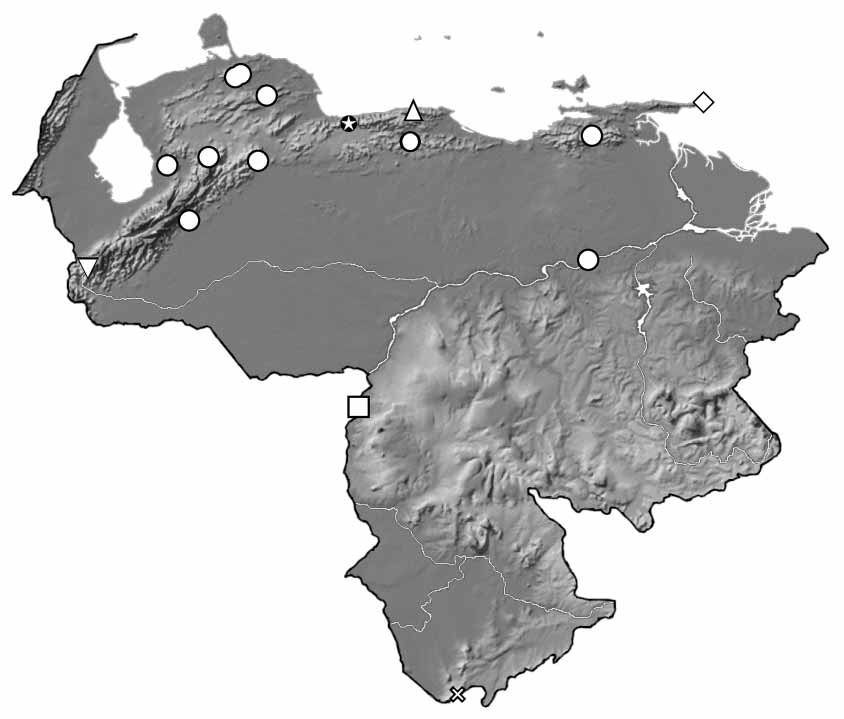
Distribution. Venezuela (Districto Federal).
Etymology. The name camuriensis is derived from the type locality, Río Camuri Grande. The name is to be treated as a noun in the genitive case.
No known copyright restrictions apply. See Agosti, D., Egloff, W., 2009. Taxonomic information exchange and copyright: the Plazi approach. BMC Research Notes 2009, 2:53 for further explanation.