
Helicopsyche (Feropsyche) neblinensis, Johanson, Kjell Arne & Holzenthal, Ralph W., 2004
|
publication ID |
https://doi.org/ 10.5281/zenodo.169460 |
|
publication LSID |
lsid:zoobank.org:pub:5403C332-6597-48D3-BD4D-7AD6D4CDC2CA |
|
DOI |
https://doi.org/10.5281/zenodo.6271564 |
|
persistent identifier |
https://treatment.plazi.org/id/03B1879D-FFB9-DE5A-FE98-FA847C7DF9EB |
|
treatment provided by |
Plazi |
|
scientific name |
Helicopsyche (Feropsyche) neblinensis |
| status |
sp. nov. |
Helicopsyche (Feropsyche) neblinensis , new species
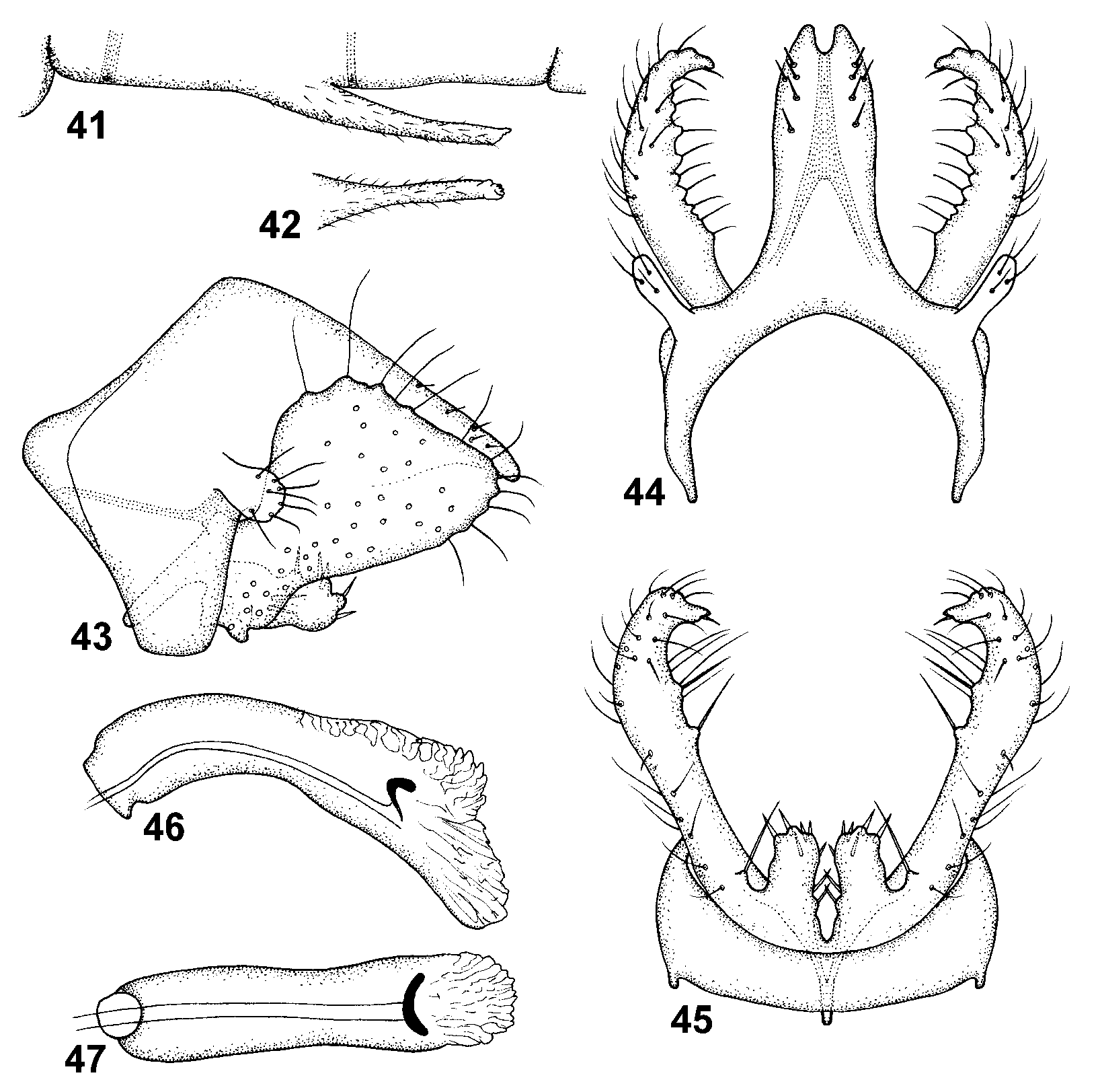
Fig. 41–47 View FIGURES 41 – 47 , 88 View FIGURE 88
Together with H. lara , new species, H. neblinensis has a rounded, triangular gonocoxite, the form being unique for the American Helicopsyche . Helicopsyche neblinensis can be separated from H. lara , in dorsal view, by the slightly more slender tergum X having only five pairs of dorsal setae, and in lateral view, by the slightly wider gonocoxite.
Male. Head: Antennal scape about as long as eye diameter. Maxillary palp segments equally long, each segment slightly shorter than eye diameter. Cephalic warts oval, slightly convex, with golden brown and grey setae. Forewing golden browngrey, length 3.1–3.5 mm. Sternum VI process ( Fig. 41, 42 View FIGURES 41 – 47 ) nearly 2/5 its segment length, covered by microtrichiae; in lateral view ( Fig. 41 View FIGURES 41 – 47 ), slightly curving posteriorly and tapering before apex, oriented posteriorly; distally parallelsided in ventral view ( Fig. 42 View FIGURES 41 – 47 ); apex bearing minute ventral lamellae and spines ( Fig. 42 View FIGURES 41 – 47 ).
Male genitalia ( Fig. 43–47 View FIGURES 41 – 47 ). Segment IX, in lateral view ( Fig. 43 View FIGURES 41 – 47 ), with anterior lobe hyperboloid, oriented anteriad, present ventrolaterally; anterodorsal and anteroventral margins shallowly concave; in dorsal view ( Fig. 44 View FIGURES 41 – 47 ), with inner margin widely ellipsoid; in ventral view, ( Fig. 45 View FIGURES 41 – 47 ) without posterior process; lateral apodeme present as anterodorsally oriented, nearly straight line ( Fig. 43 View FIGURES 41 – 47 ) nearly reaching anterior margin; submarginal line present; tergal transverse apodeme absent; sternal transverse apodeme absent. Segment X, in lateral view ( Fig. 43 View FIGURES 41 – 47 ), oriented posteroventrad, nearly straight; strongly tapering along its length, apex slightly pointed and curving ventrally; in dorsal view ( Fig. 44 View FIGURES 41 – 47 ), slightly narrowing toward apex; apex deeply and narrowly notched ( Fig. 44 View FIGURES 41 – 47 ); with 5 pairs of about equally long, weak megasetae in longitudinal group starting at distal half. Superior appendage ( Fig. 43 View FIGURES 41 – 47 ) tubular in dorsal view, slightly clubshaped in lateral view, oriented posterolaterally. Primary branch of gonocoxite, in lateral view ( Fig. 43 View FIGURES 41 – 47 ), generally widely triangular, with slightly undulate dorsal margin; apex rounded in lateral view, produced into hooklike process visible in dorsal view ( Fig. 44 View FIGURES 41 – 47 ); central part of primary branch wider than height of central part of tergum X ( Fig. 43 View FIGURES 41 – 47 ); anterodorsal margin convex, smooth; posteroventral margin weakly sigmoid.; basimesal lobe large, tubular, protruding primary branch and with undulating apex ( Fig. 43, 45 View FIGURES 41 – 47 ); with slightly converging median margins armed with strong setae; with about 6 long megasetae on dorsal margin; basal plate, in lateral view ( Fig. 43 View FIGURES 41 – 47 ), nearly straight, oriented anteroventrally, apex rounded; in ventral view ( Fig. 45 View FIGURES 41 – 47 ), strongly narrowing toward apex. Phallus, in lateral view ( Fig. 46 View FIGURES 41 – 47 ), with anterior half curving posteriorly, gently bent ventrad at midlength, dorsal margin convex along its length; ventral margin curving; anterior 1/5 about as broad as its central part ( Fig. 46 View FIGURES 41 – 47 ); in ventral view ( Fig. 47 View FIGURES 41 – 47 ), tubular along its length; phallobase absent; endotheca slightly produced; sperm channel undivided; sclerotized posteroventral part narrow.
Holotype male: VENEZUELA: T.F.A. [Territorio Federal Amazonas = Estado Amazonas]: Cerro de la Neblina, Basecamp, 0°51'N, 66°10'W, 140 m, 13–15.iii.1984, O.S. Flint, Jr. & J. Louton ( NMNH, pinned).
Paratypes: same data as holotype — 8 males, 9 females ( NMNH, pinned); same data as holotype, except 20–24.iii.1984, O.S. Flint, Jr. & J. Louton — 30 males, 50 females ( NMNH, alcohol), 2 males, 2 females ( UMSP, alcohol), 2 males, 2 females ( NRM, alcohol), 2 males, 2 females ( IZAM, alcohol); same data as holotype, except 0°50'N, 66°9'44''W, 140 m, 13–20.ii.1984, D. Davis & T. McCabe — 1 male, 6 females ( NMNH, alcohol); same data as holotype, except 0°50'N, 66°9'44''W, 140 m, 21–29.ii.1984, D. Davis & T.McCabe — 3 males, 4 females ( NMNH, alcohol); same data as holotype, except 0°50'N, 66°9'44''W, 140 m, 1–10.iii.1984, D. Davis & T. McCabe — 14 males, 19 females ( NMNH, alcohol); same data as holotype, except 8–9.ii.1985, W.E.Steiner — 7 males, 10 females ( NMNH, pinned); same data as holotype, except 10–20.ii.1985, P.J. & P.M. Spangler, R.A. Faitoute, & W.E. Steiner — 4 females ( NMNH, pinned); same data as holotype, except Camp IV, 0°58'N, 65°57'W, 760 m, 15–18.iii.1984, O.S. Flint, Jr. — 2 males ( NMNH, alcohol).
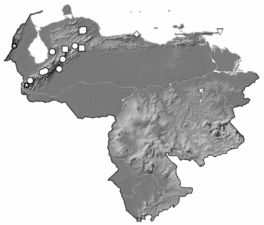
Distribution. Venezuela (Amazonas).
Etymology. neblinensis , derived from the type locality, Cerro de la Neblina. The name is to be treated as a noun in the genitive case.
No known copyright restrictions apply. See Agosti, D., Egloff, W., 2009. Taxonomic information exchange and copyright: the Plazi approach. BMC Research Notes 2009, 2:53 for further explanation.
|
Kingdom |
|
|
Phylum |
|
|
Class |
|
|
Order |
|
|
Family |
|
|
Genus |