
Polydrepanum fissum, Sankaran & Sebastian, 2018
|
publication ID |
https://doi.org/10.11646/zootaxa.4471.1.8 |
|
publication LSID |
lsid:zoobank.org:pub:5C14EA87-03AC-4BBC-B622-AB69EF6786C2 |
|
DOI |
https://doi.org/10.5281/zenodo.5970300 |
|
persistent identifier |
https://treatment.plazi.org/id/03AFD369-390B-1014-FF11-F93EF97BF80F |
|
treatment provided by |
Plazi |
|
scientific name |
Polydrepanum fissum |
| status |
sp. nov. |
Polydrepanum fissum View in CoL sp. nov.
( Figs 1–4 View FIGURE 1 View FIGURE 2 View FIGURE 3 View FIGURE 4 , 7 View FIGURE 7 )
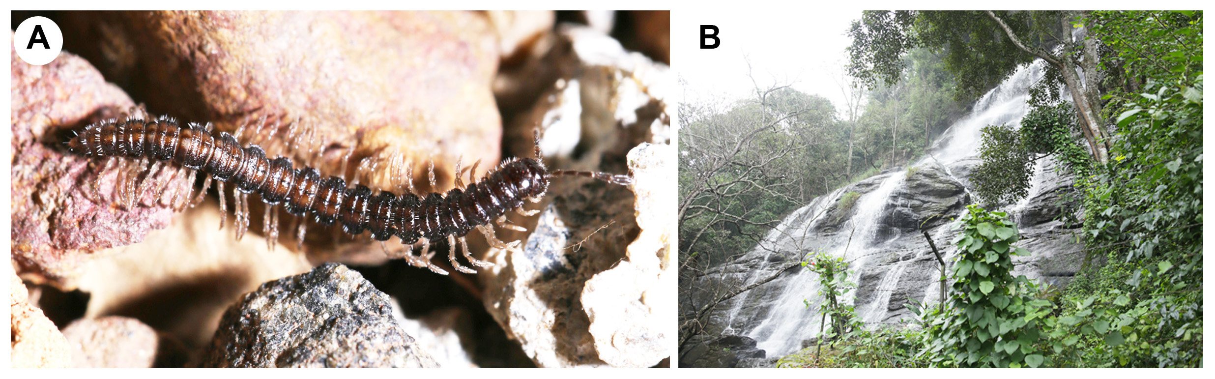
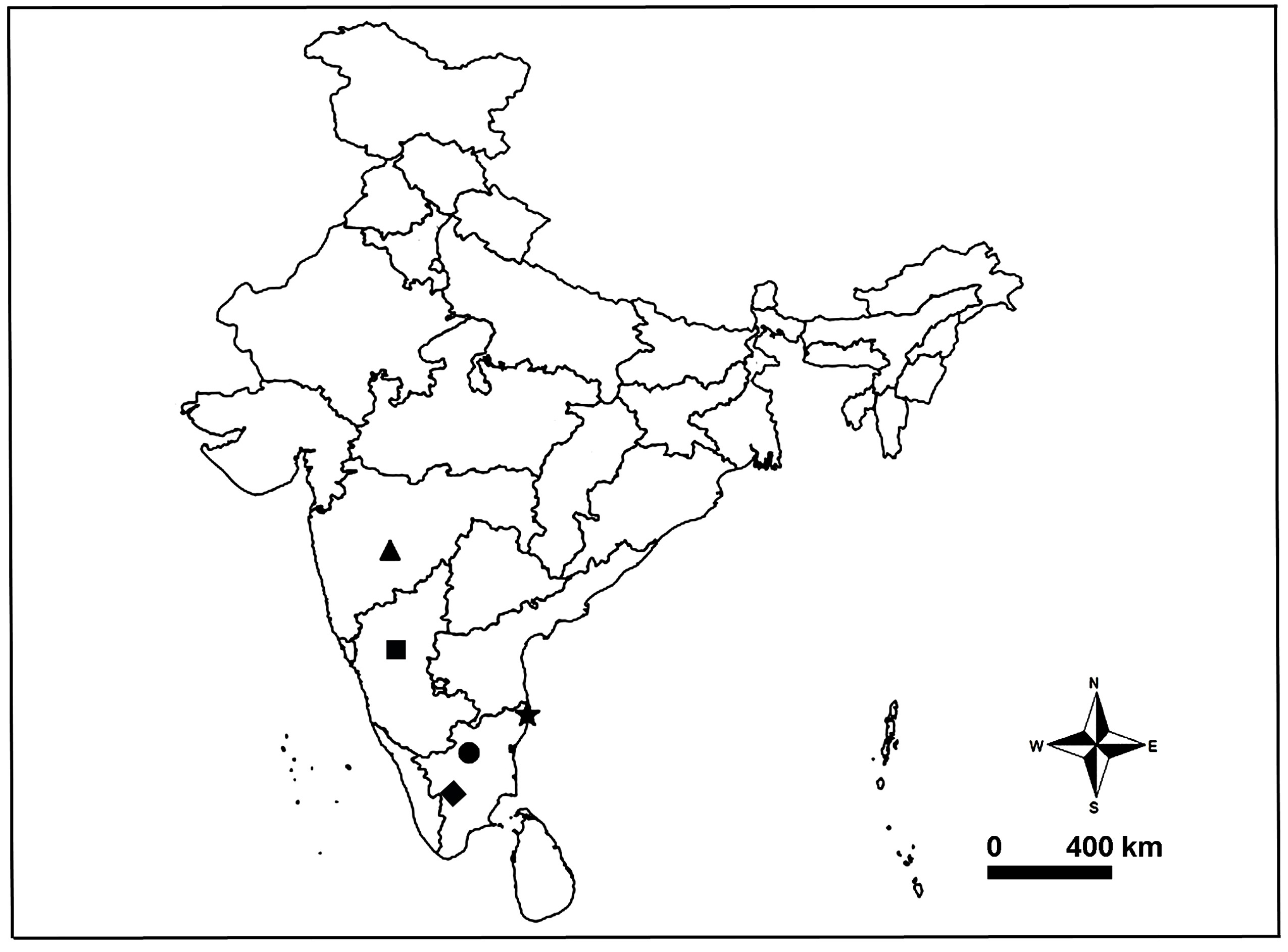
Type material: Holotype: Male (MILLI-ADSH0005), INDIA: Tamil Nadu: Salem, Yercaud , near the foothills of Kiliyur Falls , 11o47'45.1''N, 78o12'02.6''E, 1167 m alt., 3 December 2017, M. S. Pradeep leg., from litter, by hand GoogleMaps . Paratypes: 2 males (MILLI-ADSH0006), same data as holotype GoogleMaps .
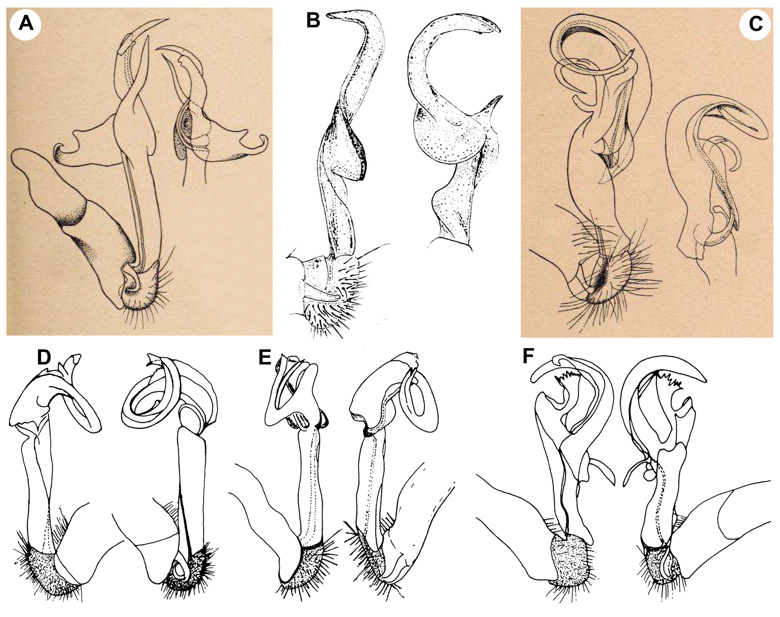
Etymology. The specific epithet refers to the bifurcated solenophore tip of the new species ( Fig. 4A View FIGURE 4 , 1 View FIGURE 1 st and 2nd arrows). Latin fissum = split, neuter.
Diagnosis. Polydrepanum fissum sp. nov. seems to be especially similar to P. tamilum , at least concerning the complexity of the gonopods, but can be distinguished from the latter by the following combination of characters: femorite with a lateral thumb-like process and paired apical folds (femorite of P. tamilum lacks such modifications), solenophore with a moderately and regularly curved distal part (solenophore of P. tamilum with a strongly curved/coiled distal part) and solenophore without mesal process (solenophore of P. tamilum with paired mesal processes) (compare Figs 3E–F View FIGURE 3 , 4A–C View FIGURE 4 with Carl 1932: figs 17–18). The occurrence of paired apical demarcation sulci/folds of gonopodal femorite relates P. fissum sp. nov. with the Polydrepanum sp. 1 illustrated by Bano and Murthy (1997). However the complexity of tibiotarsus of the former clearly distinguishes it from the latter (compare Fig. 4A–B View FIGURE 4 with Bano & Murthy 1997: fig. 10A–B and herein Fig. 6D View FIGURE 6 ).
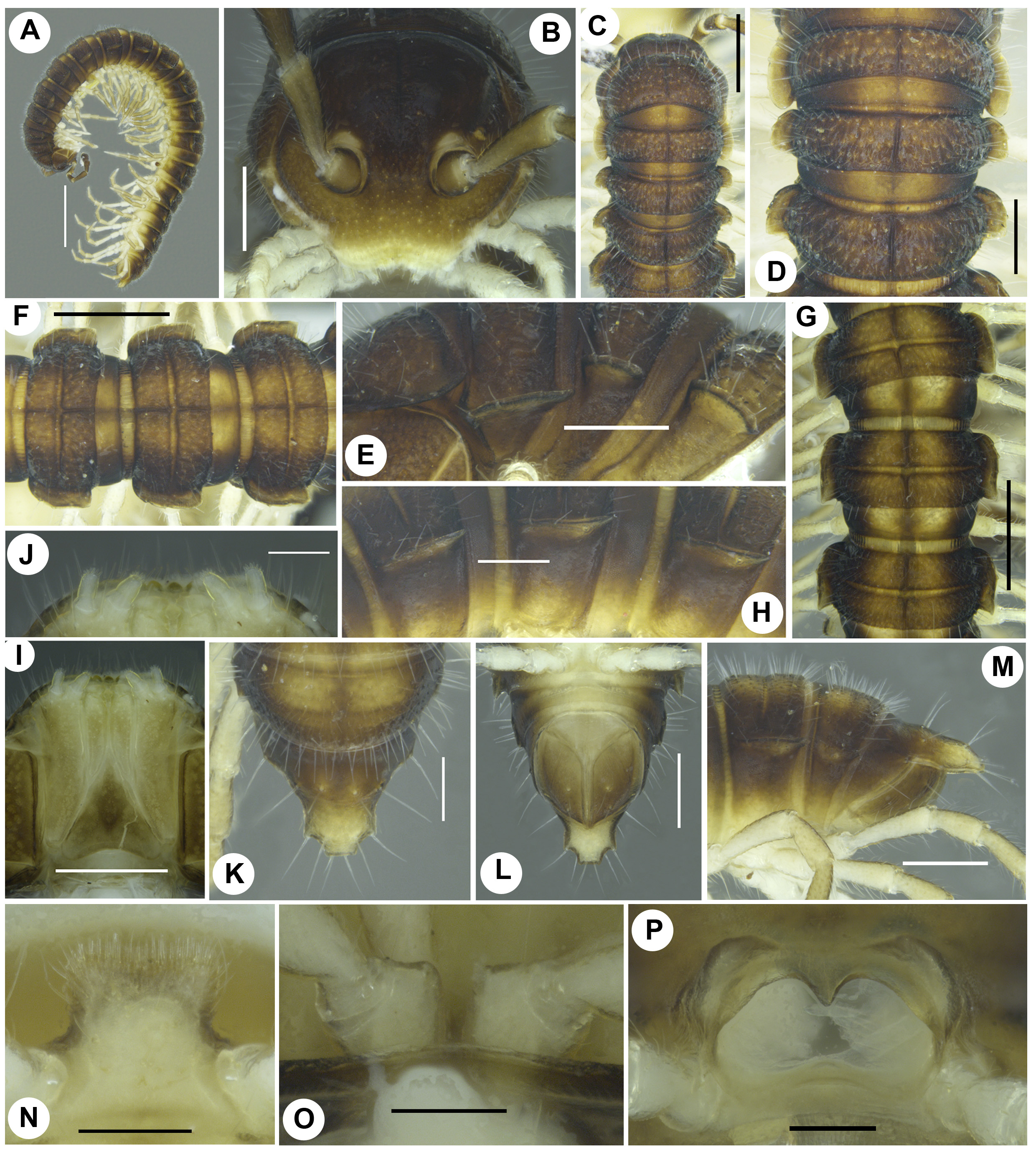
Description. Male. Body with 20 segments. Length ca 15 mm, width of midbody pro- and metazonites 1.1 and 1.2 mm, respectively. Width of midbody paranota 0.1 mm. Colouration of live animals uniformly chocolate brown; antennae, postero-lateral part of paranota and surface below paranota all chocolate brown; legs and sternites coffee brown. Colouration of alcohol material after preservation faded to brownish, postero-lateral part of paranota pale brown, sternites and legs straw coloured.
Head slightly wider than collum with paranota (1.4/ 1.3 mm) in dorsal view ( Fig. 2C View FIGURE 2 ); relative ring widths: collum ± 1 ~ 2 <3 <(4 = 16)> 17> 18> 19. Vertex shiny, sparsely setose; frons, stipes, cardo, clypeus moderately setose; epicranial sulcus distinct, ending at midanterior level of antennal sockets. Antennae moderately long (3.0 mm), filiform, reaching dorsally until waist of ring 4; relative antennomere lengths: 2 <3> 4 <5> 6; antennomere 6 widest apically; post-antennal groove shallow; antennal socket diameter and isthmus between them 0.2 and 0.3 mm, respectively.
Collum with lateral margin weakly produced as a paranotum ( Fig. 2E View FIGURE 2 ), anterior, lateral and posterior margins slightly concave, without any posterior marginal setae ( Fig. 2C View FIGURE 2 ); collum dorsally bearing four transverse rows of white, short, fragile setae of nearly parallel rows.
Anterior and posterior margins of segments almost straight, with posterior marginal setae ( Fig. 2D, F–G View FIGURE 2 ).
Paranota of segments set high, lying parallel to axial line and below dorsum ( Fig. 2E, H View FIGURE 2 ), posterior corner of collum paranota and paranota of haplosegments 1–2 regularly rounded ( Fig. 2C–D View FIGURE 2 ), that of paranota of haplosegment 3 and diplosegments until 12th produced posteriorly as broad triangles ( Fig. 2D, F–G View FIGURE 2 ), that of diplosegments 13–19 strongly produced posteriorly as acute triangles ( Fig. 2L View FIGURE 2 ), posterior corner of paranota of haplo- and diplosegments widely spaced from each other ( Fig. 2E, H View FIGURE 2 ); anterior margin of paranota of collum, haplosegments 2–3 and diplosegments until 17th nearly regularly rounded, that of diplosegments 18–19 shallow ( Fig. 2D, F–G, L View FIGURE 2 ); paranota on haplosegment 1 larger than paranota of haplosegments 2–3 ( Fig. 2D View FIGURE 2 ), slightly lower than lateral margins of collum and preceding paranota ( Fig. 2E View FIGURE 2 ), lying close to that of collum, but away from that of haplosegment 2 ( Fig. 2E View FIGURE 2 ); lateral margins of paranota of haplosegments and diplosegments until 17th with paired weak marginal incisions, one anterior incision, one median ( Fig. 2D, F–G View FIGURE 2 ); lateral margins of paranota thicker on pore-bearing rings ( Fig. 2H View FIGURE 2 ).
Pleural keels well marked on haplosegments as triangular bulging, that on diplosegments less evident as narrow thickenings.
Prozonites smooth. Metazonites, surface below paranota granulate; metazonites with moderate pubescence ( Fig. 2M View FIGURE 2 ); transverse sulcus on metazonites distinct from diplosegments 1–18, reaching paranotal base ( Fig. 2F– G View FIGURE 2 ); limbus a thin, continuous ribbon. Axial line clearly traceable both on pro- and metazonites until diplosegment 18th ( Fig. 2C–D, F–G View FIGURE 2 ).
Pore formula normal (5, 7, 9, 10, 12, 13, 15–19); ozopore small, circular, opening posterolaterally on thickened paranotal margin, away from the level of transverse sulcus of metazonites ( Fig. 2H View FIGURE 2 ).
Sterna moderately setose, as wide as long, transverse impressions more distinct than longitudinal ones, without modifications except for a single chisel-shaped, setose lamella between coxae 4 ( Fig. 2N View FIGURE 2 ).
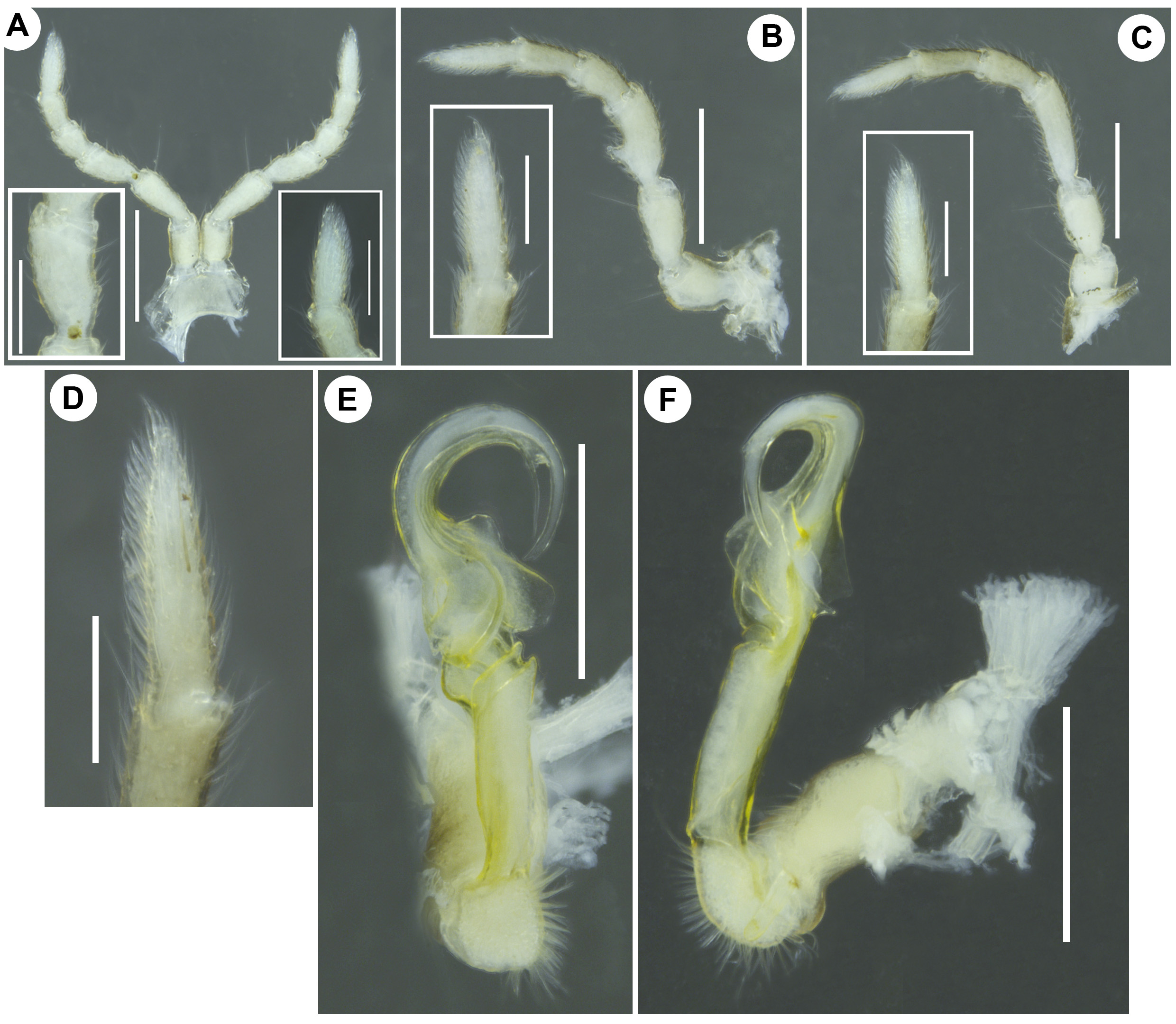
Legs slender, with well developed tarsal brushes ( Fig. 3A–D View FIGURE 3 ), with short claws; all coxae and prefemora bearing a single, ventral, stiff, long seta; relative podomere lengths: femur> tarsus> postfemur> (prefemur ~ tibia); all prefemora swollen laterally; leg pair I possesses small, rounded, midway, ventral tubercle/adentostyle ( Fig. 3A View FIGURE 3 , left inset) on femur; femora on leg pair II ventrobasally with a prominent conical tubercle/ adenostyle ( Fig. 3B View FIGURE 3 ).
Gonopore small, round, opening on short, anterolateral mound-like bulging on coxae 2 ( Fig. 2O View FIGURE 2 ).
Pre-anal ring sparsely setose; epiproct conical, flattened dorsoventrally, extending beyond anal valves, tip truncate ( Fig. 2K, M View FIGURE 2 ); hypoproct triangular ( Fig. 2L View FIGURE 2 ).
Gonopods ( Figs 3E–F View FIGURE 3 , 4A–C View FIGURE 4 ). Gonopod aperture transversely oriented, roughly triangular in outline, about 2/3 as wide as prozonite 7, with a well marked, raised lateral rim, anterior and posterior rims with median projections ( Fig. 2P View FIGURE 2 ). Gonopods high, complex, lying close to sternites. Coxite about as long as femorite, subcylindrical, distally broadened, setose distodorsally ( Figs 3F View FIGURE 3 , 4B View FIGURE 4 ). Cannula as usual ( Fig. 4B View FIGURE 4 ). Prefemur short, globose, as usual densely setose ( Figs 3E–F View FIGURE 3 , 4A–B View FIGURE 4 ), with a moderately long and stiff seta distoventrally ( Fig. 4B, 4 View FIGURE 4 th arrow), with a distal sulcus evidently demarcating acropodite ( Fig. 4A–B View FIGURE 4 ). Femorite long, subcylindrical, with incomplete torsion limited to basal 1/4th part ( Figs 3E–F View FIGURE 3 , 4A–B View FIGURE 4 ), distolaterally with a thumb-like process ( Fig. 4C View FIGURE 4 ). Seminal groove running along dorsal side of femorite, turning laterally near apical folds on femorite, then turning mesally before entering onto solenomere, strongly curved distally ( Fig. 4A View FIGURE 4 ). Postfemoral part clearly set off from femorite by two oblique demarcation sulci/folds ( Fig. 4A–B View FIGURE 4 ). Solenophore short, evidently branched, only with lamina lateralis ( Fig. 4A View FIGURE 4 ), distally curved as a sickle ( Figs 3E View FIGURE 3 , 4A View FIGURE 4 ), with bifurcated tip, lateral branch slightly longer than mesal, both directed laterally ( Fig. 4A View FIGURE 4 , 1 View FIGURE 1 st and 2nd arrows), basomesally with two membranous, triangular, wing-like processes, 1 st large , 2nd small ( Fig. 4B View FIGURE 4 ), with a widely triangular process ( Fig. 4B View FIGURE 4 ), basolaterally with a flat, lamellate process ( Fig. 4C View FIGURE 4 ); lamina lateralis well developed, with a short disolateral process ( Fig. 4C View FIGURE 4 ), with mesal triangular process ( Fig. 4A View FIGURE 4 ), distal 2/3rd folded to juxtapose with the ventral face of solenophore, proximal 1/3rd wide, lying slightly away from solenophore ( Fig. 4A View FIGURE 4 ). Solenomere simple, slender, median part enclosed within lamina lateralis, with moderately exposed, angular tip ( Fig. 4A View FIGURE 4 , 3 View FIGURE 3 rd arrow).
Female. Unknown.
Note. Polydrepanum fissum sp. nov. is quite distinct from other known Polydrepanum spp. as its leg pair I possesses adenostyle and gonopodal femorite poorly torsate, which all warrant the erection of a new genus for it. However, a distally sickle-shaped solenophore with only lamina lateralis, a flagelliform solenomere enclosed by the lamina lateralis and the seminal groove, which run along the dorsal face of the femorite fit this species into Polydrepanum . The occurrence of adenostyle on leg pair I and gonopodal femorite with less evidence of torsion of P. fissum sp. nov. suggests that it may be a derived clade within Polydrepanum , but confirmation requires phylogenetic treatment.
Distribution. Only known from the type locality ( Fig. 7 View FIGURE 7 ).

No known copyright restrictions apply. See Agosti, D., Egloff, W., 2009. Taxonomic information exchange and copyright: the Plazi approach. BMC Research Notes 2009, 2:53 for further explanation.
|
Kingdom |
|
|
Phylum |
|
|
Class |
|
|
Order |
|
|
Family |
|
|
SubFamily |
Alogolykinae |
|
Tribe |
Polydrepanini |
|
Genus |