
Atractus spinalis, Passos, Paulo, Junior, Mauro Teixeira, Recoder, Renato S., Sena, Marco Aurélio De, Vechio, Francisco Dal, Pinto, Hugo Bonfim De A., Mendonça, Sônia H. S. T., Cassimiro, José & Rodrigues, Miguel Trefaut, 2013
|
publication ID |
https://doi.org/ 10.1590/s0031-10492013000600001 |
|
persistent identifier |
https://treatment.plazi.org/id/03AF87F1-FFF9-FFC3-FEA0-FE18FDC4F891 |
|
treatment provided by |
Carolina |
|
scientific name |
Atractus spinalis |
| status |
sp. nov. |
Atractus spinalis sp. nov. Figures 1-3 View FIGURE 1 View FIGURE 2 View FIGURE 3
Holotype: An adult male ( MZUSP 20760 View Materials ) collected by M.T. Rodrigues and collaborators (all authors except the senior one) on March 26, 2011 (field number MTR 20280) at Alto do Palácio (19°15’35.5”S, 43°31’55.2”W; 1357 m above sea level = asl), Parque Nacional da Serra do Cipó , municipality of Morro do Pilar, state of Minas Gerais, Southeastern Brazil. GoogleMaps
Paratypes: Five specimens all collected by M.T. Rodrigues and collaborators (all authors except the senior one) in the Parque Nacional da Serra do Cipó , state of Minas Gerais, Southeastern Brazil: an adult female ( MZUSP 20763 View Materials ), two adult males ( MNRJ 23357 View Materials and MZUSP 20764 View Materials ) and a juvenile male ( MZUSP 20762 View Materials ), collected on November 14, 2011 (field numbers MTR 21778, 21779 , 21780 and 21777, respectively) at Currais (19°27’5.00”S, 43°29’43.78”W, 1430 m asl), municipality of Jaboticatubas ; and an adult male ( MZUSP 20761 View Materials ), collected on April 2, 2011 (field number MTR 20378) at the roadside (19°13’42.9”S, 43°30 24.4 W, 1354 m asl), in the limit between the municipalities of Morro do Pilar and Santana do Riacho GoogleMaps .
Diagnosis: Atractus spinalis is distinguished from all congeners by the following combination of characters: (1) 15-15-15 smooth dorsal scale rows; (2) two postoculars; (3) moderate loreal; (4) temporals 1+2; (5) seven supralabials, third and fourth contacting orbit; (6) seven infralabials, first four contacting chin shields; (7) usually nine maxillary teeth; (8) usually three gular scale rows; (9) three or four preventrals; (10) 149 ventrals in the single female, 136-142 in males; (11) 24 subcaudals in the female, 27-33 in males; (12) dorsum red, except for first dorsal scale rows that is red on its upper area, becoming lighter towards venter, occasionally forming conspicuous dorsolateral lines along body; (13) ventral ground color creamish white; (14) small body size, females (199 mm SVL) and males (268 mm maximum SVL); (15) moderate tail in the female (11.6%) and moderate to long in males (13.6-16.6% SVL); (16) hemipenis slightly bilobed, semicapitate, and semicalyculate. Comparisons: Among all congeners, Atractus spinalis shares 15 dorsal scale rows, white occipital region in juvenile specimens, dorsal ground color reddish pink to red with alternate black transversal spots, blotches or transversal bands in live individuals, seven upper and lower labial scales, ventrals and subcaudals creamish white and slightly bilobed, semicapitate, and semicalyculate hemipenis only with some individuals of A. paraguayensis Werner, 1924 and A. potschi Fernandes, 1995 presenting transversal black bands (both are polychromatic species; see Passos et al., 2010c). Atractus spinalis differs from banded specimens of A. paraguayensis (characters in parenthesis) in having conspicuous transversal bands along the body as long as or longer than interspaces, retracted hemipenis extending to the level of seventh subcaudal, everted hemipenis without calcareous ornamentation in the median side of the lobes and average body size of 211 mm SVL in males (vs. blotches/bands and interspaces similar in length anteriorly with the former becoming gradually shorter than interspaces posteriorly, retracted hemipenis extending to the level of 10 th subcaudal, conspicuous spinulate calyces on the medial side of lobes and average body size 304 mm SVL; Martins, 2012); it differs from banded specimens of A. potschi (characters in parenthesis) in having eight to nine maxillary teeth and 136-142 ventral scales in males (vs. seven maxillary teeth and 153-165 ventral scales in males). As far as we know there is no sympatry between the new species and any other congeners. The only other Atractus species with closer geographical distribution, A. zebrinus Jan, 1862 (found at Serra do Caraça located about 70 km far from the southern record of the A. spinalis ; see Passos et al., 2010c), can be easily distinguished from the new species by having 17 dorsal scale rows (vs. 15 in A. spinalis ).
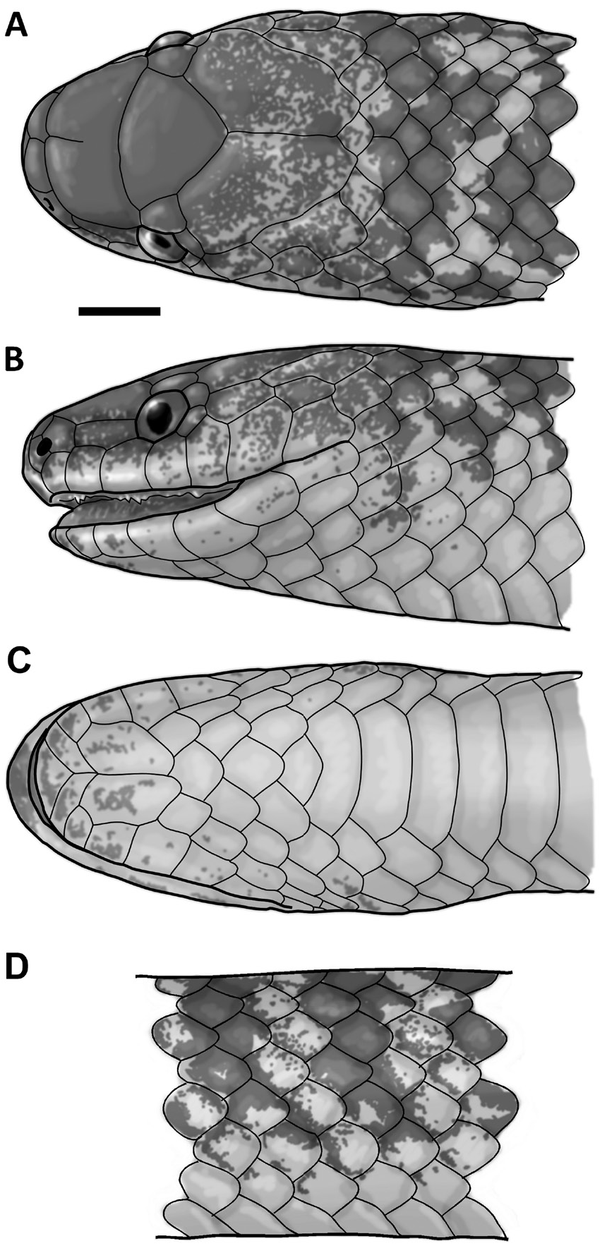
Description of the holotype ( Fig. 1 View FIGURE 1 ): Adult male, 236 mm SVL, 38 mm CL (16.1% SVL); head round- ed in dorsal view, slightly flattened in lateral view, 11.0 mm long (4.7% SVL), 5.7 mm wide (52% head length); cervical constriction poorly distinct; snout rounded in dorsal view, truncated in lateral view; rostrum-orbit distance 3.3 mm (30% head length); nasal-orbit distance 2.3 mm (20.9% head length); intraorbital distance 4.5 mm (78.9% head width); body subcylindrical, body diameter 7.3 mm (3.1% SVL); belly flattened; tail moderately long, with terminal spine moderately long and acuminate; rostral subtriangular in frontal view, wider (2.4 mm) than high (1.7 mm), visible from above; internasal slightly longer (1.0 mm) than wide (0.8 mm); internasal suture (1.0 mm long) sinistral to prefrontal median suture; prefrontal as wide (2.2 mm) as long (2.1 mm); prefrontals fused mesoposteriorly; frontal triangular, wider (3.4 mm) than long (2.9 mm); supraocular subretangular, about twice as long (1.3 mm) as wide (0.7 mm); parietal about twice as long (4.6 mm) as wide (2.5 mm); nasal divided; prenasal about twice as high (0.8 mm) as long (0.5 mm), contacting rostral, internasal, first supralabial, and postnasal; postnasal about as high (0.8 mm) as long (0.9 mm), contacting prenasal, prefrontals, loreal, and second supralabial; long loreal (1.6 mm long, 0.5 mm high); loreal contacting eyes, prefrontals, nasals, and second to third supralabials; eye diameter 1.5 mm; pupil round; two postoculars, upper postocular as high (0.6 mm) and longer (0.6 mm) than lower postocular (0.6 mm high, 0.4 mm long); upper postocular contacting eye, supraocular, parietal and anterior temporal; lower postocular, contacting eye, fourth and fifth supralabial, and anterior temporal; 1+2 temporals; anterior temporal twice as long (1.4 mm) as high (0.7 mm), contacting parietal, fifth to sixth supralabials, postoculars, and posterior temporals; upper posterior temporal elongate (2.8 mm long, 0.9 mm high), almost three times as long as high; seven supralabials, third and fourth contacting orbit; second supralabial high- er than first and lower than third; sixth supralabial (1.6 mm long, 1.6 mm high) higher and longer than remaining supralabials; symphyseal triangular, almost twice as wide (1.9 mm) as long (1.1 mm); seven infralabials, first four pairs contacting chin shields; first pair of infralabials in contact behind symphyseal, preventing symphyseal/chin shield contact; chin shields about three times as long (2.7 mm) as wide (1.1 mm); 15-15-15 smooth dorsal scale rows, lacking apical pits and supra-anal tubercles; three gular scale rows between last supralabial and preventral; three preventral scales; 142 ventral scales; anal plate single; 31/31 subcaudal scales. Maxillary bone arched upward anteriorly in lateral view, ventral portion curved on anterior and nearly flattened on median to posterior portion; maxillary arch with nine teeth; teeth angular in cross section, robust at base, narrower at apices, curved posteriorly; first five teeth large, moderately spaced, similar in size (even offsetting the level of curvature of the maxillary); sixth to ninth teeth gradually reduced in size and spacing; maxillary diastema absent or indistinct from interspaces between sixth and seventh teeth; last two teeth (topographically homologous to postdiastemal tooth) smaller and less spaced than the anterior teeth; lateral process of maxilla moderately developed.
Color in preservative of the holotype ( Fig. 2 View FIGURE 2 ): Dorsum of head dark brown, with pale brown spots dispersed on internasals, anterolateral area of prefrontals, supraoculars, anterolateral and mesodorsal regions of parietal; parietals mostly dark brown with two ill defined black round blotches (about equivalent to nostril size) positioned above each side of the posterior end of interparietal suture; lateral surface of head dark brown until dorsal edges of supralabials, ventral area of loreal and temporal region invaded with dispersed pale brown pigment; lateral sides of head with dark brown pigments concentrated around orbit (supraocular, posterior area of loreal, upper margins of third and fourth supralabials and postoculars); first four supralabials mostly creamish white, with irregular brown dots not attaining ventral border of each scale; fifth to seventh supralabials mostly pale brown, with disperse brown dots reaching ventral border of scales; anterior temporal pale brown scattered with cream pigment; posteri- or temporal and occipital series pale brown with fuzzy cream pigmentation; symphyseal, first three infralabials and anterior chin shields creamish white dotted with brown concentrated on dorsolateral margins of the scales; last four infralabials, gulars, preventrals and posterior chin shields creamish white with few disperse brown dots; ventral ground color uniformly creamish white; ventral surface of tail creamish white suffused with few dark brown dots concentrated on suture regions of posterior subcaudals; dorsal ground color of body beige, with 56 well-defined transversal black blotches (two scale long in dorsal view and up to three in lateral view) on the body and 14 on the tail; beige interspaces one scale long dorsally and possibly two scales long laterally; black blotches on the anterior region of body with variegated pattern when each blotch connects to adjacent blotch in inverted “Y” shape and not to the opposite one; at the level 30 th ventral scale dorsal marks become almost a regular transversal band; blotches or bands on body flanks usually reaching second scale row; body flanks (second and third scale rows) with dispersed irregular spots generally connected to transversal blotches or bands; paraventral region scattered with brown dots; sometimes bands are not symmetrically connected to the band on the other side along body and tail; first dorsal scale rows with brown pigment restricted to dorsal scale margins.
Color in life of the holotype ( Fig. 3 View FIGURE 3 ): Dorsal and lateral surfaces of head brown with invasion of scattered red pigment; supralabials reddish cream with dispersed brown dots; infralabials and mental region mostly creamish white discretely invaded by reddish brown pigment; ventrals and subcaudals uniformly creamish white; dorsal ground color red scattered with alternate black blotches or conspicuous transversal bands; first dorsal scales rows almost entirely red.
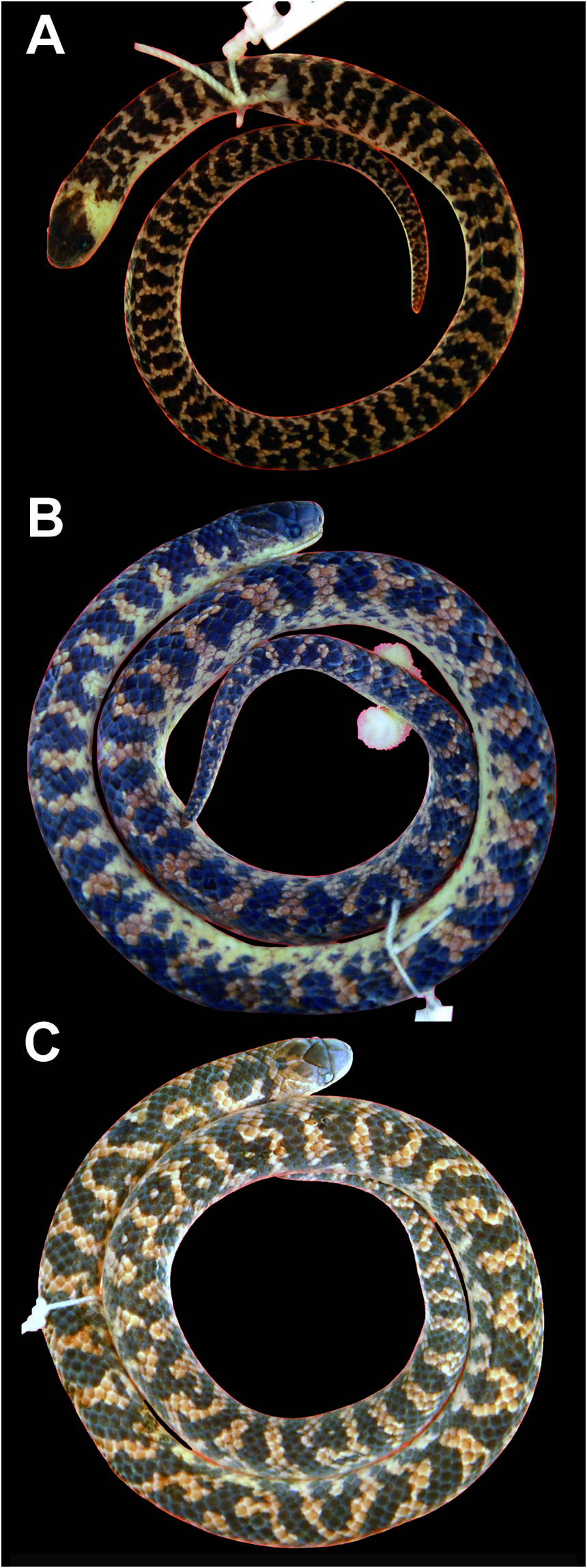
Juvenile color in preservative ( Fig. 4A View FIGURE 4 ): Conspicuous white nuchal collar covering occipital and temporal regions, entirely separating the black cephalic cap from the first black transversal body band.
Color variation in preservative ( Fig. 4 View FIGURE 4 B-C): Dorsum of head with cephalic cap region brown with a few pale brown to uniformly dark brown dots or spots; temporal and occipital regions become darker along species ontogeny; supralabials creamish white variably invaded by disperse brown dots; ventrals and subcaudals immaculate creamish white or with few brown dots; dorsal ground color of body beige to pale brown with 43-56 (x = 49.4; SD = 5.4; n = 5) body blotches or transversal bands in males and 40 in the only known female; dorsal marks comprising alternate black blotches (2-3 scales long), sometimes connected to posterior or anterior blotch on vertebral region; laterally, blotches sometimes displaying an inverted “Y” shape laterally with ventral ramification; ventral ramification occurs in blotches as well as in posterior transversal bands ( MZUSP 20761); dorsum with irregular marks displays a barely variegate pattern ( MZUSP 20763, MZUSP 20764, MNRJ 23357); sometimes specimens show irregular blotches restrict- ed to anterior portion of body and posteriorly there are conspicuous transversal bands ( MZUSP 20760 and 20761); body flanks with interspaces ranging from one to three scales long.
Meristic and morphometric variation (Table 1): Largest male 268 mm SVL, 40 mm CL, female 199 mm SVL, 23 mm CL; tail 13.6-16.6% SVL (x = 15.5; SD = 1.3; n = 5) in males, 11.6% SVL in the single female; 136-142 (x = 139.8; SD = 2.4; n = 5) ventrals in males, 149 in the female; 26-33 (x = 30.5; SD = 2.0; n = 5) subcaudals in males, 24 in the female; 3-4 (x = 3.3; SD = 0.5; n = 6) preventrals; 3-4 (x = 3.2; SD = 0.3; n = 12 sides) gular series; 6-8 (x = 7.7; SD = 0.6; n = 6) dorsal scale rows around tail at the level of second subcaudal; 8-9 (x = 8.9; SD = 0.2; n = 11 sides) maxillary teeth; 6.4-7.5 mm (x = 7.0; SD = 0.4; n = 4) adult body diameter; retracted hemipenis bifurcates and extends to the level of seventh subcaudal (n = 6 sides from three specimens).
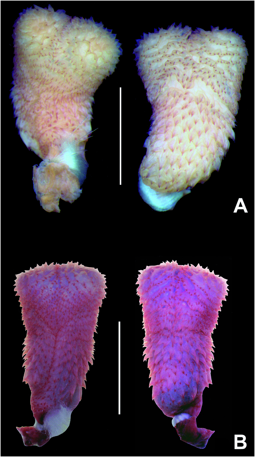
Hemipenis morphology (entirely everted organs n = 2) ( Fig. 5 View FIGURE 5 ): Fully everted and almost maximally expand- ed hemipenis ( MZUSP 20760; Fig. 5A View FIGURE 5 ) renders a slightly bilobed, semicapitate and semicalyculate organ; lobular branches almost indistinct in frontal view and restricted to distal portion of capitulum; lobes nearly flattened on the apices and with a median and relatively shallow invagination separating each lobular ramus; lobes uniformly covered by well-defined spinulate calyces on the sulcate, lateral and asulcate sides of capitulum; calyces large with conspicuous vertical walls on the sulcate and lateral sides of the capitulum; calyces on the asulcate side lacking vertical walls and horizontal walls connected forming regular spinulate flounces; from above lobular branches are medially delimited by vertical (= longitudinal) calyculate flounces; medial surface of both lobes almost indistinct due to median contact of lobular ramus; median-distal (= internal surface) of lobes only visible in frontal view through mechanical separation of lobes, revealing irregular large fringes without calcareous ornamentation (without red pigmentation); capitulum positioned immediately above sulcus spermaticus bifurcation, similar in size to hemipenial body; sulcate side of capitulum scattered with small spinulate calyces laterally connected, forming four irregular flounces on the intrasulcar region; capitular groove slightly evident on sulcate and well defined on the lateral and asulcate sides of the organ; lateral portion of capitulum (below the lobular region), with calyces forming barely defined spinulate flounces; capitular crotch with a “M-shaped” notch on median region of asulcate side of organ; non-lobular region of capitulum with four irregular spinulate flounces on the asulcate side of the hemipenis; sulcus spermaticus divides on distal portion of hemipenial body (nearly half the length of organ); branches of sulcus spermaticus centrifugally orientated, running towards tips of lobes; margins of the sulcus spermaticus narrow and stout, bordered by spinules on the base and papillae directed upwards on the extreme tip of lobes; hemipenial body subcylindrical, narrower than capitulum densely covered by moderate to relatively large hooked-shaped spines; largest spines located on lateral portion of sulcate side and basal region of the asulcate side; naked pocket restricted left side of proximal region of hemipenial, attaining nearly half the length of hemipenial body; basal portion of hemipenial body part nude, part ornamented by longitudinal plicae or disperse spinules ( Fig. 5A View FIGURE 5 ). The hemipenis of the holotype ( MZUSP 20760) was not filled by full in order to observed the median-distal face of the lobes. The organ of the paratype ( MNRJ 23357), fully everted and filled by full, is very similar to holotype’s organ, differing from it by nearly straight capitular crotch on median region of asulcate side of hemipenis ( Fig. 5B View FIGURE 5 ).
Etymology: The specific epithet “ spinalis ” is a Latin adjective derived from Spina, meaning the vertebrate backbone. The use of spina referring to backbone dates back to the Roman poet Virgil (70-19 before Christ). This name is herein used in reference to the Espinhaço Mountains Range (= Cadeia do Espinhaço), where the type series of Atractus spinalis was collected and the species appears to be endemic.
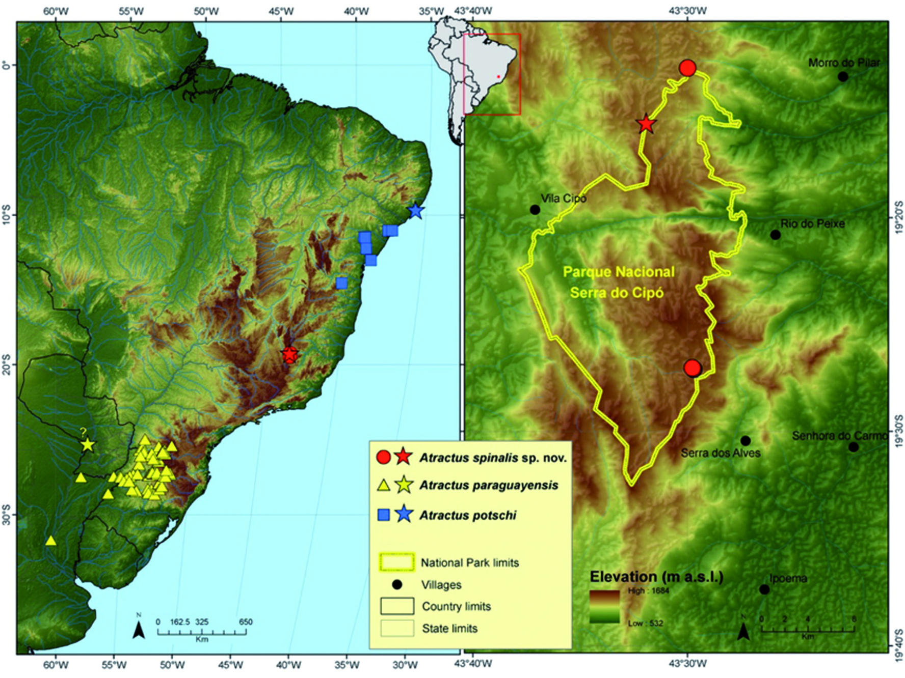
Distribution and natural history ( Fig. 6 View FIGURE 6 ): Currently known only from a few localities, Atractus spinalis seems to be endemic to the Serra do Cipó mountains, the local name for the southern section of the meridional segment of Espinhaço Range, an extensive quartzitic sandstone mountainous range stretching from central Minas Gerais state to northern Bahia. This section comprises elevated mountain ranges reaching up to 1660 m asl, where the habitats are dominated by rocky meadows (Campos Rupestres), interspersed with grasslands and a characteristic open vegetation with many endemic elements. The slopes and lower areas at its eastern versant and western side respectively contact the Atlantic Forest and the Cerrados (= savannas) (Chemale Jr. et al., 2011). All specimens of Atractus spinalis were found in typical Campos Rupestres, inside termite nests or under rocks, between 1340 and 1430 m asl ( Fig. 7 View FIGURE 7 ).
Remarks: Although suggestive, we do not anticipate the possible inverted sexual dimorphism in Atractus spinalis , since the only female has the smallest SVL (considering adult specimens) and, as consequence, fewer dorsal marks (Table 1). We cannot establish if the only female represents an outlier, but its presents the high number of ventral scales which is usually correlated with snakes body size (see Lindell, 1994). Otherwise, A. spinalis will be the only congener exhibiting inverted sexual dimorphism (males longer than females), but this need to be confirmed by more representative samples.


No known copyright restrictions apply. See Agosti, D., Egloff, W., 2009. Taxonomic information exchange and copyright: the Plazi approach. BMC Research Notes 2009, 2:53 for further explanation.