
Morobea Carvalho, 1987
|
publication ID |
https://doi.org/10.1206/0003-0082(2007)3558[1:ANGANS]2.0.CO;2 |
|
persistent identifier |
https://treatment.plazi.org/id/03AF87C7-FC52-FFAA-FF18-BAF0291DF943 |
|
treatment provided by |
Carolina (2021-08-30 06:24:15, last updated 2021-09-02 15:04:21) |
|
scientific name |
Morobea Carvalho |
| status |
|
TYPE SPECIES: Morobea longipes Carvalho, 1987 (original designation) .
DIAGNOSIS: Recognized among other Orthotylini by the following combination of characters: elongate parallel-sided body ( fig. 1a, b, d View Fig ); projecting clypeus ( fig. 2c View Fig ); strongly carinate posterior margin of head projecting over anterior margin of pronotum ( fig. 2a, c View Fig ); disc of pronotum coarsely rugulose ( fig. 2a View Fig ); posterior margin of pronotum broadly emarginate; metathoracic legs relatively long. Males with second antennal segment with species specific arrangement of short setae at base ( fig. 3 View Fig ); genital capsule enormous ( fig. 4a View Fig , 5a View Fig ), subequal to one-half abdominal length; opening of genital capsule disproportionately large, clearly exposing complex aedeagus; aedeagus with remarkably convoluted and heavily sclerotized phallotheca ( figs. 4d View Fig , 5e View Fig ); ductus seminis with thickened heavily sclerotized ring proximal to secondary gonopore.
REDESCRIPTION: Male: General Aspect: Macropterous, elongate, parallel-sided, total length 5.88–6.57; length from apex to clypeus to cuneal fracture 4.42–4.75; width across humeral angles of pronotum 0.83–0.96. Coloration and Vestiture ( figs. 1a, d View Fig ): Body yellowish brown with red and green maculations; antennae and legs yellowish brown with red maculation on apices of metafemora; membrane of forewing moderately suffused with brown; veins red to undifferentiated. Dorsum with sparse fine, short, hyaline setae; mesepisternum and metepisternum with field of dense microchaetae; legs and antennae with fine, distally orientated short, red, setae. Structure: Head ( fig. 2a–c View Fig ) moderately projecting in front of eyes; posterior margin convex; posterior margin strongly carinate, projecting over anterior margin of pronotum; frons flat posteriorly becoming steeply declivent anteriorly; clypeus distinctly projecting in front of frons; gena and maxillary plates extending anteroventrally; buccal cavity obovate, short; eyes occupying about one-half of total head height in lateral view, posterolateral margin slightly separated from margin of pronotum; antennae inserted midway between ventral and dorsal margins of eye. Antennae elongate, gracile; segment I subequal to length of pronotum, vasiform; segment II cylindrical, elongate with species specific arrangement of short setae at base ( fig. 3 View Fig ), three times longer than segment I; segment III and IV gracile, often coiled in dry specimens. Labium reaching just beyond posterior margin of metacoxae. Pronotum elongate trapeziform, slightly longer than broad, coarsely punctate posteriorly; pronotal collar thin, slightly overlapped dorsally by weakly carinate margin of disc; lateral margins slightly emarginate; posterior margin distinctly sinuate with medial emargination; disc weakly convex, vaguely separated into anterior and posterior lobes by shallow sulcus; calli weakly raised, undifferentiated. Mesoscutum weakly exposed. Scutellum triangular, slightly convex. Hemelytra elongate, parallel-sided to posterior margin of cuneus; cuneus two to three times longer than broad. Metathoracic spiracle large with evaporative areas ( fig. 2e View Fig ); ostiole lenticular, placed anteriorly on metepisternum; peritreme swollen anteriorly and posteriorly ( fig. 2f View Fig ); evaporative area extending to posterior margin of metepisternum, deflexed posteriorly. Legs extremely elongate; claws moderately curved with apically convergent parempodia ( fig. 2d View Fig ). Genitalia ( figs. 4–5 View Fig View Fig ): Genital capsule large; genital opening disproportionately large; ventral margin of aperture with dorsal and ventral projections. Left paramere elongate, bent near base with a sinuous apical process. Right paramere relatively short, robust, bent at midlength. Phallotheca complex, highly dissected, projecting well outside of genital capsule. Aedeagus relatively simple; ductus seminis with heavily sclerotized ring basal to secondary gonopore; secondary gonopore weakly defined.
FEMALE: Ovipositor approximately onehalf length of abdomen; subgenital plate narrowly triangular with acute distal margin, roughly one-third length of ovipositor.
Carvalho, J. C. M. 1987. New genera and new species of Miridae from Papua New Guinea (Hemiptera). Revista Brasileira de Biologia 47: 177 - 187.
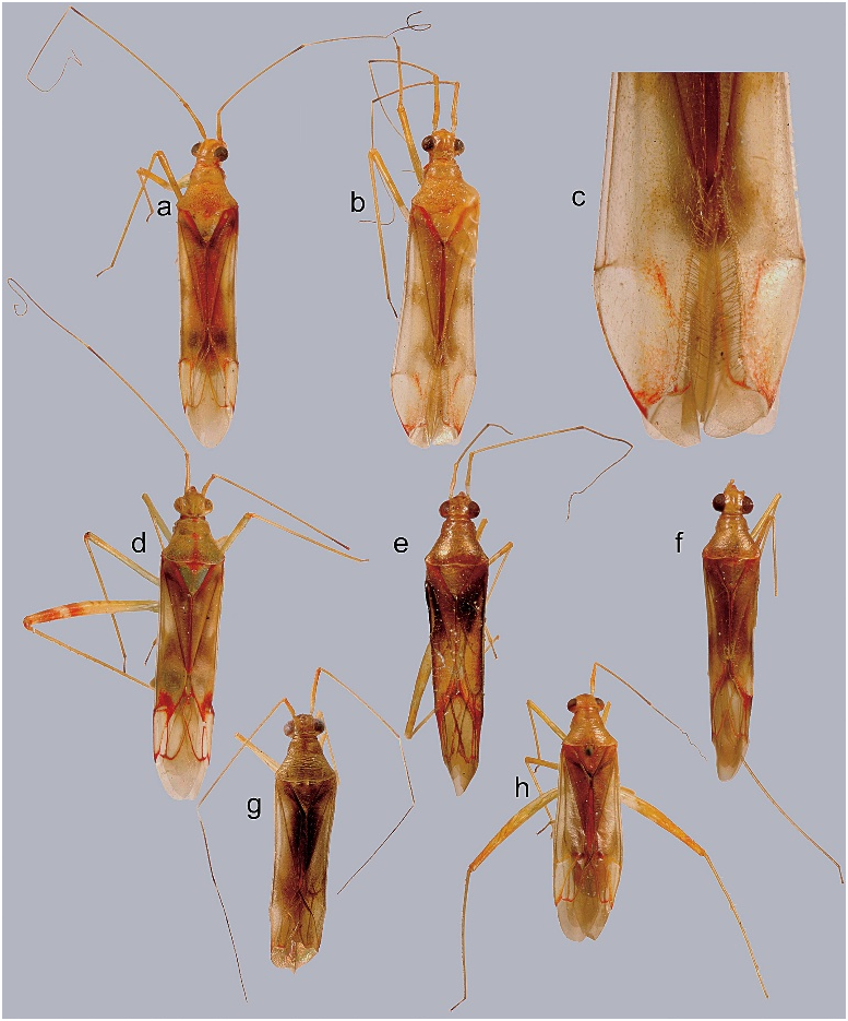
Fig. 1. Dorsal habitus photographs. a. Morobea longipes (male). b. M. longipes (female). c. close-up of cuneus of M. longipes (female). d. M. spectabilis. e. Wumea cassisi. f. W. cylpealis. g. Sagittacopula carvalhoi. h. S. rufescens.
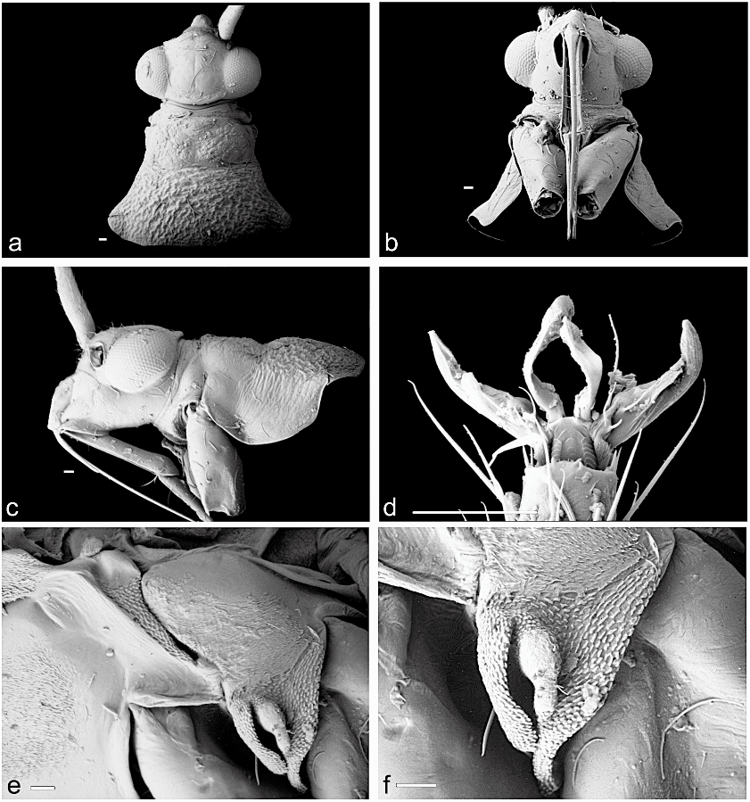
Fig. 2. Scanning electron micrographs of Morobea longipes. a. Head and pronotum, dorsal view. b. Head and pronotum, ventral view. c. Head and pronotum, lateral view. d. Pretarsus, ventral view. e. Meso- and metathorax, lateral view. f. Evaporative area (scale bars 5 50 mm).

Fig. 3. Scanning electron micrographs of the base of second antennal segment in Morobea. a. M. longipes. b. M. spectabilis (scale bars 5 30 mm).

Fig. 4. Male genitalia of Morobea longipes. a. Genital capsule, ventral view. b. Right paramere, ventral view. c. Left paramere, ventral view. d. Aedeagus, left lateral view. e. Distal portion of aedeagus, right lateral view (dvp dorsal projection of ventral margin; sgp secondary gonopore; sr sclerotized ring; vvp ventral projection of ventral margin; scale bars 5 0.1 mm).

Fig. 5. Male genitalia of Morobea spectabilis. a. Genital capsule, ventral view. b. Right paramere, ventral view. c. Left paramere, ventral view. d. Portion of aedeagus, left lateral view. e. Aedeagus, ventral view (lsc lobal sclerite; thl thecal lobe; vvp ventral projection of ventral margin; scale bars 5 0.1 mm).
No known copyright restrictions apply. See Agosti, D., Egloff, W., 2009. Taxonomic information exchange and copyright: the Plazi approach. BMC Research Notes 2009, 2:53 for further explanation.
|
Kingdom |
|
|
Phylum |
|
|
Class |
|
|
Order |
|
|
Family |
Morobea Carvalho
| WALL, MICHAEL A. 2007 |
Morobea
| Carvalho, J. C. M. 1987: 181 |