
Morobea longipes Carvalho, 1987
|
publication ID |
https://doi.org/10.1206/0003-0082(2007)3558[1:ANGANS]2.0.CO;2 |
|
persistent identifier |
https://treatment.plazi.org/id/03AF87C7-FC51-FFAF-FF03-BD70297AF940 |
|
treatment provided by |
Carolina (2021-08-30 06:24:15, last updated by Plazi 2023-11-05 21:58:53) |
|
scientific name |
Morobea longipes Carvalho |
| status |
|
Morobea longipes Carvalho View in CoL figures 1a–c View Fig , 2 View Fig , 3a View Fig , 4 View Fig
Morobea longipes Carvalho 1987: 181 View in CoL (n.sp.).
DIAGNOSIS: Differentiated from similar taxa by characters listed in generic diagnosis and discontinuous red maculations along dorsal margin of propleuron, most prominent anteriorly; genital capsule without lateral tubercle; aedeagus with relatively small, serrated, hatchetlike lobal sclerite ( fig. 4d–e View Fig ). Females with long medially oriented setae on posterior margin of corium ( fig. 1c View Fig ).
REDESCRIPTION: Coloration ( fig. 1a View Fig ): Head yellowish brown with vague red maculations on clypeus and mandibular plates. Antennae yellowish brown becoming darker distally. Prothorax yellowish brown, tinged with green in some specimens; disc and collar undifferentiated; dorsal margin of propleuron with red maculation at anterolateral margin, undifferentiated ventrally. Scutellum yellowish brown, sometimes suffused with blue-green. Hemelytra predominately yellowish brown; margin of clavus weakly suffused with red along scutellar margin; cuneus ringed with red. Abdomen yellow with dorsal surface of male genital capsule dark brown. Legs predominately yellow, becoming brown distally, ventral surface suffused with blue-green; metafemur with pale brown spots on anterior surface and red distally. Structure: As in generic description with the following additions. Head with weak sulcus along length of vertex and frons; antennal segment II weakly swollen as base, swollen portion with dense field of short hairs oriented perpendicular to antennal surface ( fig. 3a View Fig ). Cuneus 2.5 times longer than broad, subtriangular. Small cell of hemelytral membrane less than one-third length of cuneus. Genitalia ( fig. 4 View Fig ): Genital capsule broadly subconical with large aperture; dorsal projection of ventral rim roughly symmetrical, slightly skewed to left, broadly rounded; ventral projection of ventral rim asymmetrical skewed left, acute. Parameres as in generic description; right paramere with small apical lobe. Theca sclerotized basally, distally convoluted into several large flanges and hooks; apical serrate process relatively small and hatchetlike. Aedeagus as in generic description.
FEMALE ( fig. 1b View Fig ): Similar to males except antennal segment II simple; cuneus 1.5 times longer than broad, subrectangular; posterior margin of corium with large medially oriented setae, hairs longer than width of large cell ( fig. 1c View Fig ); small cell of hemelytral membrane minute, almost obsolete; medial portion of membrane with large field of short velutinous hairs. Genitalia: Ovipositor as in generic description. Internal genitalia unknown due to damaged material.
MEASUREMENTS: Males (N 5 2): L-Tot 5.88–6.11, Cly-Cun 4.42–4.44, L-Hd 0.59– 0.62, L-Prn 0.83–0.93, L-Sct 0.59–0.62, L- Cun 0.92–1.03, W-Hd 0.57–0.79, W-Prn 1.18– 1.21, W-Sct 0.57–0.61, IOD 0.35–0.37, L-AI 0.86–1.01, L-AII 2.88–3.19, L-AIII 1.67–1.67, L-AIV 2.11, L-mtf 3.55, L-mtt 5.87. Females (N 5 2): L-Tot 6.06, Cly-Cun 4.73, L-Hd 0.62, L-Prn 0.83–0.90, L-Sct 0.55–0.65, L-Cun 1.05– 1.13, W-Hd 0.79, W-Prn 1.17–1.29, W-Sct 0.61–0.64, IOD 0.41, L-AI 1.02, L-AII 3.18, L- AIII 1.72 , L-AIV 1.65, L-mtf 3.30, L-mtt 5.61 .
HOSTS: Collected from Pipturus sp. (Urticaceae) .
DISTRIBUTION: Eastern New Guinea.
SPECIMENS EXAMINED: PAPUA NEW GUINEA: Morobe: Wau, 7.3333 ° S 146.71667 ° E, 1100 m, 16 Aug 1972, G. G. E. Scudder, 1♀ (AMNH_PBI 00053260) (BPBM); 12 Aug 1977, W. C. Gagne, 1♀ (AMNH_PBI 00053259) (BPBM); 20 Jul 1974, A.D. Hart, Pipturus sp. (Urticaceae) , 1♀ (AMNH_PBI 00053258) (BPBM); 21 Apr 1979, W. C. Gagne, 13 (AMNH_PBI 00053261) (BPBM).
Carvalho, J. C. M. 1987. New genera and new species of Miridae from Papua New Guinea (Hemiptera). Revista Brasileira de Biologia 47: 177 - 187.
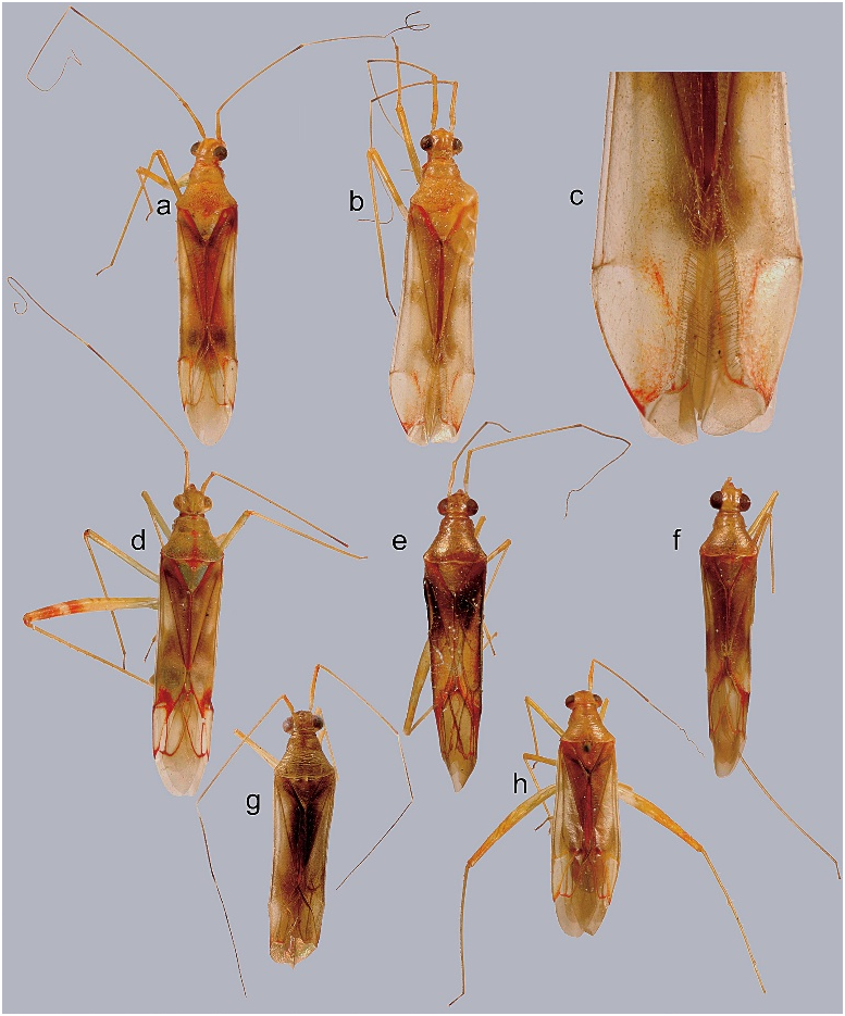
Fig. 1. Dorsal habitus photographs. a. Morobea longipes (male). b. M. longipes (female). c. close-up of cuneus of M. longipes (female). d. M. spectabilis. e. Wumea cassisi. f. W. cylpealis. g. Sagittacopula carvalhoi. h. S. rufescens.
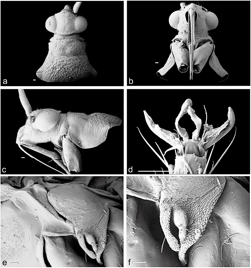
Fig. 2. Scanning electron micrographs of Morobea longipes. a. Head and pronotum, dorsal view. b. Head and pronotum, ventral view. c. Head and pronotum, lateral view. d. Pretarsus, ventral view. e. Meso- and metathorax, lateral view. f. Evaporative area (scale bars 5 50 mm).

Fig. 3. Scanning electron micrographs of the base of second antennal segment in Morobea. a. M. longipes. b. M. spectabilis (scale bars 5 30 mm).

Fig. 4. Male genitalia of Morobea longipes. a. Genital capsule, ventral view. b. Right paramere, ventral view. c. Left paramere, ventral view. d. Aedeagus, left lateral view. e. Distal portion of aedeagus, right lateral view (dvp dorsal projection of ventral margin; sgp secondary gonopore; sr sclerotized ring; vvp ventral projection of ventral margin; scale bars 5 0.1 mm).
No known copyright restrictions apply. See Agosti, D., Egloff, W., 2009. Taxonomic information exchange and copyright: the Plazi approach. BMC Research Notes 2009, 2:53 for further explanation.