
Prolivatis Emeljanov
|
publication ID |
https://doi.org/ 10.5281/zenodo.196171 |
|
DOI |
https://doi.org/10.5281/zenodo.6203210 |
|
persistent identifier |
https://treatment.plazi.org/id/03AF0519-027B-C94B-FF3C-FB6BF743FF24 |
|
treatment provided by |
Plazi |
|
scientific name |
Prolivatis Emeljanov |
| status |
|
Prolivatis Emeljanov View in CoL View at ENA
Prolivatis Emeljanov, 1995: 781 View in CoL ; 1996: 138–139. Type species: P. gorochovi Emeljanov, 1995 View in CoL , by original designation.
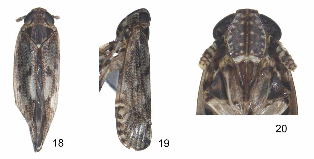
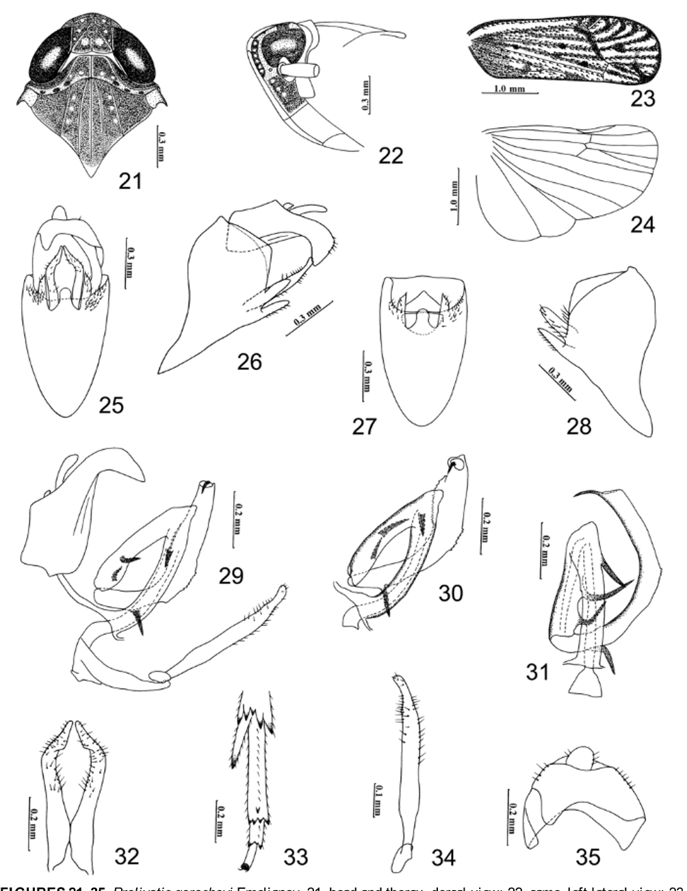
Diagnosis. Small, brown to dark brown delphacids. Vertex quadrate, slightly wider at base than long, anterior margin of vertex rounded, projecting in front of eyes ( Figs 18 View FIGURES 18 – 20 , 21 View FIGURES 21 – 35 ), in profile rounded to frons ( Figs 19 View FIGURES 18 – 20 , 22 View FIGURES 21 – 35 ), submedian carinae originating from near base of lateral carinae, converging and meeting before apex of vertex, forming isosceles triangle at base of vertex ( Figs 18 View FIGURES 18 – 20 , 21 View FIGURES 21 – 35 ), Y-shaped carina with stem faint ( Figs 18 View FIGURES 18 – 20 , 21 View FIGURES 21 – 35 ). Median carinae of frons simple ( Fig. 20 View FIGURES 18 – 20 ). Lateral carinae of pronotum sinuate, not reaching posterior margin ( Figs 18 View FIGURES 18 – 20 , 21 View FIGURES 21 – 35 ). Mesonotum with five carinae ( Figs 18 View FIGURES 18 – 20 , 21 View FIGURES 21 – 35 ). Antennal segments distinctly elongate ( Figs 18–20 View FIGURES 18 – 20 ). Forewings with more or less continuous transverse veins before apical area, with well-defined bend of the membrane when wings are folded ( Figs 18, 19 View FIGURES 18 – 20 ), forewings with 12 closed apical cells, Sc+R with two branches before subapical transverse nodal line ( Fig. 23 View FIGURES 21 – 35 ). Post tibial spur spine-like, without teeth on inner margin ( Fig. 33 View FIGURES 21 – 35 ), hind tibiae with 3 lateral teeth, metatarsomere I with five apical teeth, metatarsomere II with row of teeth, marginal teeth considerably longer than others. Male pygofer strongly excavated ventrocaudally, with large median process on midventral margin, lateroventral margin with lanceolate lobes ( Figs 25–28 View FIGURES 21 – 35 ). Aedeagus 3-segmented, distal segment arched clockwise ( Fig. 31 View FIGURES 21 – 35 ). Parameres slender and narrow, convergent apically ( Figs 25, 32 View FIGURES 21 – 35 ). Anal segment with anterior margin sinuate, strongly produced at left side ( Figs 25, 35 View FIGURES 21 – 35 ).
Remarks. Prolivatis is similar to Punana by the mesonotum having 5 longitudinal carinae, by the hind tibiae having 3 lateral teeth, by the forewings having 12 closed apical cells and by the Sc+R having two branches before subapical transverse nodal line. However, Prolivatis differs from Punana by its distinctly elongate antennae (reaching near apex of postclypeus, segment II about 2.3 times longer than I) (in Punana antennal segments short, at most reaching frontoclypeal suture, segment II a little longer than I); male pygofer having lanceolate lobe at each side of lateroventral margins (in Punana without lanceolate lobes) and laterodistal angle of male anal segment strongly produced at left side (in Punana not produced). Distribution. Southern China (Hainan Province) new record, Vietnam.
No known copyright restrictions apply. See Agosti, D., Egloff, W., 2009. Taxonomic information exchange and copyright: the Plazi approach. BMC Research Notes 2009, 2:53 for further explanation.
|
Kingdom |
|
|
Phylum |
|
|
Class |
|
|
Order |
|
|
Family |
Prolivatis Emeljanov
| Qin, Dao-Zheng & Zhang, Ya-Lin 2010 |
Prolivatis
| Emeljanov 1995: 781 |