
Atypena acutala, Irfan & Zhang & Peng, 2022
|
publication ID |
https://doi.org/10.11646/megataxa.8.1.1 |
|
DOI |
https://doi.org/10.5281/zenodo.7541375 |
|
persistent identifier |
https://treatment.plazi.org/id/03AE87CE-BD5C-FF80-FF2E-3CBEFD72F787 |
|
treatment provided by |
Plazi (2023-01-11 20:01:21, last updated 2024-11-28 12:31:43) |
|
scientific name |
Atypena acutala |
| status |
sp. nov. |
Atypena acutala sp. nov. (ḰẌṞƉƦ)
Figures 11–13 View FIGURE 11 View FIGURE 12 View FIGURE 13 , 16 View FIGURE 16
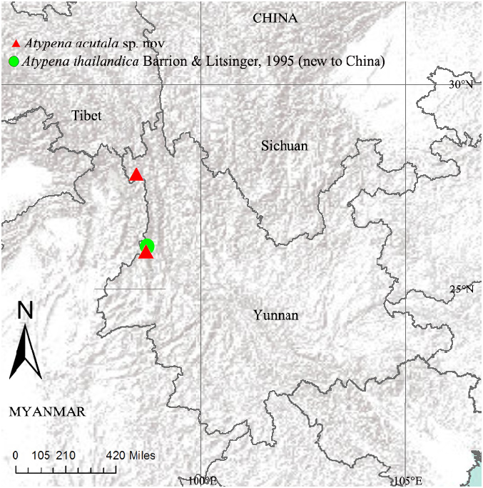
Types. Holotype ♂, CHINA, Yunnan; Lushui County, 53 km W of Nujiang Road on Pianma Road, 25.97182°N, 98.70047°E, alt. 3140m, 15 October 2002, D. H. Kavanaugh leg. ( DHK –2002–49) GoogleMaps . Paratypes: 4♀, same data as holotype GoogleMaps ; 1♂ 1♀, Nujiang Prefecture , Nujiang State Nature Reserve, No. 12 bridge Camp area, 16.3 km W of Gongshan, 27.71503°N, 98.50244°E, alt. 2775m, 15–19 July 2000, Heng-mei Yan, D. H. Kavanaugh, Charles Griswold, Hong-bin Liang, Darrell Ubick and Da-zhi Dong leg. (00–QD) GoogleMaps .
Etymology. This epithet derives from the Latin adjective “ acutalis ”, meaning “pointed” and referring to pointed terminal apophysis in male palp.
Diagnosis. The new species resembles Atypena cirrifrons (Heimer, 1984) in having the similar cephalic lobe,long terminal apophysis with pointed end and epigyne with a broad median plate ( Figs 11A–C View FIGURE 11 , 12A–C View FIGURE 12 , 13A, C, D View FIGURE 13 ; Heimer, 1984, fig. 2, 7), but can be distinguished by the distal suprategular apophysis basally broad with blunt end and pointing towards the terminal apophysis in A. acutala sp. nov. ( Fig. 11A–C View FIGURE 11 ), whereas curved and pointing towards the cymbium in A. cirrifrons ( Song, Zhu & Chen, 1999, fig. 114Q). Embolus slightly curved and distal end bifurcated in A. acutala sp. nov. ( Fig. 11A– C View FIGURE 11 ), whereas straight and not bifurcated in A. cirrifrons (Heimer, 1984, fig. 5, 6). Epigyne: The copulatory ducts forming two loops and located close to each other in A. acutala sp. nov. ( Fig. 12A, B View FIGURE 12 ), whereas in A. cirrifrons the CD form one loop and are located further from each other (Heimer, 1984, fig. 7, 8).
Description. Male (holotype, Fig. 13A–D View FIGURE 13 ): Total length: 1.63. Carapace 0.72 long, 0.59 wide, the cephalic region pale with cephalic lobe 0.14 high; fovea, cervical and radial grooves distinct. Carapace with distinct cephalic lobe. Cephalic pits present in between the posterior lateral and posterior median eyes. Ocular quad densely covered with macrosetae. Clypeus 0.14 high. Sternum wider than long, pale, sparsely covered with microsetae. Labium wider than long. Maxillae long, distal end broad with scopulae. Chelicerae with two promarginal and four retromarginal teeth. Eye region narrow, AER recurved, PER procurved and slightly wider than AER. Posterior median eyes located on the cephalic lobe. Eye sizes and interdistances: AME 0.06, ALE 0.07, PME 0.05, PLE 0.06, AME–AME 0.02, PME–PME 0.11, AME–ALE, 0.03, PME–PLE 0.12, ALE–ALE 0.30, PLE–PLE 0.32, ALE–PLE contiguous. Length of legs: I 2.16 (0.61, 0.73, 0.48, 0.34), II 2.08 (0.59, 0.69, 0.47, 0.33), III 1.7 (0.53, 0.54, 0.37, 0.26), IV 2.31 (0.64, 0.74, 0.58, 0.35). Leg formula IV-I-II-III. Tm I and Tm IV present. Tibial spine formula: 1-1-1-1. Abdomen 0.91 long, 0.63 wide, oval, dorsally with two black longitudinal bands, ventral side pale without any marking.
Palp ( Fig. 11A–D View FIGURE 11 ): Tibia conic, with two retrolateral trichobothria and some short or long setae on all surfaces; reterolateral tibial apophysis with blunt end and a small sclerotized tooth at the base; paracymbium highly sclerotized, hook-shaped, the inner side excavated to a shallow groove, distal end with a short projection; tegulum with a transparent protegulum; distal suprategular apophysis highly sclerotized and basally broad with blunt end; radix highly sclerotized giving rise to long pointed terminal apophysis, median membrane partially covering the embolus; dorsal projection of embolic plate with broad round distal end and overlapping the suprategulum in prolateral view; embolus highly sclerotized, slightly curved with bifurcated tip giving rise to embolic tip and a sclerotized lobe.
Female (one of the paratypes, Fig. 14A, B View FIGURE 14 ): Total length: 1.81. Carapace 0.66 long, 0.63 wide, the cephalic region slightly elevated, pale; fovea, cervical and radial grooves distinct. Clypeus 0.13 high. Sternum wider than long, pale, sparsely covered with microsetae. Labium wider than long. Maxillae long, distal end broad with scopulae. Chelicerae with three promarginal and five retromarginal teeth. Eye region narrow, AER recurved, PER straight and slightly wider than AER. Eye sizes and interdistances: AME 0.05, ALE 0.07, PME 0.06, PLE 0.06, AME–AME 0.04, PME–PME 0.05, AME–ALE, 0.03, PME–PLE 0.04, ALE–ALE 0.29, PLE–PLE 0.32, ALE–PLE contiguous. Length of legs: I 2.45 (0.70, 0.81, 0.55, 0.39), II 2.33 (0.66, 0.79, 0.51, 0.37), III 1.96 (0.57, 0.64, 0.48, 0.27), IV 2.66 (0.79, 0.88, 0.65, 0.34). Leg formula IV-I-II-III. Tm I and Tm IV present. Tibial spine formula: 1-1-1-1. Abdomen 1.15 long, 0.81 wide, cylindrical, dorsally with two black longitudinal bands; ventral side pale without any marking.
Epigyne ( Fig. 12A–C View FIGURE 12 ): Simple, with a broad median plate. copulatory openings located on the base of the median plate at the posterior end of the epigyne. Copulatory ducts forming two loops before joining to the spermathecae. Fertilization ducts directed mesally. Spermathecae globular, separated from each other by about 1.5 their diameters.
Distribution. Known only from the type locality ( Fig. 16 View FIGURE 16 ).
Atypena thailandica Barrion & Litsinger, 1995 (new to China)
Figures 15 View FIGURE 15 , 16 View FIGURE 16
Atypena thailandica Barrion & Litsinger, 1995: 480 View in CoL , figs 293a–c, 294a–e (Dmf); World Spider Catalog, 2020.
Material examined. 1♀, CHINA, Yunnan, Lushui County, Pianma Township, Chanyan He , 9.3 km ESE Pianma, 25.99363°N, 98.66651°E, alt. 2470m, mixed broad leaf deciduous and evergreen forest; beating understory vegetation, 14 May 2005, Charles Griswold leg. ( CGY107 ) GoogleMaps .
Distribution. China (Yunnan), Thailand.
Barrion, A. T. & Litsinger, J. A. (1995) Riceland spiders of South and Southeast Asia. CAB International, Wallingford, UK, xix + 700 pp.
Song, D. X., Zhu, M. S. & Chen, J. (1999) The spiders of China. Hebei Science and Technology Publishing House, Shijiazhuang, 640 pp.
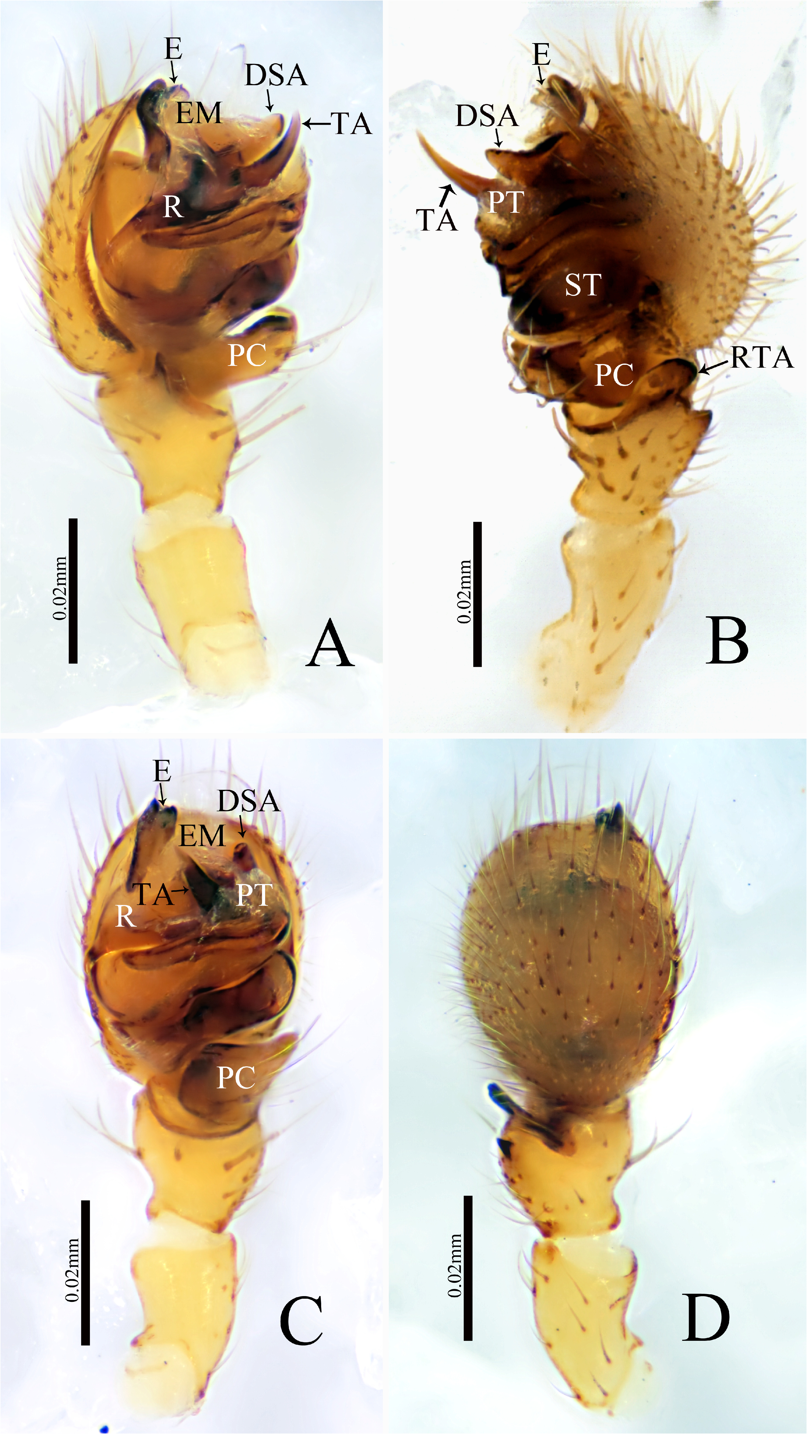
FIGURE 11. Atypena acutala sp. nov., male palp of holotype. A prolateral view B retrolateral view C ventral view D dorsal view.
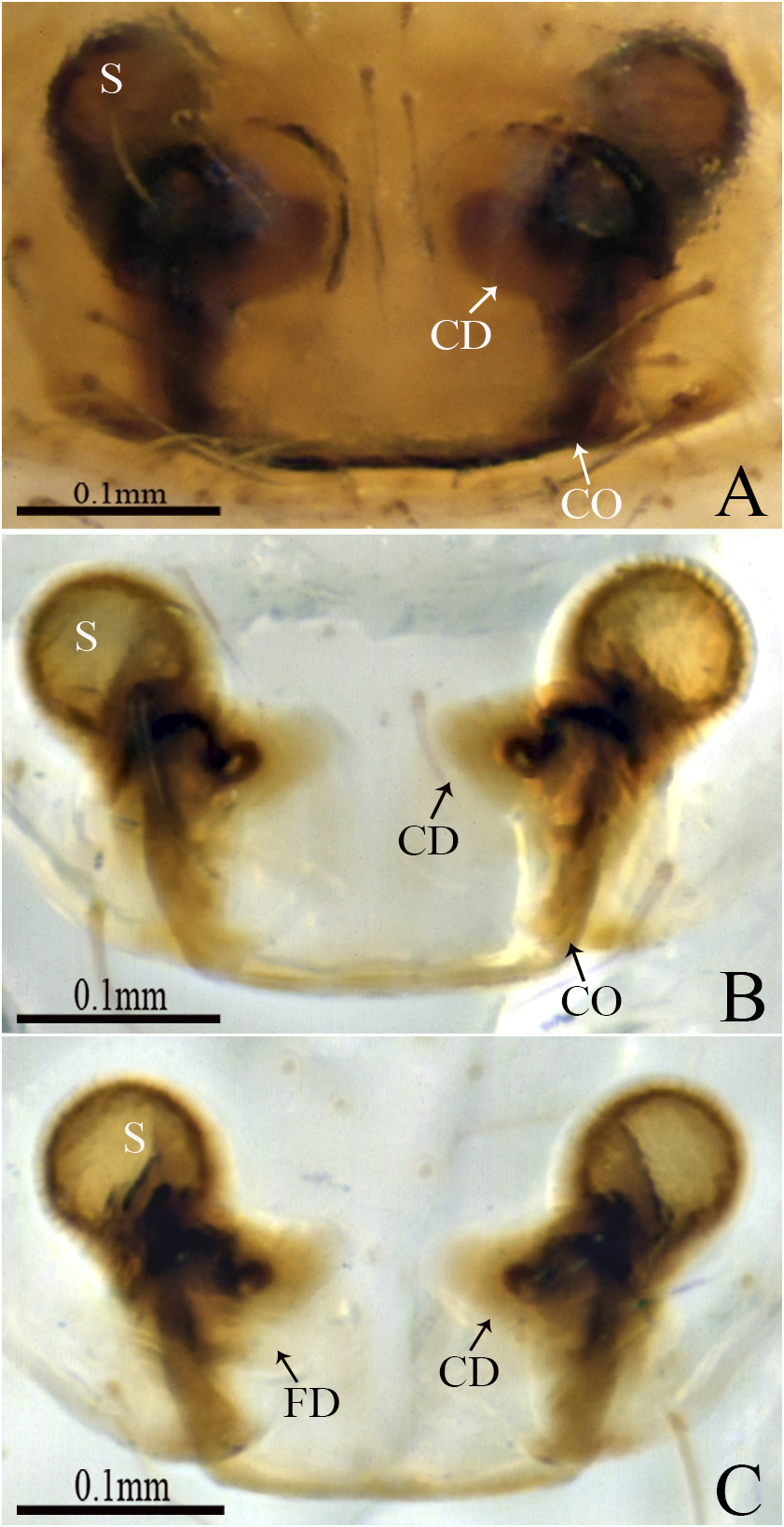
FIGURE 12. Atypena acutala sp. nov., female (one of paratypes). A, B Epigyne, ventral view C Vulva, dorsal view.
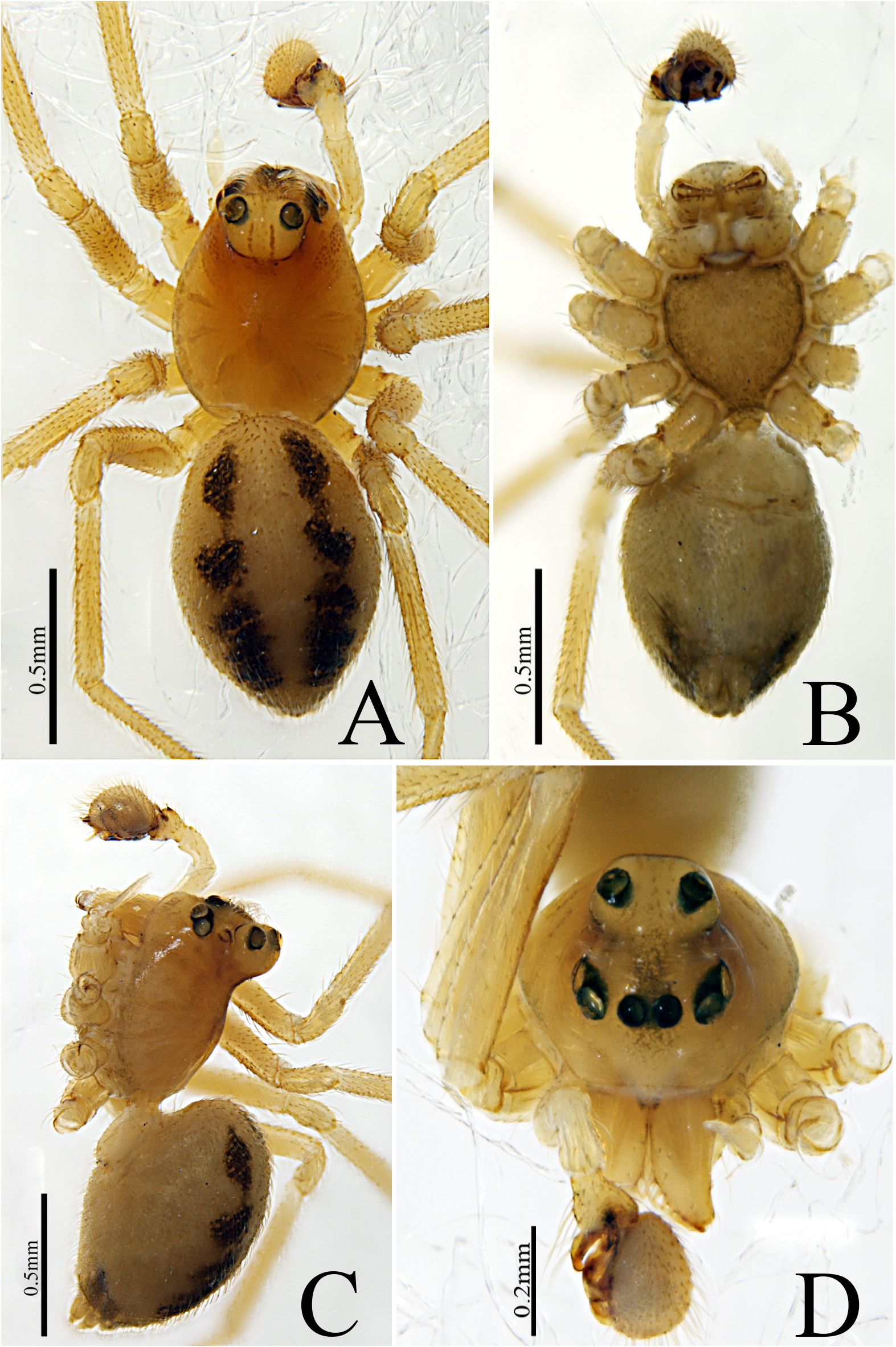
FIGURE 13. Atypena acutala sp. nov., male holotype. A Habitus, dorsal view B Habitus, ventral view C Habitus, lateral view D Habitus, frontal view.
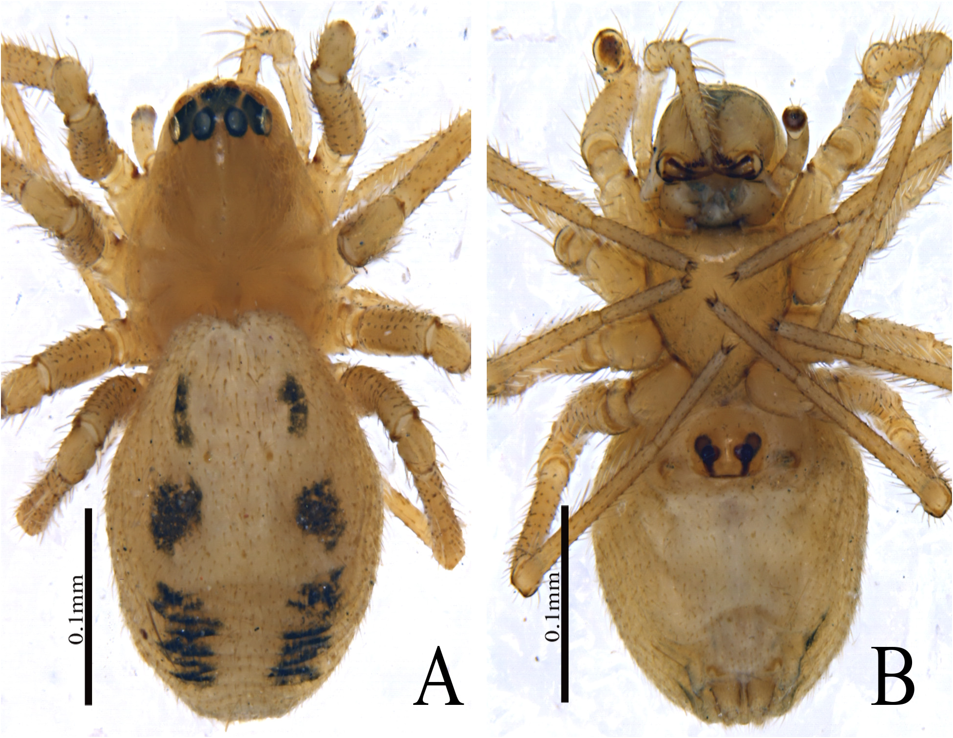
FIGURE 14. Atypena acutala sp. nov., female (one of paratypes). A Habitus, dorsal view B Habitus, ventral view.
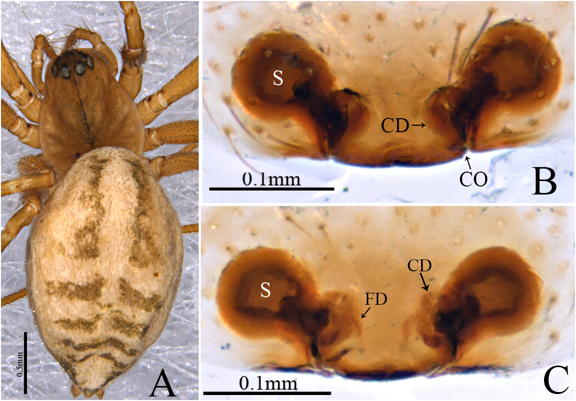
FIGURE 15. Atypena thailandica Barrion & Litsinger, 1995 f, female. A Habitus, dorsal view B Epigyne, ventral view C Vulva, dorsal view.
No known copyright restrictions apply. See Agosti, D., Egloff, W., 2009. Taxonomic information exchange and copyright: the Plazi approach. BMC Research Notes 2009, 2:53 for further explanation.
|
Kingdom |
|
|
Phylum |
|
|
Class |
|
|
Order |
|
|
Family |
|
|
Genus |
Atypena acutala
| Irfan, Muhammad, Zhang, Zhi-Sheng & Peng, Xian-Jin 2022 |
Atypena thailandica
| Barrion, A. T. & Litsinger, J. A. 1995: 480 |
1 (by plazi, 2023-01-11 20:01:21)
2 (by ExternalLinkService, 2023-01-11 20:42:56)
3 (by valdenar, 2023-01-16 15:17:22)
4 (by ExternalLinkService, 2023-01-16 16:09:26)
5 (by valdenar, 2023-01-20 17:51:04)
6 (by ExternalLinkService, 2023-01-26 17:19:48)
7 (by ExternalLinkService, 2023-01-26 18:48:55)
8 (by plazi, 2023-11-08 12:27:32)
9 (by ExternalLinkService, 2023-11-08 20:03:52)