
Phylloicus obliquus Navás 1932
|
publication ID |
https://doi.org/ 10.11646/zootaxa.4748.2.8 |
|
publication LSID |
lsid:zoobank.org:pub:4E2A3042-3B8B-4288-92D3-1B3E92ECDCB2 |
|
DOI |
https://doi.org/10.5281/zenodo.3705018 |
|
persistent identifier |
https://treatment.plazi.org/id/03AC6910-FFBB-971B-2FF7-4D2CFBFB6FA0 |
|
treatment provided by |
Plazi |
|
scientific name |
Phylloicus obliquus Navás 1932 |
| status |
|
Phylloicus obliquus Navás 1932 View in CoL
Figs. 2–5 View FIGURE 2 View FIGURE 3 View FIGURE 4 View FIGURE 5 .
Description. Larva: body length 1 cm (n = 4).
Head: About 1.5x longer than wide, dark brown with yellowish-brown rounded spots (muscular scars) on vertex and postgenal and genal areas ( Figs. 2A, 2C View FIGURE 2 ). Chaetotaxy as in Fig. 2A View FIGURE 2 . Labrum and regions around stemmata yellowish brown. Labrum trapezoidal; anterior margin rounded with dorsal row with a maximum of 14 bristle-like setae of varying sizes; brushes of yellow setae on anterolateral margins ( Fig. 2A View FIGURE 2 ). Frontoclypeal apotome triangular, broader anteriorly, with median constriction ( Fig. 2A View FIGURE 2 ). Ventral apotome triangular, not reaching posterior region of head; ventral ecdysial line well-marked ( Fig. 2B View FIGURE 2 ). Gena with conspicuous band of scabrous tegument anteriorly ( Fig. 2B, 2C View FIGURE 2 ). Mandibles each with two well-developed teeth, apical tooth acute and pointed apicad and subapical tooth blunt and obtuse; inner margin bearing dense row of yellow stout setae; dorsolateral margin with two stout setae near base ( Fig. 2D View FIGURE 2 ).
Thorax: Pronotum yellowish-brown, with light-brown muscular scars posterolaterally and posteriorly, posterior margin dark brown ( Fig. 2E View FIGURE 2 ); anterolateral margins strongly projecting, with apices hooked anteroventrad ( Fig. 2C View FIGURE 2 ), each with three long bristle-like setae on posterior half of inner margin and one short, mesal bristle-like seta dorsally ( Fig. 2E View FIGURE 2 ). pronotum mesolaterally with 8–10 bristle-like setae ( Fig. 2E View FIGURE 2 ). Mesonotum membranous, with pair of irregular central sclerites, weakly sclerotized, their posterior borders not clearly delimited; lateral sclerites small, slightly sclerotized; sa 1 each with short seta, sa 2 without setae, and each sa 3 consisting of anterolateral sclerite with six long setae ( Figure 2E View FIGURE 2 ). Metanotum fully membranous; sa 1 without setae, each sa 2 with long seta, each sa 3 consisting of anterolateral sclerite with five long setae ( Fig. 2E View FIGURE 2 ). Legs golden-brown, with few setae (chaetotaxy as in Figs. 2 View FIGURE 2 F–2H); tarsal claws simple, well developed and more prominent on legs I and II; foretrochantin prominent, sinuous, tapering towards apex and curved dorsad with pointed apex ( Fig. 2F View FIGURE 2 ).
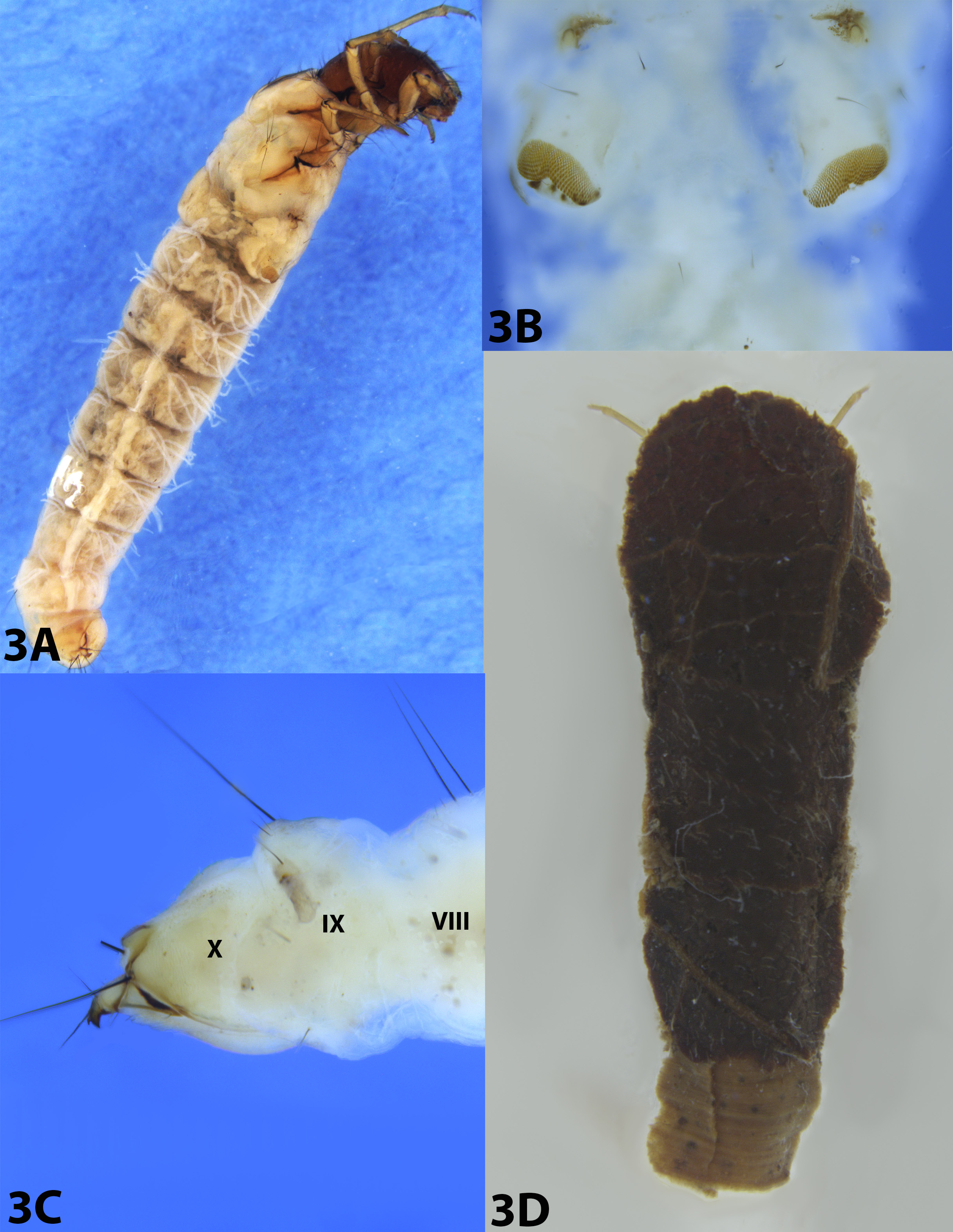
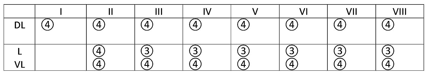
Abdomen: Gills with 3-4 filaments present on segments I–VIII ( Fig. 3A View FIGURE 3 ); shape and position of gills as indicated in Fig. 4 View FIGURE 4 . Segment I with ventrolateral pair of membranous lobes bearing oval sclerotized plates covered by many minute spines ( Fig. 3B View FIGURE 3 ). Dorsal hump often fully retracted. Tergites VIII and IX each with pair of long, dark dorsal setae ( Fig. 3C View FIGURE 3 ); tergite X with five pairs of long, black dorsolateral setae. Anal prolegs each with pair of long, stout, dark setae dorsally; terminal claw strongly sclerotized, almost right-angled, with short accessory claw on dorsal margin ( Fig. 3C View FIGURE 3 ).
Case: Total length 1–1.3 cm (n = 4); flattened, with anterior portion slightly wider than posterior one, 3.5 times longer than wide anteriorly, 5.0 times longer than wide posteriorly; dorsal and ventral surfaces formed by leaf fragments cut into quadrangular shape, overlapped in layers, fastened with silk; dorsal surface with anterior margin convex, larger than the ventral surface, ventral surface with anterior margin straight ( Fig. 3D View FIGURE 3 ).
Description. Pupa (exuviae): Total length 1 cm (n = 1).
Head: Mandibles sickle-shaped, well developed, symmetrically curved mesad, without teeth, acute apically, bearing pair of long basolateral channels and two long, stout, dark setae basolaterally ( Fig. 5A View FIGURE 5 ). Maxillary palps 5-segmented, exceeding bases of anterior coxae. Labial palps 3-segmented, 0.4 times length of maxillary palps. Labrum subtrapezoidal; lateral margins each bearing row of 12–14 long setae on each side ( Fig. 5A View FIGURE 5 ). Clypeus short, bearing three pairs of lateral setae ( Fig. 4B View FIGURE 4 ). Eyes large, two pairs of setae, one on genal area anterior to eye and another on frontal area ( Fig. 5B View FIGURE 5 ). Frons with two pairs of long, dark setae on frontal area between antennae ( Fig. 5B View FIGURE 5 ). Antennae long; scapes long, each with one tuft of four setae ( Fig. 5B View FIGURE 5 ); flagellum about twice as long as body.
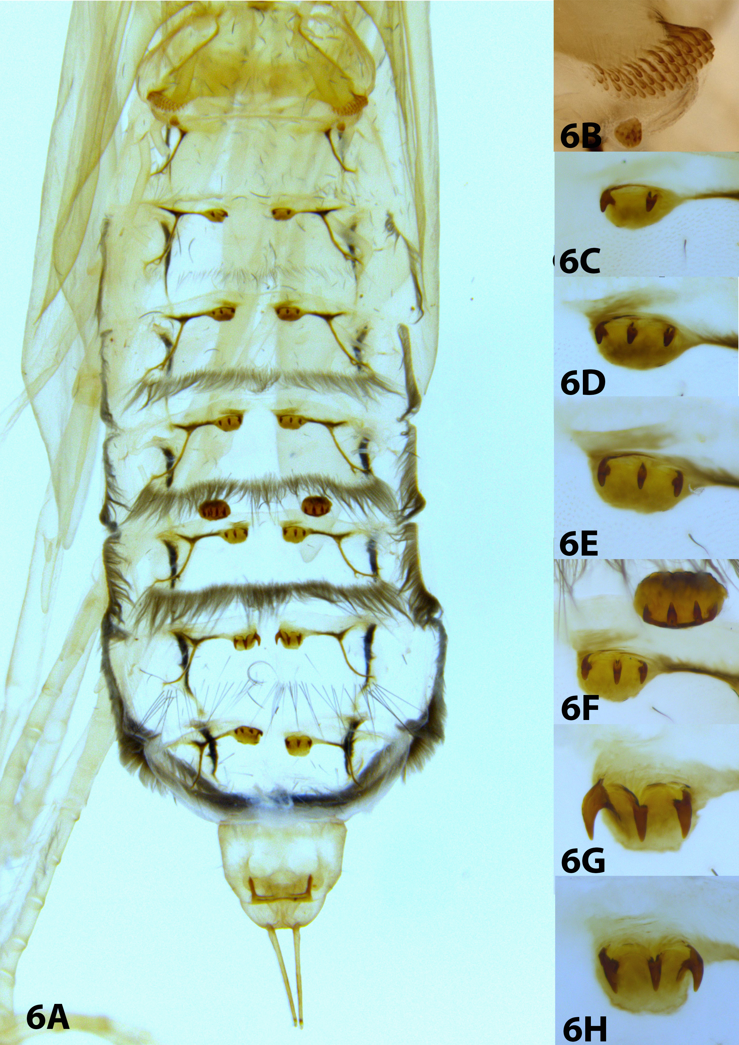
Thorax: Tarsus of each middle leg with row of fine setae; wing pads reaching posterior margin of abdominal segment VI ( Fig. 6A View FIGURE 6 ).
Abdomen: Terga I, III–VIII with paired anterior hook plates, terga I and V with paired posterior hook plates ( Fig. 6A View FIGURE 6 ). Segment I with central prominent subtriangular region; anterior hook plates larger, elliptical, with 3-4 rows of small hooks directed posterad; posterior hook plates elevated, small, rounded, with tiny hooks ( Fig. 6B View FIGURE 6 ). Segment III with pair of oval anterior hook plates with two hooks directed posterad ( Fig. 6C View FIGURE 6 ). Segments IV and V each with pair of oval anterior hook plates, each with three hooks directed posterad ( Figs. 6D and 6E View FIGURE 6 ). Segment V with pair of oval posterior hook plates with four or five hooks directed anterad (one specimen with five hooks on left, four on right) ( Fig. 6F View FIGURE 6 ). Segments VI and VII each with pair of oval anterior hook plates, each with three hooks directed posterad ( Figs. 6F and 6G View FIGURE 6 ). Segment VIII with pair of oval anterior hook plates with three to four hooks directed posterad (one specimen with four hooks on left, three on right) ( Fig. 6H View FIGURE 6 ). Segment IX with apical processes narrow, approximately as long as segment IX, apparently glabrous ( Fig. 6A View FIGURE 6 ).
Material examined. Brazil: Rio de Janeiro: Parque Nacional da Tijuca, Rio Humaitá , 22°57’45.2”S, 043°17’36.5”W, 12-viii- 2016, 494 m, JL Nessimian, LL Dumas, BM Silva & FQ Machado leg., 4 larvae, 4 pupae GoogleMaps , 1 pupal exuviae ( DZRJ).
Taxonomic notes. The color pattern, presence of head muscle scars and their distribution, and the shape of mandibles can be used to distinguish the larva of P. obliquus from those of other species in the genus. The larva of P. obliquus , as well as that of P. abdominalis Ulmer 1905 ( Huamantinco et al. 2005), does not have dark patches on the head as in P. cubanus Banks 1924 ( Botosaneanu & Sykora 1973), nor clear spots on the occipital suture, as in P. aeneus (Hagen 1861) ( Bowles & Flint 1997; Prather 2003). The mandibles of P. obliquus are similar to those of P. pulchrus Flint 1964 , both with dense rows of yellow setae on the inner margins. However, the mandibles of P. obliquus have two teeth, while those of P. pulchrus have three teeth, as in P. camargoi ( Flint 1964; Quinteiro et al. 2011). Despite the head similarities among P. lituratus Banks 1920 ( Rueda Martín 2013), P. camargoi ( Quinteiro et al. 2011) , and P. obliquus , the general shapes of the head, the anterior ventral apotome (anterior margin distinctively concave in P. camargoi and P. lituratus , almost straight in P. obliquus ), and the patterns of distribution of muscle scars in ventral view are different among these species.
Cases dorsally are always arranged in overlapping layers with the anterior margin always convex and the posterior one straight. The cases of P. abdominalis and P. obliquus are more nearly rectangular than in other species, but in the case of P. obliquus the rectangular leaf fragments have a more uniform shape. In P. abdominalis , the case is larger, made with larger fragments and usually with two large leaf fragments forming lateral apices (see illustration by Huamantinco et al. 2005).
The pupa of P. obliquus is similar to that of P. abdominalis in the presence of 2–4 hooks on each abdominal hook plate (in P. obliquus ) and 2–5 (in P. abdominalis ), whereas in P. pulchrus and P. lituratus the plates each have two hooks and in P. camargoi the number ranges from 1 to 4 hooks ( Rueda Martín 2013; Quinteiro & Calor 2011). In addition, in comparison with the species that have the anal processes described or represented in the illustrations (e.g., P. abdominalis , P. pulchrus and P. camargoi ), P. obliquus has no setae on these structures, whereas in these other species there are different numbers and positions of long setae.
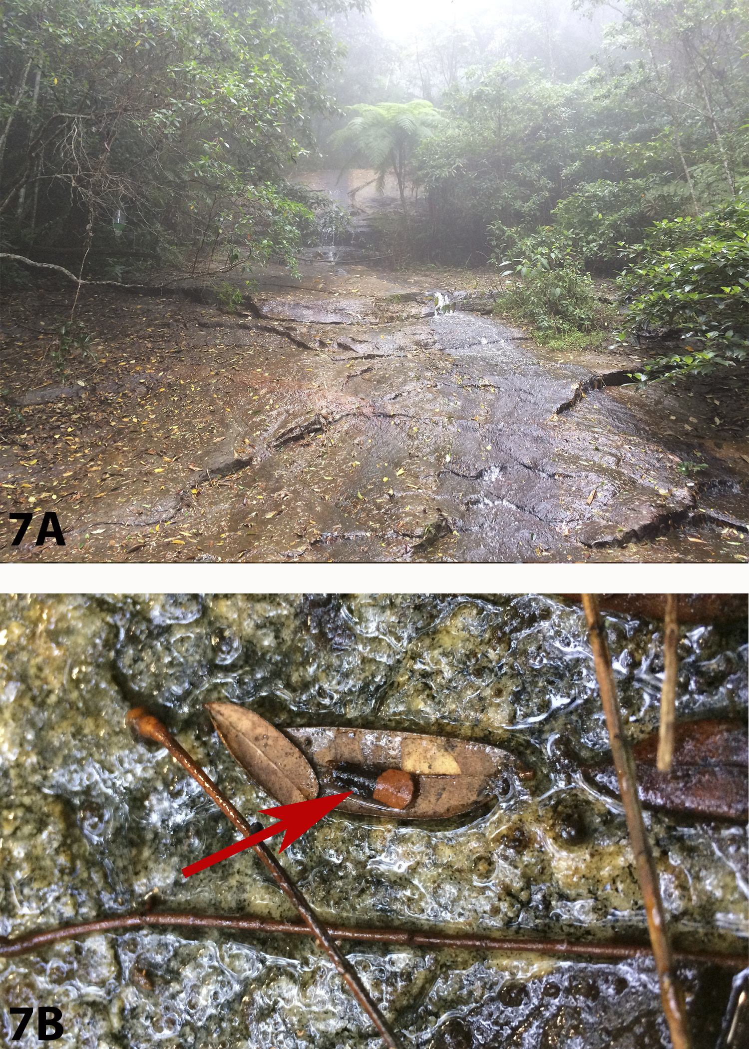
Bionomics: The specimens were collected in Rio Humaitá, a small, shallow, second order stream in reforested Atlantic Forest reserve, the Parque Nacional da Tijuca (Rio de Janeiro state), at 494 m a.s.l. The stream has cold and clean water over a rocky slope, with abundant riparian vegetation ( Fig. 7A View FIGURE 7 ). The larvae were found in small backwater areas with accumulations of litter. However, specimens also were always on the leaves in humid regions out of the water ( Fig. 7B View FIGURE 7 ). Prior to pupation, larvae fasten their cases in rocky substrate or underneath leaves or twigs in hygropetric environments, each closing the case entrance with a leaf fragment.


| BM |
Bristol Museum |
No known copyright restrictions apply. See Agosti, D., Egloff, W., 2009. Taxonomic information exchange and copyright: the Plazi approach. BMC Research Notes 2009, 2:53 for further explanation.
|
Kingdom |
|
|
Phylum |
|
|
Class |
|
|
Order |
|
|
Family |
|
|
Genus |