
Pseudoameiropsis suphankaraytugi, Sönmez, 2019
|
publication ID |
https://doi.org/ 10.3906/zoo-1803-41 |
|
DOI |
https://doi.org/10.5281/zenodo.11130435 |
|
persistent identifier |
https://treatment.plazi.org/id/03AB87A4-227E-F859-537C-FD1F5DC9FCF7 |
|
treatment provided by |
Felipe |
|
scientific name |
Pseudoameiropsis suphankaraytugi |
| status |
sp. nov. |
Pseudoameiropsis suphankaraytugi View in CoL sp. nov. ( Figures 1–4 View Figure 1 View Figure 2 View Figure 3 View Figure 4 )
3.2. Material examined
Holotype: Adult ♀ (ZMADYU 2007/267) dissected and mounted on eight slides, collected from the type locality, 24.11.2007, leg. Serdar Sak, Serdar Sönmez.
Paratypes: One adult ♀ (ZMADYU 2007/268) dissected and mounted on four slides and one whole adult ♀ (ZMADYU 2007/269) preserved in alcohol, collected from the type locality (36°08.315′N, 35°54.598′E), 24.11.2007, leg. Serdar Sak, Serdar Sönmez. One adult ♀ (ZMADYU 2007/270) dissected and mounted on one slide, collected from Samandağ Beach / Hatay (36°05.783′N, 35°56.182′E), 24.11.2007, leg. Serdar Sak, Serdar Sönmez. GoogleMaps
Type locality: Intertidal zone of Viranşehir Beach / Mersin, Turkey (36°08.315′N, 35°54.598′E), interstitial GoogleMaps .
3.3. Description
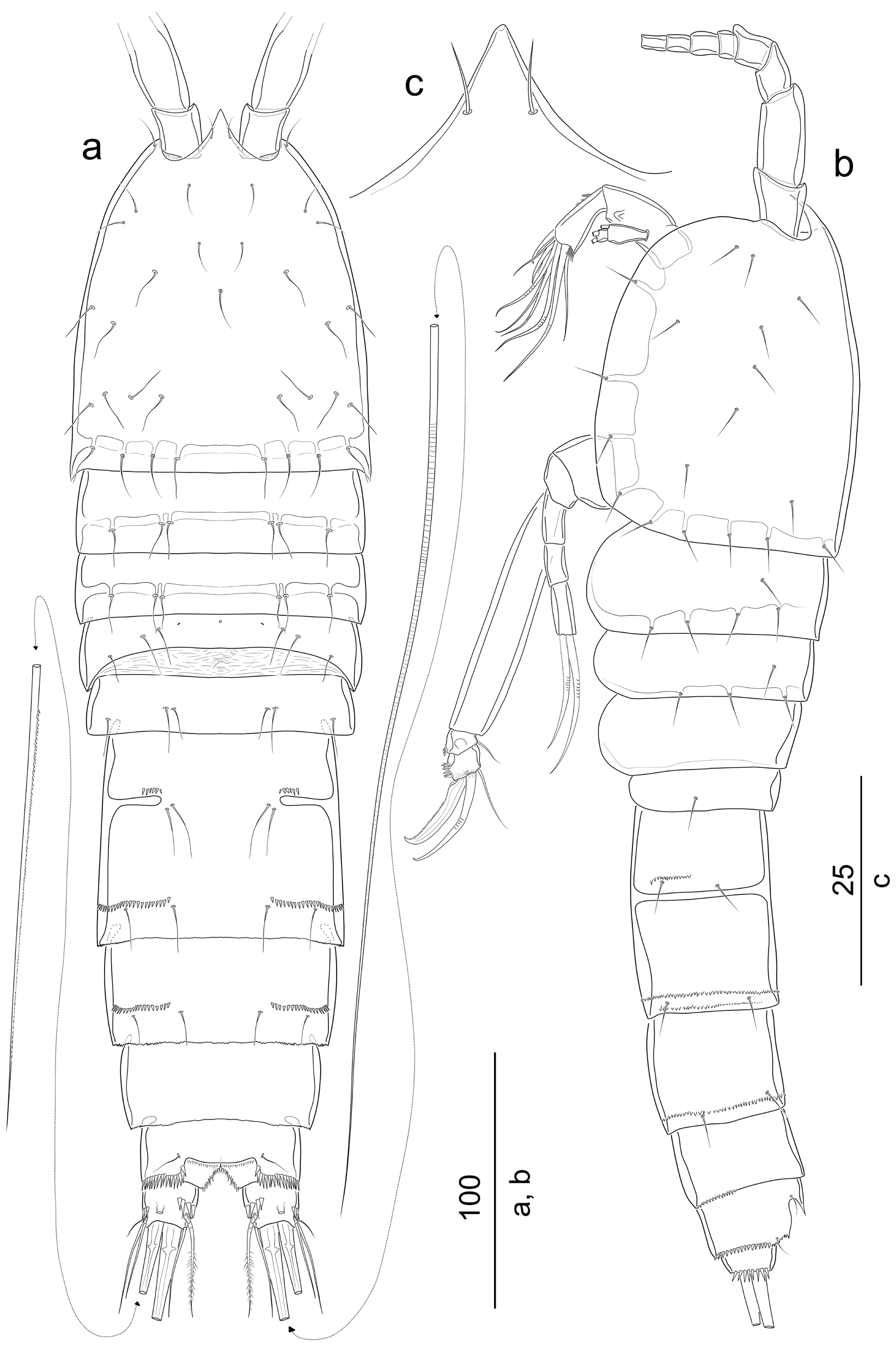
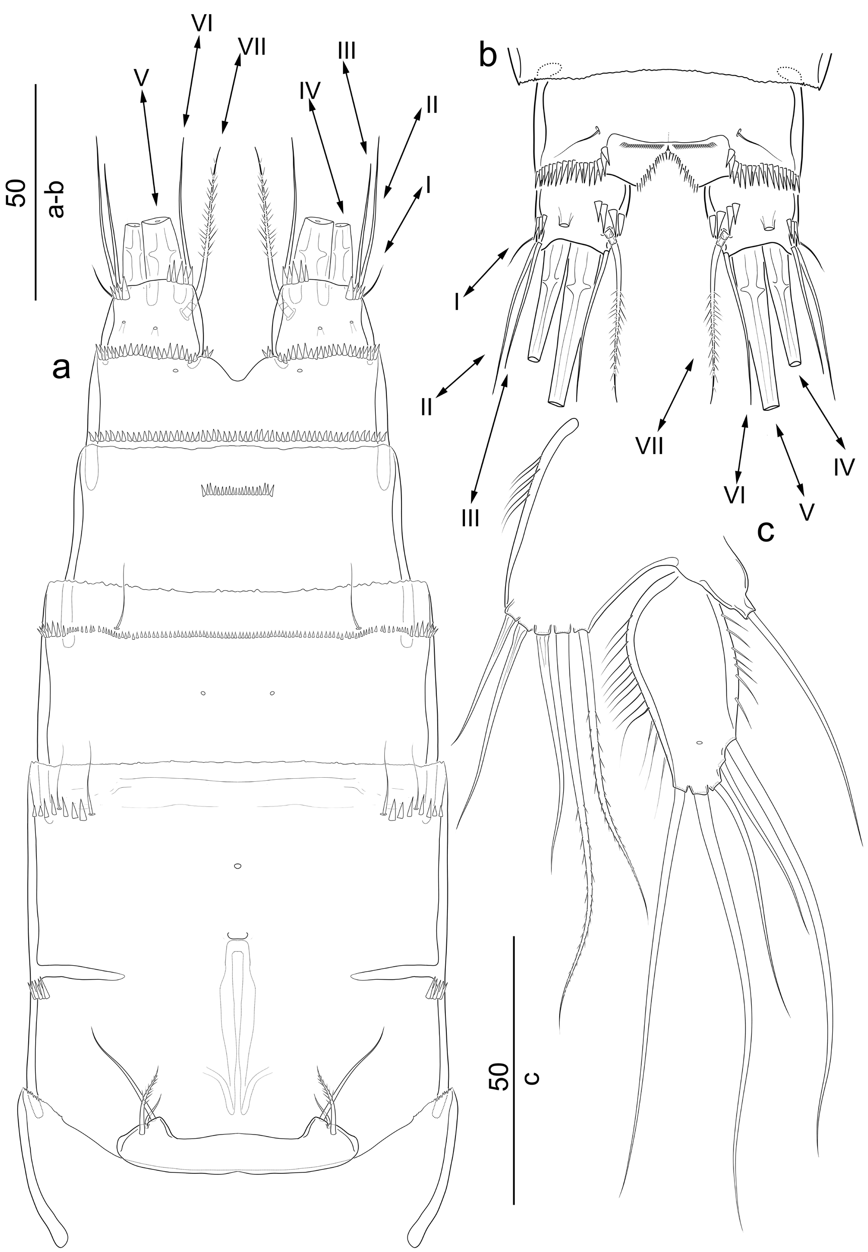
Female: Body ( Figures 1a and 1b View Figure 1 ) semicylindrical; total body length measured from tip of rostrum to posterior end of caudal rami, 393 µm (mean = 421,5 n = 2), widest at posterior part of cephalothorax in dorsal view; gradually tapering posteriorly. Rostrum ( Figure 1c View Figure 1 ) fused to cephalothorax, triangular, with pointed tip, reaching about end of first antennulary segment, with two sensilla. Surface of somites with sensilla as figured except penultimate somite. All somites with plain hyaline frill. Genital doublesomite ( Figures 1a, 1b View Figure 1 , 2a View Figure 2 ) completely fused dorsally and ventrally, with suture laterally; about as long as wide; with 2 rows of spinules dorsally ( Figure 1a View Figure 1 ) and ventrally ( Figure 2a View Figure 2 ), with 1 ventral pore medially. Genital field as figured ( Figure 2a View Figure 2 ). Anal somite ( Figures 2a, 2b View Figure 2 ) with 1 slightly convex unornamented operculum; dorsally and ventrally with transverse row of coarse spinules close to caudal rami, dorsally with additional short spinules near anal cleft.
Caudal rami ( Figures 1a, 1b View Figure 1 , 2a, 2b View Figure 2 ) short, about as long as wide in dorsal view, slightly depressed laterally, with 1 tube pore dorsally, and 2 relatively small tube pores ventrally; with 1 medial transverse row of spinules laterally; dorsally with 1 medial row of coarse inner spinules; ventrally with 1 transverse row of spinules close to distal inner corner; with 7 elements. Seta I very small and naked, located near outer distal corner of dorsal surface; seta II about 4 times as long as seta I, located distally on lateral surface; seta III slightly shorter than seta II, located at outer distal corner ventrally, naked; seta IV about half length of seta V, unipinnate, spinulose; seta V longest, about 0.8 times as long as total body length; seta VI located at inner distal corner, about as long as seta II, naked; dorsal seta VII located on distal third of ramus close to inner margin, triarticulated, spinulose.
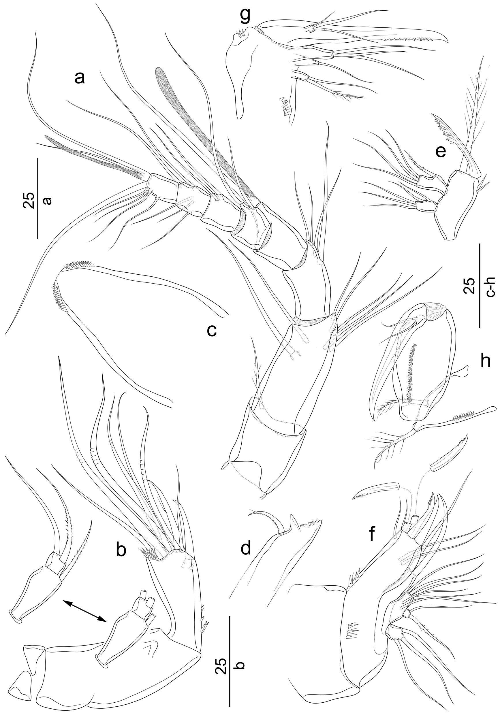
A1 ( Figure 3a View Figure 3 ) 8-segmented and slender; segment 2 longest. All elements naked except seta on first segment. Segment 4 with 1 aesthetasc fused basally to a very long naked seta. Segment 8 with 1 acrothek consisting of 1 short aesthetasc fused basally to 1 relatively short and 1 long, naked seta. Armature formula: 1-[1 plumose], 2-[7], 3-[4], 4-[1+ae], 5-[2], 6-[2], 7-[5], 8-[5+acrothek].
A2 ( Figure 3b View Figure 3 ) with unarmed allobasis, about 2.5 times as long as wide, fusion of basis and first endopodal segment marked at inner margin. Exopod 2-segmented; first segment elongated, vase-shaped, with 1 strong spinulose inner distal seta; second segment short, about as long as wide, with 1 naked and 1 spinulose seta. Free endopodal segment elongated, about 2 times as long as wide, with short inner spinules proximally and at base of subdistal spine, with 1 transverse row of spinules close to outer distal corner, with 2 strong naked spines, 3 bare and 4 geniculate setae.
Labrum ( Figure 3c View Figure 3 ) arch-shaped, with coarse spinules as figured.
Mandible ( Figure 3d, 3e View Figure 3 ). Cutting edge of coxal gnathobase ( Figure 3d View Figure 3 ) with several teeth as figured and with 1 short curved unipinnate seta. Basis ( Figure 3e View Figure 3 ) about 2.4 times as long as wide, with 2 outer subapical spinules, distally with 1 strong spinulose spine and 1 plumose seta; with well-developed rami. Exopod unisegmented, with 3 naked distal setae and 2 naked lateral elements. Endopod unisegmented, with 1 lateral spinulose element, and 4 distal naked setae.
Maxillule ( Figure 3f View Figure 3 ) well developed. Praecoxal arthrite with 2 rows of spinules, with 2 naked seta, 3 spinulose and 1 naked spine, and 2 surface setae. Coxal endite with 1 long naked spine-like element, and 1 long naked seta. Basis with 6 naked setae. Exopod unisegmented, small, squarish, with 1 naked and 1 spinulose setae. Endopod unisegmented, with 3 long naked setae.
Maxilla ( Figure 3g View Figure 3 ). Syncoxa with 2 short rows of spinules on anterior surface as figured. With 3 endites; proximal endite with 1 plumose and 1 naked seta; middle endite with 1 naked seta; distal endite with 3 naked setae. Allobasis transformed into 1 very strong spinulose claw. Endopod very short, incorporated to allobasis, with 1 minute naked seta, and 2 relatively long naked elements fused basally.
Maxilliped ( Figure 3h View Figure 3 ). Syncoxa with inner row of spinules, with 2 plumose setae. Basis about 2 times as long as wide, with row of inner spinules at anterior surface, unarmed. Endopod transformed into 1 strong curved claw, with 2 accessory naked setae near its base.
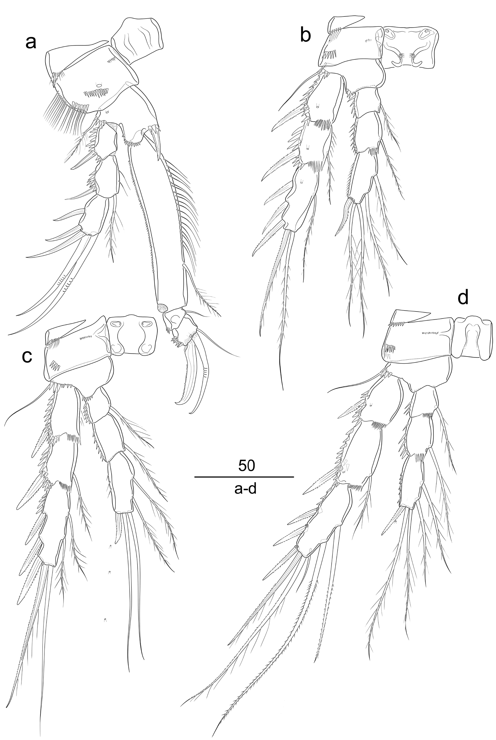
P1–P4 ( Figures 4a–4d View Figure 4 ). Praecoxa triangular, with 1 row of spinules near outer corner. Intercoxal sclerites as figured.
P1 ( Figure 4a View Figure 4 ). Coxa squarish, with long outer setules, 3 rows of spinules as figured, and 1 pore on anterior surface. Basis with 1 pore on anterior surface, with strong spinules at base of outer and inner elements and with smaller spinules between rami, with 1 plumose outer seta and 1 unipinnate inner spine. Endopod 3-segmented, prehensile; first segment elongated, about 5.3 times as long as wide, with small outer spinules and with long setules along 2/3 of inner margin proximally, with 1 inner plumose seta; second segment very short, with 1 naked inner seta; third segment slightly longer than previous segment, squarish, with coarse outer spinules, distally with 1 strong outer claw, 1 geniculate medial seta, and 1 inner element. Exopod three-segmented, reaching about half of enp-1; first segment with coarse outer spinules, with 1 strong outer spine, without inner armature; second segment with coarse outer spinules, with a few inner setules, and 1 plumose inner seta; third segment without ornamentation, with 3 outer spines and 2 geniculate distal setae.
P2–P4 ( Figures 4b–4d View Figure 4 ). Coxa rectangular, with row of coarse spinules near outer distal corner, and with very short medial spinules. Basis with spinules as figured, outer seta naked. Endopod 3-segmented; first and second segment with coarse inner spinules, with 1 short plumose inner seta; third segment slightly elongated, with inner spinules, distally with 1 spinulose outer spine and 2 plumose (P2 and P4) or naked (P3) setae, and 2 plumose inner setae. Exopod 3-segmented; all segments with a small pore at anterior surface; first segment with coarse outer spinules, with 1 bipinnate spinulose outer spine, inner margin naked (P2, P4) or with a few fine setules (P3); second segment with coarse outer spinules, with 1 bipinnate spinulose outer spine, and with 1 short inner plumose seta; third segment elongated, about 2.7 (P2) or 3.3 (P3, P4) times as long as wide, with 3 bipinnate spinulose outer spines; and 1 long plumose seta (P2), 2 relatively short plumose elements (P3), or 2 relatively long spinulose and 1 minute naked setae (P4) at inner margin, and with 1 long unipinnate spinulose spiniform element and 1 long plumose (P2 and P4) or naked (P3) seta apically.
Armature formula of the swimming legs as follows:
P5 ( Figure 2c View Figure 2 ). Baseoendopod with proximal inner long setules, outer basal seta naked; endopodal lobe slightly elongated, with 3 naked and 2 spinulose setae. Exopod not fused to baseoendopod, elongated, about 2 times as long as wide at midline of anterior surface, with long inner setules, and long outer spinules, with 1 subdistal anterior pore, with 3 outer setae (proximal one visibly longer), 1 apical and 1 inner seta subequal in length; all setae naked.
P6 ( Figure 2a View Figure 2 ). Both legs fused, forming a short and wide symmetric plate. Each pair with 1 short and plumose seta, 1 relatively long and naked element, and 1 minute seta.
Male: Unknown.
3.4. Variability
The anal operculum of one female is furnished with spinules distally.
3.5. Etymology
The species was named after Prof Dr Süphan Karaytuğ for his great contribution to copepod taxonomy.
No known copyright restrictions apply. See Agosti, D., Egloff, W., 2009. Taxonomic information exchange and copyright: the Plazi approach. BMC Research Notes 2009, 2:53 for further explanation.
|
Kingdom |
|
|
Phylum |
|
|
Class |
|
|
Order |
|
|
Family |
|
|
Genus |