
Petta williamsonae, Zhang & Hutchings & Kupriyanova, 2019
|
publication ID |
https://doi.org/ 10.11646/zootaxa.4614.2.3 |
|
publication LSID |
lsid:zoobank.org:pub:3053533C-BDDE-4321-95B2-D557F3CF048D |
|
DOI |
https://doi.org/10.5281/zenodo.5625087 |
|
persistent identifier |
https://treatment.plazi.org/id/03A887E7-D43C-A022-60CE-D992306DFDA3 |
|
treatment provided by |
Plazi |
|
scientific name |
Petta williamsonae |
| status |
sp. nov. |
Petta williamsonae View in CoL n. sp.
Figs 12–14 View FIGURE 12 View FIGURE 13 View FIGURE 14 , Table 2 View TABLE 2
Material examined. Holotype AM W.50667, broken between segments 16–17 : Paratype AM W.51459 (mounted for SEM), complete, Bass Strait , Australia: 39º27.72´S 149º16.56´E, 2760– 2692 m, coll. R/V “ Investigator ” Voyage 3 St. 022, Beam Trawl, May 2017 GoogleMaps .
Description. Preserved specimens pale reddish in colour, dumb-bell shaped with broad anterior end and scaphe ( Figs 13A View FIGURE 13 ; 14A View FIGURE 14 ). Body length 14.5–20.0 mm (holotype about 14.5 mm) including paleae and scaphe, width 2.3–2.9 mm (2.3 mm) at cephalic regions.
Cephalic veil heart-shaped, free operculum, with smooth margin and short triangular anterior end ( Figs 12B View FIGURE 12 ; 13A, D View FIGURE 13 ; 14B, D View FIGURE 14 ). Pair of lateral ear-shaped lobes (palps) adjacent to dorsal base of cephalic veil ( Fig. 12C View FIGURE 12 ). Buccal tentacles with deep longitudinal grooves, arising from around buccal cavity, posterior to cephalic veil ( Figs 12B View FIGURE 12 ; 13A View FIGURE 13 ; 14D View FIGURE 14 ). Lower lip not visible between buccal cavity and segment 1.
Operculum semicircular; dorsal and lateral margins well developed, smooth; ventral margin (opercular ridge) with 14 pairs of amber-coloured, stout, long notopodial paleae, curved dorsally, with blunt tips ( Figs 12A View FIGURE 12 ; 13B View FIGURE 13 ; 14C View FIGURE 14 ).
First pair of tentacular cirri not extending beyond tips of paleae, annuli not distinct, arising from connection of opercular margin and paleal ridge ( Figs 12 View FIGURE 12 A–B; 13B; 14D). Pair of narrow triangular lappets present behind tentacular cirri, on both sides of segment 1, partly covered by ventral lobes of segment 2 ( Figs 12B View FIGURE 12 ; 13E View FIGURE 13 ; 14D View FIGURE 14 ). Ventral region of segment 1 covered by ventral lobes of segment 2 ( Figs 12B View FIGURE 12 ; 13A View FIGURE 13 ; 14B, D View FIGURE 14 ).
Second pair of tentacular cirri almost same length as first, annulation indistinct, and displaced dorsally, on almost mid-lateral connecting ridge of segment 2 ( Figs 12 View FIGURE 12 A–B; 13B; 14D). Segment 2 with pair of broad ventrolateral lobes separated from each other by a narrow deep mid-ventral groove, each lobe with 7–8 continuous row of triangular lappets ( Figs 12B View FIGURE 12 ; 13E View FIGURE 13 ; 14B, D View FIGURE 14 ).
Two pairs of equal sized comb-like branchiae on segments 3–4, consisting of large basal hump and series of loose flat lamellae ( Figs 12D View FIGURE 12 ; 13F View FIGURE 13 ; 14C View FIGURE 14 ). First pair of branchiae on segment 3 inserted more ventrally than those on segment 4.
Pair of dorso-lateral pads small and smooth, arising from dorsal side of notopodia on segment 5 ( Figs 12A View FIGURE 12 ; 13 View FIGURE 13 B–C; 14C).
Distinct ventral glandular lobes (pads) present on segments 2–7, becoming progressively more dorsal and broader on segments 3–5 ( Figs 12B View FIGURE 12 ; 13 View FIGURE 13 A–B; 14B). Humps near branchiae absent on ventral lobes ( Figs 12B View FIGURE 12 ; 13B View FIGURE 13 ; 14B View FIGURE 14 ). Segment 3 with a pair of broad ventro-lateral lobes and a pair of mid-ventral lappets, separated from those by deep notches; ventro-lateral lobes smooth with rounded ventral margin; mid-ventral lappets oblong and narrow about 1/5 width of ventro-lateral lobes, and more posterior than ventro-lateral lobes ( Figs 12B View FIGURE 12 ; 13A View FIGURE 13 ; 14B View FIGURE 14 ). Segments 4–6 with a pair of broad ventro-lateral lobes separated by a shallow median groove becoming progressively broader on segments 4–6. Segment 7 with a pair of broad ventro-lateral lobes separated from each other by median swelling about 1/5 width of ventro-lateral lobes.
Notopodia of segment 1 with paleae, segments 5–21 (17 pairs) with two rows of chaetae; anterior row of shorter chaetae with distal serrated wings, anterior surface below wing to about mid-basal portion of chaeta covered with numerous minute spines; posterior row with about 1.4 times longer capillary chaetae, straight and stout, tapering to acute tips, anterior surface covered with numerous spines from mid-length to tip ( Figs 12 View FIGURE 12 H–I; 14F–G). Neuropodia on segments 8–21 (14 pairs), each with slightly raised torus bearing a transverse row of uncini. Each uncinus with one rounded anterior peg with blunt tip embedded into torus, followed by several rows of minor teeth on a swelling, a longitudinal row of two major teeth, each covered with many small teeth basally ( Figs 12G View FIGURE 12 ; 14H View FIGURE 14 ). Neuropodia of segment 21 with enlarged posterior lobe ( Figs 12 View FIGURE 12 E–F; 13G–I).
Scaphe long ovoid, flattened dorsally, well separated by a constriction from posterior segments. Lateral margins of scaphe dorsally rolled, with six pairs of lobes; first pair of lobes largest, connecting with dorsal margin of scaphe; posterior lobes narrow triangular, almost same size; dorsal margin of scaphe smooth with a median notch ( Figs 12 View FIGURE 12 E–F; 13G–I; 14E). Anal flap vestigial with oblong swollen area distally bearing long anal cirrus ( Figs 12F View FIGURE 12 ; 13G, I View FIGURE 13 ). Anus located behind anal cirrus, between last pair of lateral lobes on scaphe. Scaphal hooks amber-coloured, nine pairs arising from both sides of dorsal margin of scaphe, with blunt tips slightly curved dorsally ( Figs 12F View FIGURE 12 ; 14I View FIGURE 14 ).
Tube broken, made of concretions and shell fragments.
Distribution. Bass Strait, Australia ( Fig. 1 View FIGURE 1 ). Known only from type locality.
Habitat. 2760– 2692 m. No substrate data available.
Etymology. The species is named after Associate Professor Jane Williamson, Biological Sciences, Macquarie University, Australia who faciliatated the research.
Remarks. Petta williamsonae n. sp. can be distinguished from other species of Petta by the following characters: cephalic veil with smooth margin and short narrow triangular anterior end; 14 pairs of paleae; pair of narrow triangular lappets on segment 1; pair of short ventro-lateral lobes on segment 2, separated from each other by a narrow deep mid-ventral groove, with 7–8 triangular lappets per lobe; ventro-lateral lobes smooth on segment 3, with rounded inner angle; neurochaetae on segments 8–21; distinct constriction between the abdomen and the scaphe; anal flap vestigial with long anal cirrus; scaphal hooks 9 pairs, slightly curved. Petta williamsonae n. sp. is similar to P. tenuis from Sulu Sea, Phillipines and P. pellucida from Santarem Channel, Caribbean Sea as they all have smooth anterior end on cephalic veil and smooth ventro-lateral lobes on segment 3. However, Petta williamsonae n. sp. differs from the last two species as P. williamsonae n. sp. has the distinct constriction between the abdomen and the scaphe ( Table 2 View TABLE 2 ). Petta williamsonae n. sp. differs from P. investigatoris n. sp. from Australian waters as the latter has continuous papillae on the ventro-lateral lobes of segment 3 and the scaphe is not clearly separated from posterior segments, and also P. williamsonae n. sp. has a smooth margin on the ventro-lateral lobes of segment 3 and a distinct constriction between the abdomen and the scaphe.
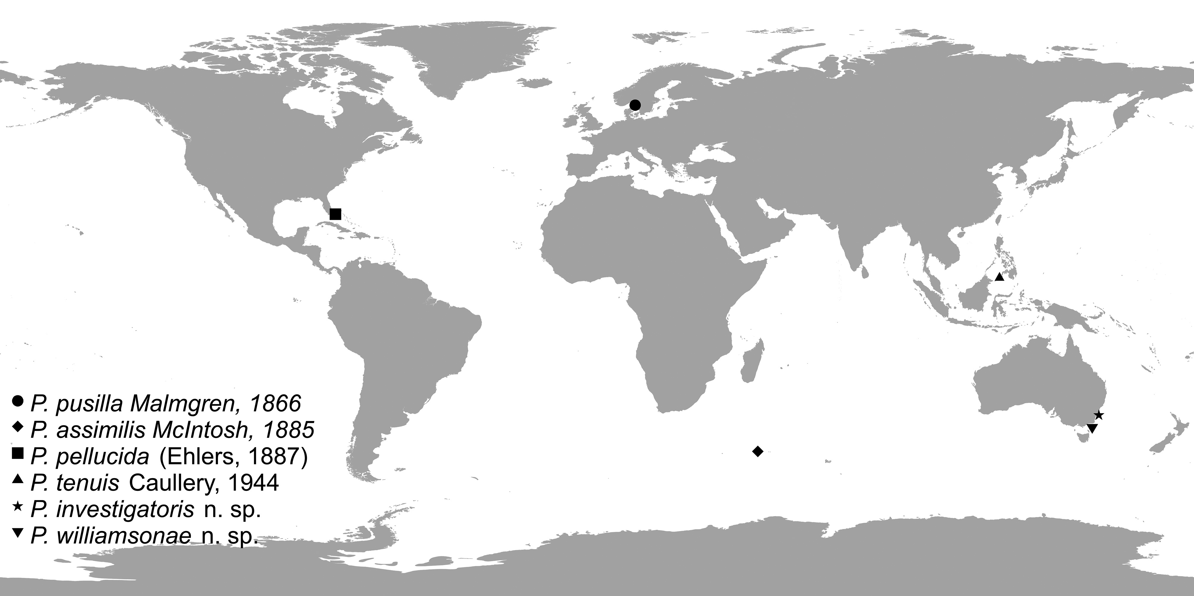

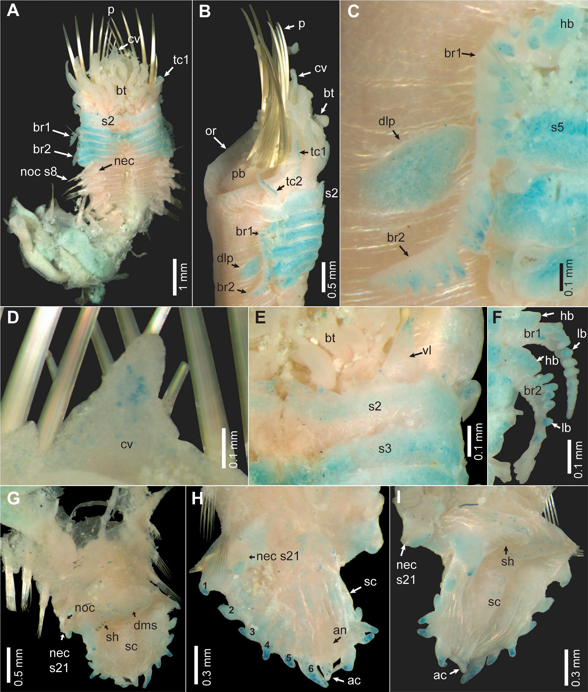

TABLE 2. A summary of comparative diagnostic characters for the species of the genus Petta species as used in this study.
| Characters | P. pusilla Malmgren, 1866 | P. assimilis McIn- tosh, 1885 | P. pellucida (Ehlers, 1887) | P. tenuis Caullery, 1944 | P. investigatoris n. sp. | P. williamsonae n. sp. |
|---|---|---|---|---|---|---|
| Holotype and topotypi- cal material | Holotype | Types | Types | Types | Types | |
| Anterior tip of cephalic veil | 3–4 lappets | smooth | smooth | smooth | smooth | smooth |
| Pairs of paleae | 10–11 | 14 | 12 | 11 | 12–13 | 14 |
| Lower lip behind buccal cavity | broad lobe not covered with ventral lobes of segment 2 | covered with ventral lobes of segment 2 | covered with ventral lobes of segment 2 | covered with ventral lobes of segment 2 | covered with ventral lobes of segment 2 | covered with ventral lobes of segment 2 |
| Lappets on segment 1 | short acute | triangular | triangular | triangular | triangular | triangular |
| Ventro-lateral lobes on segment 2 | elongated with 4–5 lappets | short 4–5 lappets | short with 6 lappets | short with 4–5 lap- pets | short with 7–8 lappets | short with 7–8 lappets |
| Ventro-lateral lobes on segment 3 | smooth | with papillae | smooth | smooth | with papillae | smooth |
| Ventral margin of ventro- lateral lobes on segment 3 | rounded | rounded | rounded | with triangular pro- jection | rounded | rounded |
| Mid-ventral lappets on segment 3 | rounded | ? | rectangular | rectangular | oblong | oblong |
| Neurochaetae on segments | 8–21 | 8–21 | 7–21 | 8–21 | 8–21 | 8–21 |
| Constriction before scaphe | indistinct | absent | absent | absent | absent | distinct |
| Shape of anal flap | vestigial oblong | short conical | vestigial oblong | vestigial oblong | vestigial oblong | vestigial oblong |
| Anal cirri | long | absent | long | long | long | long |
| Pairs of scaphal hooks | 8 | ? | 7 | 8 | 9–12 | 9 |
| Shape of scaphal hooks | curved | curved | curved | curved | straight | curved |
| AM |
Australian Museum |
No known copyright restrictions apply. See Agosti, D., Egloff, W., 2009. Taxonomic information exchange and copyright: the Plazi approach. BMC Research Notes 2009, 2:53 for further explanation.
|
Kingdom |
|
|
Phylum |
|
|
Class |
|
|
Order |
|
|
Family |
|
|
Genus |