
Microcotyle pacinkar, Kamio & Nitta, 2023
|
publication ID |
https://doi.org/ 10.12782/specdiv.28.263 |
|
publication LSID |
lsid:zoobank.org:pub:3076FC46-D67C-42CC-AD9E-3BEA7B29CCD7 |
|
persistent identifier |
https://treatment.plazi.org/id/03A587AE-290E-FFA7-AB83-F892FB4241DB |
|
treatment provided by |
Felipe |
|
scientific name |
Microcotyle pacinkar |
| status |
sp. nov. |
Family Microcotylidae Taschenberg, 1879 View in CoL Subfamily Microcotylinae Taschenberg, 1879 Genus Microcotyle van Beneden and Hesse, 1863 Microcotyle pacinkar n. sp.
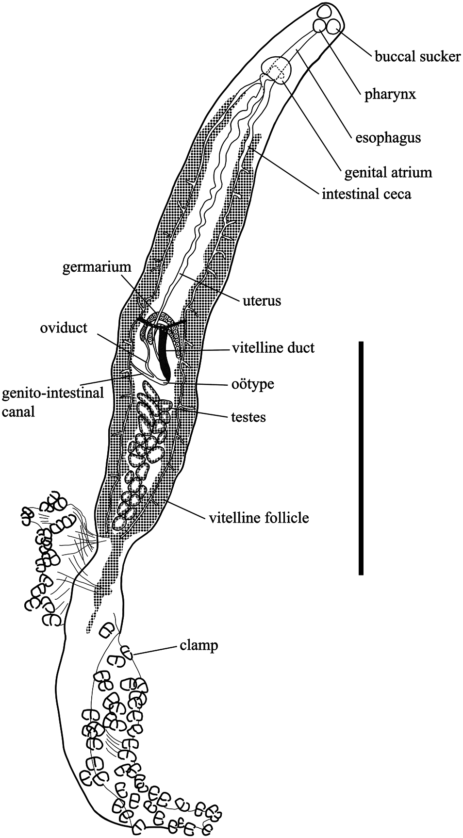
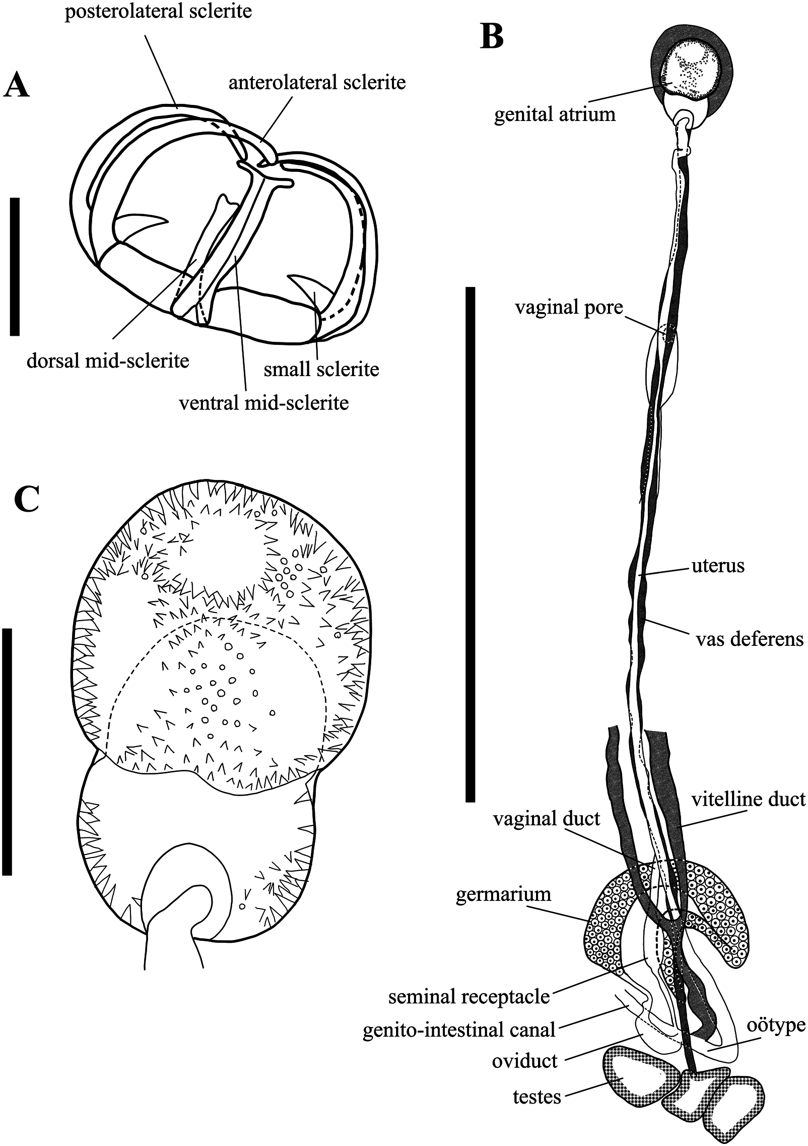
( Figs 1 View Fig , 2 View Fig )
Type material. Holotype ( MPM Coll.-No. 25226) and 21 paratypes ( MPM Coll.-Nos 25227, 25228).
Description. Body ( Fig. 1 View Fig ) elongated 2690–6250 (4788, n = 15) long including haptor, 300–774 (472, n = 21) wide at level of germarium. Haptor wedge-shaped, sub-symmetrical, with 34–67 (49, n = 16) clamps, arranged in 2 subequal lateral rows. Clamps ( Fig. 2A View Fig ) of equal structure, each clamp 48–76 × 68–100 (58 × 84, n = 21) in diameter. Clamps of Microcotyle - type, formed by two jaws. Ventral arm of median spring long, thin, ends distally in an inverted T, with short branches. Lateral sclerites of ventral jaw approaching midline distally. Dorsal arm of median spring inverted Y- shaped. Posterolateral sclerite and anterolateral sclerites curved toward inside.
Pair of buccal suckers ( Fig. 1 View Fig ) septate, elliptical, 50–80 × 45–90 (68× 64, n = 17). Pharynx ( Fig. 1 View Fig ) globular, immediately posterior to buccal sucker, sometimes overlapping buccal sucker, 45–95 × 37.5–85 (66 × 61, n = 18). Esophagus ( Fig. 1 View Fig ) short, without lateral diverticula. Intestinal bifurcation ( Fig. 1 View Fig ) behind genital atrium. Intestinal ceca ( Fig. 1 View Fig ) blind, extending to haptor, with numerous lateral diverticula, not united posteriorly.
Testes ( Figs 1 View Fig , 2B View Fig ) with irregular shape, 14–28 (21, n = 20) in number, post-ovarian, intercaecal, in posterior half of body. Vas deferens ( Figs 1 View Fig , 2B View Fig ) long, narrow, coming from anterior of testes, ventral to germarium, extending anteriorly, dorsal to uterus along its dorsal side, entering base of genital atrium. Genital atrium ( Figs 1 View Fig , 2C View Fig ) inverted heart-shaped, 45–175 × 60–160 (124 × 106, n = 17), located posterior to pharynx, with lateral expansions, surrounded by muscle. Genital atrium proper bearing numerous small spines. Edge of genital atrium opening and its inner walls armed with numerous conical spines, 4–7 (5, n = 10) in length. Spines more dense in central anterior region, less dense in lateral pockets; no spines present in center of posterior pockets.
Germarium complex ( Figs 1 View Fig , 2B View Fig ) begins at level of anteriormost testes, continuing anteriorly in midline, reflexing approximatively at level of confluence of vitelline ducts, reflexing again toward anterior extremity, forming an equally thin anterior curve and reflexing a last time posteriorly before ending as oviduct, 37.5–200 (101, n = 21) long, 50–200 (102, n = 21) wide. Oviduct ( Figs 1 View Fig , 2B View Fig ) arising from distal end of germarium, extending towards anterior of testes, connected to genito-intestinal canal. Genito-intestinal canal ( Figs 1 View Fig , 2B View Fig ) originates from right intestinal cecum and connected to oviduct and vitelline duct. Vitelline duct ( Figs 1 View Fig , 2B View Fig ) Yshaped, ventral, extending from genito-intestinal canal and bifurcating to either side near the germarium. Oötype ( Figs 1 View Fig , 2B View Fig ) extending from genito-intestinal canal to uterus. Mehlis’ gland not observed. Uterus ( Figs 1 View Fig , 2B View Fig ) originating from oötype, extending anteriorly along body midline, ventral to vas deferens, to the opening of the genital atrium. Vaginal pore ( Fig. 2B View Fig ) unarmed, ventral in mid-body, posterior to genital atrium. Vaginal duct ( Fig. 2B View Fig ) dorsal to uterus and vas deferens, arising from vaginal pore, connecting to seminal receptacle. Seminal receptacle ( Fig. 2B View Fig ) located ventral to germarium in center of body, oval-shaped. Eggs fusiform 120–180 × 50–65 (150 × 58, n = 2) excluding filament, with filaments at both ends broken. Vitelline follicles ( Fig. 1 View Fig ) coextensive with intestinal branches, extending from behind genital atrium to posterior end of body, fused posterior to testes.
Type locality. Usujiri port (41°56′10.6″N, 140°56′39.4″E) in Usujiri-cho , Hakodate City, Hokkaido, northern Japan, the North Pacific Ocean GoogleMaps .
Type host. Sebastes taczanowskii Steindachner, 1880 ( Scorpaeniformes : Sebastidae ).
Site of infection. Gill filament.
Etymology. The new specific name “ pacinkar ” refers to the Ainu name of the type host, S. taczanowskii , and thus is treated as indeclinable.
Representative DNA sequences. INSD accession numbers LC753264 and LC753265 (cox1) from two paratypes ( MPM Coll.-No. 25228) .
Remarks. Microctyle pacinkar n. sp. is distinguished from other congeners by the following characters of the new species: the inverted heart-shaped genital atrium, the genital atrium surrounded by muscle, the arrangement of the spines in the genital atrium, the shapes of the posterolateral sclerites of the clamps, the number of testes, and the long and narrow body (length/width ratio: 6.7–16.0).
Molecular data analysis
The trimmed multiple sequence alignment of cox1 fragments consisted of 379 base pairs. A sequence of Bivagina pagrosomi (Murray, 1931) ( Microcotylidae ) was used as the outgroup ( Fig. 3 View Fig ). In the phylogenetic tree based on the cox1 gene, M. pacinkar n. sp. and M. sebastis Goto, 1894 formed a clade with a strong support. The intraspecific pairwise sequence difference was 0.8%, and the differences between M. pacinkar n. sp. and M. sebastis were between 5.8%–7.0%. The pairwise sequence differences of Microcotyle species in the cox1 gene are provided in Table 3.

| MPM |
Milwaukee Public Museum |
No known copyright restrictions apply. See Agosti, D., Egloff, W., 2009. Taxonomic information exchange and copyright: the Plazi approach. BMC Research Notes 2009, 2:53 for further explanation.
|
Kingdom |
|
|
Phylum |
|
|
Class |
|
|
Order |
|
|
Family |
|
|
Genus |