
Martensiellus tenuipalpus, Schwendinger, Peter J., 2006
|
publication ID |
https://doi.org/ 10.5281/zenodo.174076 |
|
DOI |
https://doi.org/10.5281/zenodo.6260942 |
|
persistent identifier |
https://treatment.plazi.org/id/03A5879D-753C-FF94-FE98-5BDAD9F02E6B |
|
treatment provided by |
Plazi |
|
scientific name |
Martensiellus tenuipalpus |
| status |
sp. nov. |
Martensiellus tenuipalpus View in CoL n. sp.
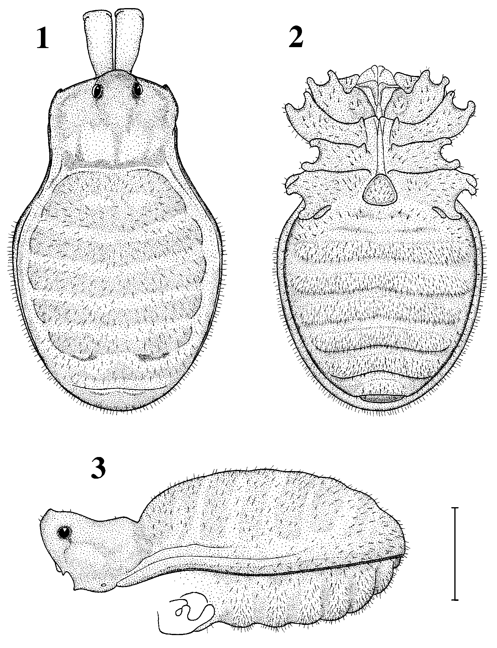
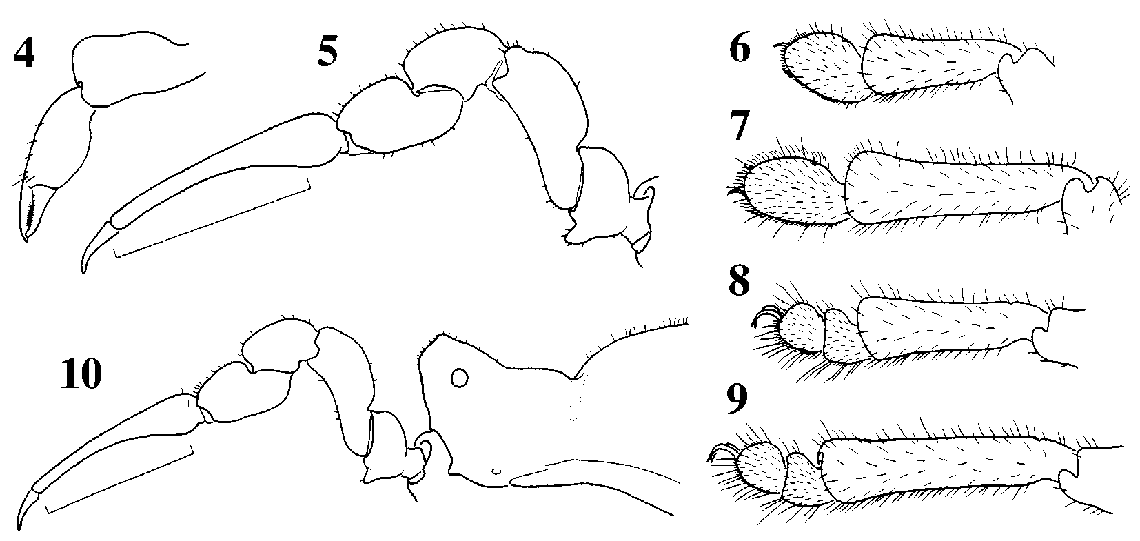
Figs 1–17 View FIGURES 1 – 3 View FIGURES 4 – 10 View FIGURES 11 – 17
Oncopodidae g. sp.: Schwendinger & Martens 2002b: figs 50–51, 59.
Types. Male holotype (AMNH) and male paratype (ventral scutum broken; MHNG; ex AMNH, donated by L. Prendini) from Eastern Malaysia, Sarawak, Baleh River, north of Kapit, 40 m, collected 25 June 1976 by Walter C. Sedgwick.
Etymology. The specific epithet, a noun in apposition formed from the Latin tenuis (= thin, slender) and palpus (= feeler), refers to the unusually slender palpal tarsus of males in this species.
Diagnosis. Externally similar to Caenoncopus cuspidatus (see Schwendinger 1992: 190–192, figs 76–80; Martens & Schwendinger 1998: 507, figs 4c–f, 5–8), but with stronger pilosity; no carapaceopisthosoma bridge; eye mound (tubercle) distinctly elevated; carapace region fairly large; ventral scutal areas distinctly elevated; anteroproximal process on coxa III present; genital operculum small ( Figs 1–3 View FIGURES 1 – 3 ); palpus with distinct ventral process on trochanter, no process on femur, tarsus exceptionally thin in distal portion; leg tarsi/tarsomeres ( Figs 4–10 View FIGURES 4 – 10 ) much longer and larger than in C. cuspidatus . Penis similar to that of Biantoncopus fuscus Martens & Schwendinger (see Martens & Schwendinger 1998: 521–525, figs 58–70), but glans wider, situated more distally, with indistinct membranous socket, wider lateral sclerites and narrower median plate ( Figs 11–17 View FIGURES 11 – 17 ).
Description. Male (holotype). Colouration (in alcohol) and surface texture: Ground colour light orange, with indistinct darker pattern on dorsal scutal elevations (probably lightened by longtime preservation in alcohol; Fig. 1 View FIGURES 1 – 3 ). A finely granular surface on prosoma, genital operculum, chelicera (except for fingers), legs and palpus (except for tarsi).
Carapace region with rounded, slightly anteriadinclined eye tubercle carrying a small pointed tip; carapaceopisthosoma bridge absent; no lateral tubercles in posterior part of carapace region. Dorsal scutal areas of opisthosoma region only indistinctly elevated; ventral scutal areas moderately swollen and pallid ( Fig. 3 View FIGURES 1 – 3 ). Ventral scutum and opisthosoma region of dorsal scutum densely covered by short hairs ( Figs 1–3 View FIGURES 1 – 3 ). Palpal coxa with small ventral process; leg coxa I with indistinct anterolateral process; leg coxae II and III with distinct anteroproximal processes, no posteroproximal process on coxa II ( Fig. 2 View FIGURES 1 – 3 ); dorsal side of leg coxa IV without tubercles. Genital operculum somewhat triangular, wider than long; posterior margin of stigmatic pit without tubercle ( Fig. 2 View FIGURES 1 – 3 ).
Chelicera ( Fig. 4 View FIGURES 4 – 10 ): Hand weak, with a few small hairs dorsally; proximal article with a higher dorsodistal and a lower dorsomedian boss, no ventral process.
Palpus ( Fig. 5 View FIGURES 4 – 10 ): Trochanter with slightly distadinclined ventral process; femur short, with indistinct, broadly rounded ventroproximal boss, no process; tarsus cylindrical, exceptionally thin in distal half, there about as deep and wide as claw (possibly a sexual dimorphism).
Legs ( Figs 6–9 View FIGURES 4 – 10 ) 1342 (from shortest to longest), tarsal formula 1122; tarsi I and II somewhat ovoid ( Figs 6–7 View FIGURES 4 – 10 ), with a deep pore dorsally; tarsus of leg II about 1.6 times longer than deep ( Fig. 7 View FIGURES 4 – 10 ).
Penis ( Figs 11–14 View FIGURES 11 – 17 ): Truncus fairly robust, slightly constricted midway, from there widening to a second moderate constriction below glans; distal margin of truncus widely rounded; several subapical setae laterally, two on each side separated from other setae and flanking proximal part of glans. Glans distaddirected, wider than truncus at that point, slightly projecting beyond distal margin of truncus; lateral sclerites wide, somewhat scoopshaped, their outer part flat and their inner part projecting dorsad; median plate rectangular, longer than wide (visible only in expanded state; see Fig. 16 View FIGURES 11 – 17 for paratype), lying dorsally of lateral sclerites (as in Biantoncopus , but not in Palaeoncopus ); stylus slender, with bifid tip but without pair of subapical teeth; membranous tubes wide, distally truncate and bent dorsad, lying between stylus and a somewhat pentagonal median knob. In expanded state (see Fig. 16 View FIGURES 11 – 17 for paratype) median plate and stylus of glans protruding distad, and membranous tubes folded posteriad, as in the penis of Biantoncopus fuscus Martens & Schwendinger (see Martens & Schwendinger 1998: figs 64– 65).
Measurements of male holotype (male paratype in parentheses): Body 3.51 (3.47) long, 2.18 (2.18) wide; carapace region 1.04 (1.02) long, 1.30 (1.30) wide; palpus and legs:
Female. Unknown.
Variation. The male paratype is more strongly pigmented than the holotype. Its ground colour is amber, with dark brown horizontal bands on dorsal scutum and with a dark reticulation in the carapace region; leg femora to metatarsi are darkened, leg tarsi and palpal tarsus light brown; its ventral body side is mostly amber, only the lateral margin of the ventral scutum and patches behind coxa IV and genital operculum are darkened. The ventral process on the palpal trochanter of the paratype is ventraddirected ( Fig. 10 View FIGURES 4 – 10 ) rather than slightly distaddirected as in the holotype ( Fig. 5 View FIGURES 4 – 10 ). The penis of the paratype has a slightly more rounded distal margin ( Figs 15–16 View FIGURES 11 – 17 ), but otherwise it is very similar to that of the holotype ( Fig. 13 View FIGURES 11 – 17 ).
Relationships. The presence of a distinctly derived penis with a distaddirected, expandable glans in which the median plate lies dorsally of the lateral sclerites, shows that Martensiellus n. g. from Borneo is phylogenetically closest to Biantoncopus from the Philippines (see Schwendinger & Martens 2002b: fig. 59; Martensiellus tenuipalpus n. sp. under Oncopodidae g. sp.). In its somatic morphology however, the new genus is rather conservative and more similar to Palaeoncopus (with primitive penis morphology) and Caenoncopus (with extremely derived penis morphology), which both occur in Sumatra.
Distribution. Known only from the environs of Kapit in central Sarawak.

No known copyright restrictions apply. See Agosti, D., Egloff, W., 2009. Taxonomic information exchange and copyright: the Plazi approach. BMC Research Notes 2009, 2:53 for further explanation.
|
Kingdom |
|
|
Phylum |
|
|
Class |
|
|
Order |
|
|
SubOrder |
Laniatores |
|
Family |
|
|
Genus |