
Triacrus Nordmann, 1837
|
publication ID |
https://doi.org/ 10.1649/0010-065x-69.3.514 |
|
DOI |
https://doi.org/10.5281/zenodo.5413346 |
|
persistent identifier |
https://treatment.plazi.org/id/03A58798-FFAE-391D-506F-FD63FCF7C181 |
|
treatment provided by |
Diego |
|
scientific name |
Triacrus Nordmann, 1837 |
| status |
|
Triacrus Nordmann, 1837 View in CoL
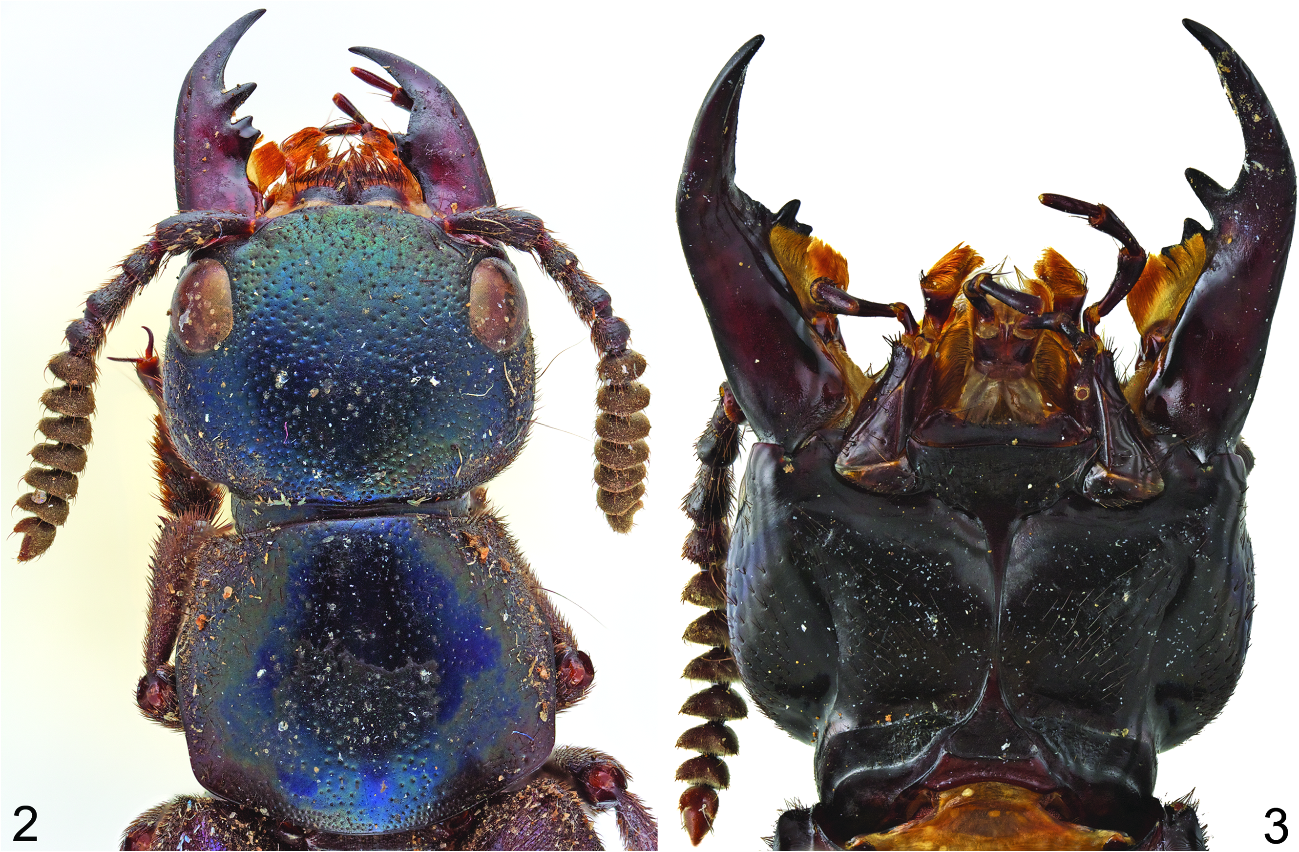
Type Species. Triacrus dilatus Nordmann View in CoL , by monotypy (but see “Nomenclature” below in the species treatment). Nordmann (1837) mentioned that the name Triacrus View in CoL means “three appendages” in Greek and can possibly refer to the structure of the left mandible ( Fig. 2 View Figs ) or abdominal apex.
Diagnosis. Due to its large size, within Xanthopygina Triacrus can only be confused with other similarly large genera such as Darwinilus Chatzimanolis or Trigonopselaphus . However, Triacrus can be easily distinguished from these two genera by the shape of the head (rounded rectangle in Triacrus ; hexagonal in Darwinilus and Trigonopselaphus ), the shape of the antennae (antennomeres 6–11 emarginate in Triacrus , not emarginate in Darwinilus and Trigonopselaphus ), and the shape of the pronotum (in Triacrus , pronotum expanding from anterolateral corners to almost middle, then converging towards posterolateral corners and becoming almost explanate, with posterolateral corners expanded and broadly rounded; in Darwinilus , posterolateral corners of pronotum not expanded and pronotum not explanate; in Trigonopselaphus , pronotum with anterolateral corners bulging and clearly extending beyond anterior margin).
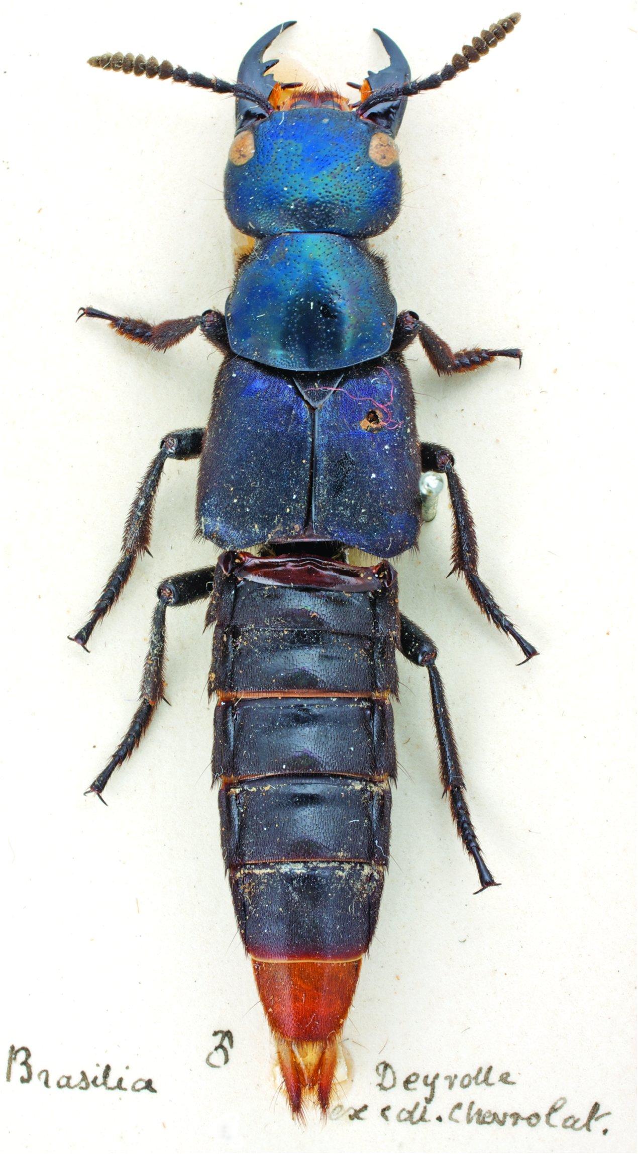
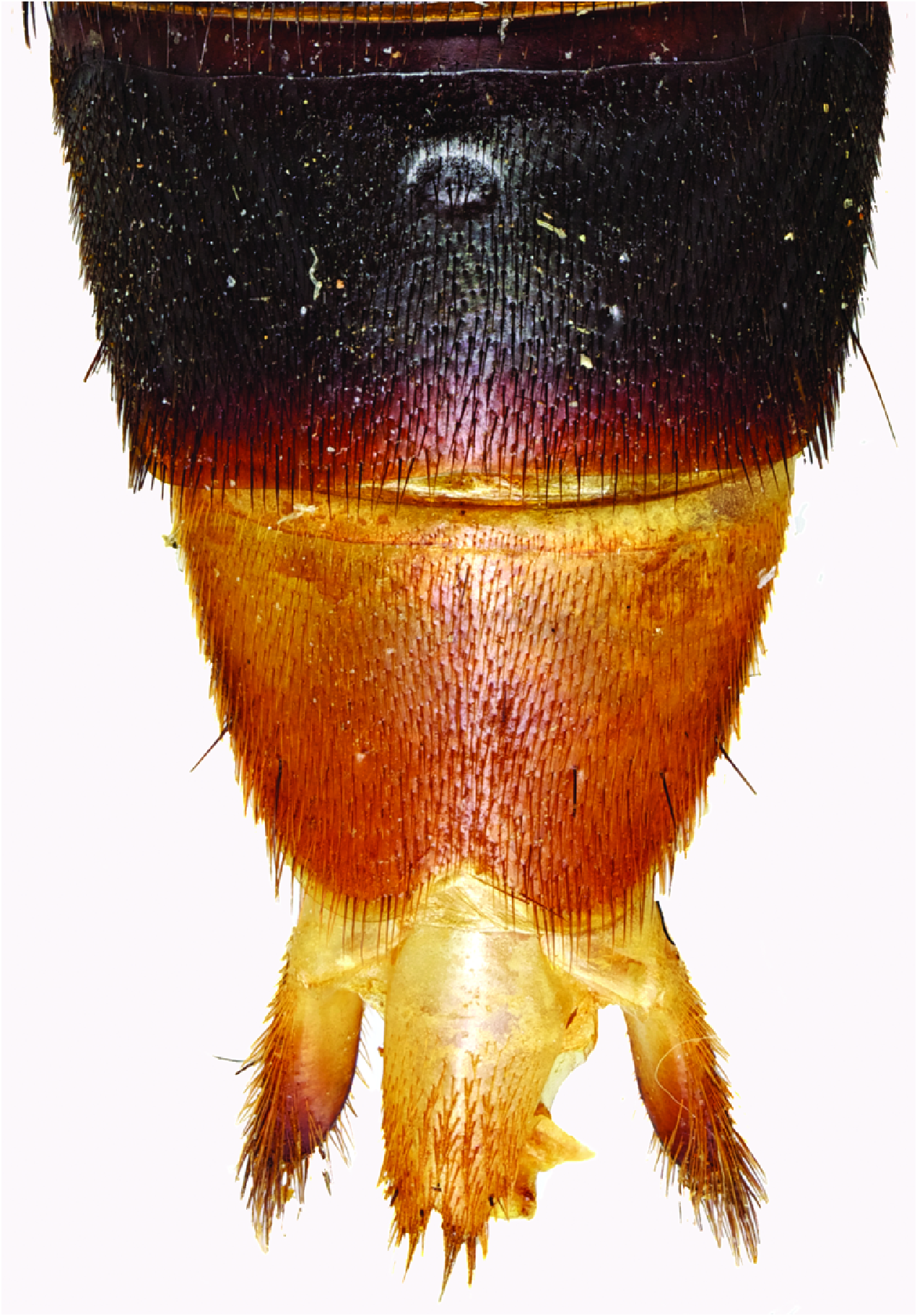
Description. Size large, body robust, habitus as in Fig. 1 View Fig . Head transverse, rounded rectangular, head size variable both in males and females; with microsculpture and small punctures. Clypeus not emarginate; anteclypeus expanded, weakly sclerotized. Eyes small to medium, positioned anteriorly in oblique angle to dorsal surface. Postoccipital suture and ventral basal ridge present; presence of infraorbital ridge not clear but ridge situated between postmandibular ridge and gular suture extends from posterior to middle part of gena; postmandibular ridge present and prominent; gular sutures converging medioposteriorly; with nuchal ridge and well-defined neck. Epicranium with large, prominent macrosetae around anterior and lateral margins. Antennae long, subserrate, with 11 antennomeres; antennomeres 1–4 with several rows of macrosetae; antennomeres 5–11 covered with microtrichiae; antennomeres 6–11 with small lateral emargination (visible in Figs. 2–3 View Figs ); antennomere 11 with 2 lobes protruding from base of antennomere (visible in Figs. 2–3 View Figs ). Mouthparts with labrum medially emarginate; mandibles ( Figs. 2–3 View Figs ) elongate, variable in size; mandibles with prominent fold extending from molar region to base; left mandible with 2 teeth, 1 with large, single molar extending laterally and smaller bicuspid molar posteriorly; right mandible with 1 bicuspid molar; with setose prostheca. Maxilla as in Fig. 3 View Figs ; galea and lacinia densely setose; maxillary palpi 4-segmented; P 1 short; P 2 curved, approximately 3 times as long as P 1; P 3 curved, slightly shorter than P 2; P 4 straight, rounded apically, longer than P 3, as long as P 2. Labium as in Fig. 3 View Figs ; labial palpi 3-segmented; P 1 as long as P 2; P 2 slightly curved; P 3 truncate, 1.5 times as long as P 2. Pronotum ( Fig. 2 View Figs ) subquadrate to transverse; slightly wider than head; pronotum expanding from anterolateral corners to almost middle, then converging towards posterolateral corners, becoming almost explanate; posterolateral corners expanded and broadly rounded; with microsculpture and small punctures. Pronotal hypomeron expanded; superior and inferior marginal lines of hypomeron separate throughout their length and superior line fully visible from above; without postcoxal process. Basisternum with microsculpture, small punctures, and macrosetae; anterior marginal depression present; sternacostal ridge present; furcasternum with large, medial, elongate carina pointed vertically. Elytra wider and longer than pronotum; with large, prominent macrosetae around lateral and posterior margins. Elytral sutures slightly elevated; elytra becoming depressed near mesoscutellum. Mesoscutellum large, triangular, with dense punctation and setation similar to elytra. Hinds wings fully developed. Mesoventrite without median carina, with small mesoventral process; metaventrite disc with narrow, elongate, impunctate line; metaventrite with 2 small, round, triangular mesoventral processes. Leg with tarsal segmentation 5-5-5; covered with dense setae; protarsus enlarged in both sexes; meso- and metatarsi not enlarged but elongate; 2 empodial setae present. Abdomen with paired protergal glands present; abdominal tergites III–V with tergal basal carina and curved (arch-like) carina. Tergites and sternites with microsculpture. Abdominal sternite VII in males with porose structure ( Fig. 4 View Fig ). Male and female genitalia typical of Xanthopygina ; spermatheca not sclerotized; aedeagus with long parameres and median lobe.
No known copyright restrictions apply. See Agosti, D., Egloff, W., 2009. Taxonomic information exchange and copyright: the Plazi approach. BMC Research Notes 2009, 2:53 for further explanation.
|
Kingdom |
|
|
Phylum |
|
|
Class |
|
|
Order |
|
|
Family |