
Gangraia anatolica, Kostopoulos & Erol & Yavuz & Mayda, 2021
|
publication ID |
https://doi.org/10.5194/fr-24-9-2021 |
|
DOI |
https://doi.org/10.5281/zenodo.11558220 |
|
persistent identifier |
https://treatment.plazi.org/id/03A4ED4E-FFAC-BD46-E02B-E96B7834FC67 |
|
treatment provided by |
Felipe |
|
scientific name |
Gangraia anatolica |
| status |
sp. nov. |
Gangraia anatolica sp. nov.
( Figs. 1–3 View Figure 1 View Figure 2 View Figure 3 )
Etymology
From the geographic area of Anatolia that makes up most of modern Turkey.
Holotype
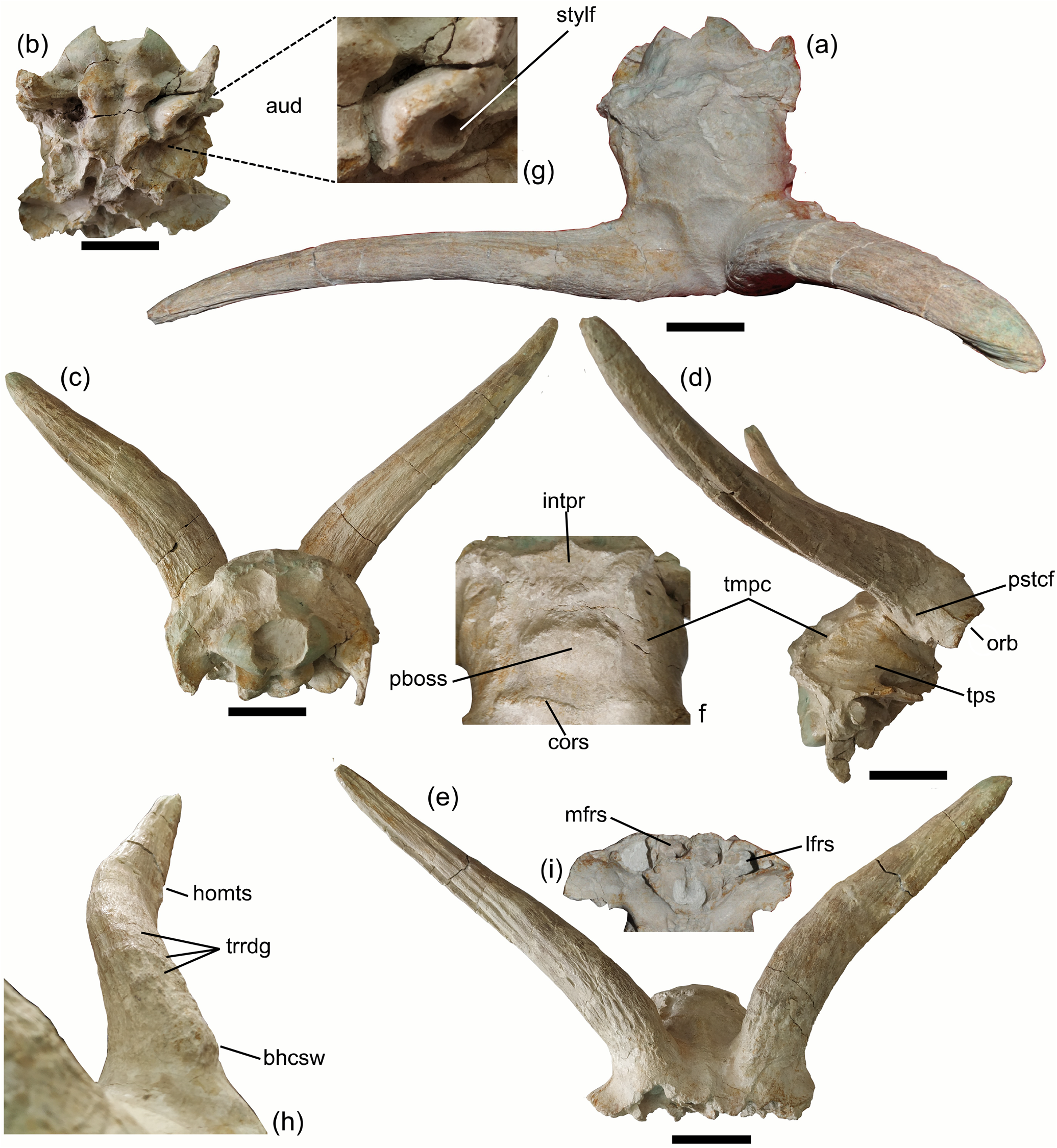
˙O 3187, partial cranium with braincase and both horn cores.
Paratypes
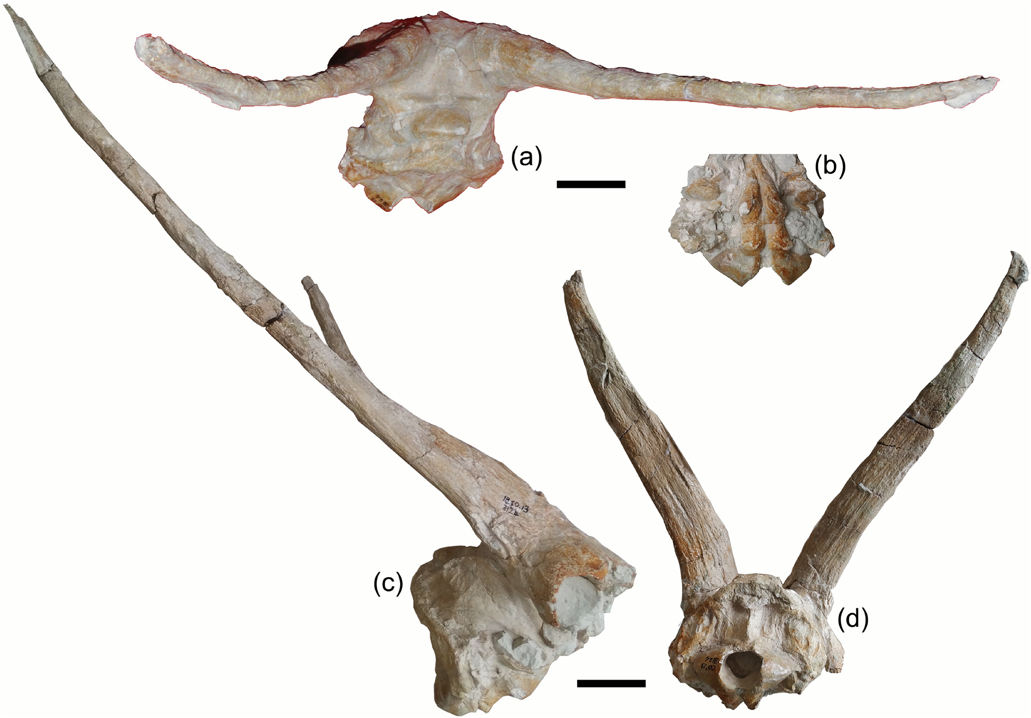
˙O 3476, partial cranium with braincase and both horn cores; ˙O 3124, partial cranium with braincase and both horn cores; ˙O 683, part of cranium with complete right and basal part of the left horn core; ˙O 1427, frontlet.
Type locality
˙orakyerler fossil site near ˙ankırı, Turkey.
Type horizon
Tüglu Formation (Upper Miocene) in ˙ankırı Basin, Turkey.
Age
Late Miocene. Geraads (2013) suggests an early Turolian age for the ˙orakyerler fauna. Magnetostratigraphic correlation by Kaya et al. (2016) frames the fauna between 8.11 and 7.64 Ma. Kostopoulos et al. (2020) suggest an age around the Vallesian–Turolian boundary (∼ 8.7 Ma).
Host institution
˙ankırı Museum, Turkey.
Diagnosis
Same as for the genus.
Description
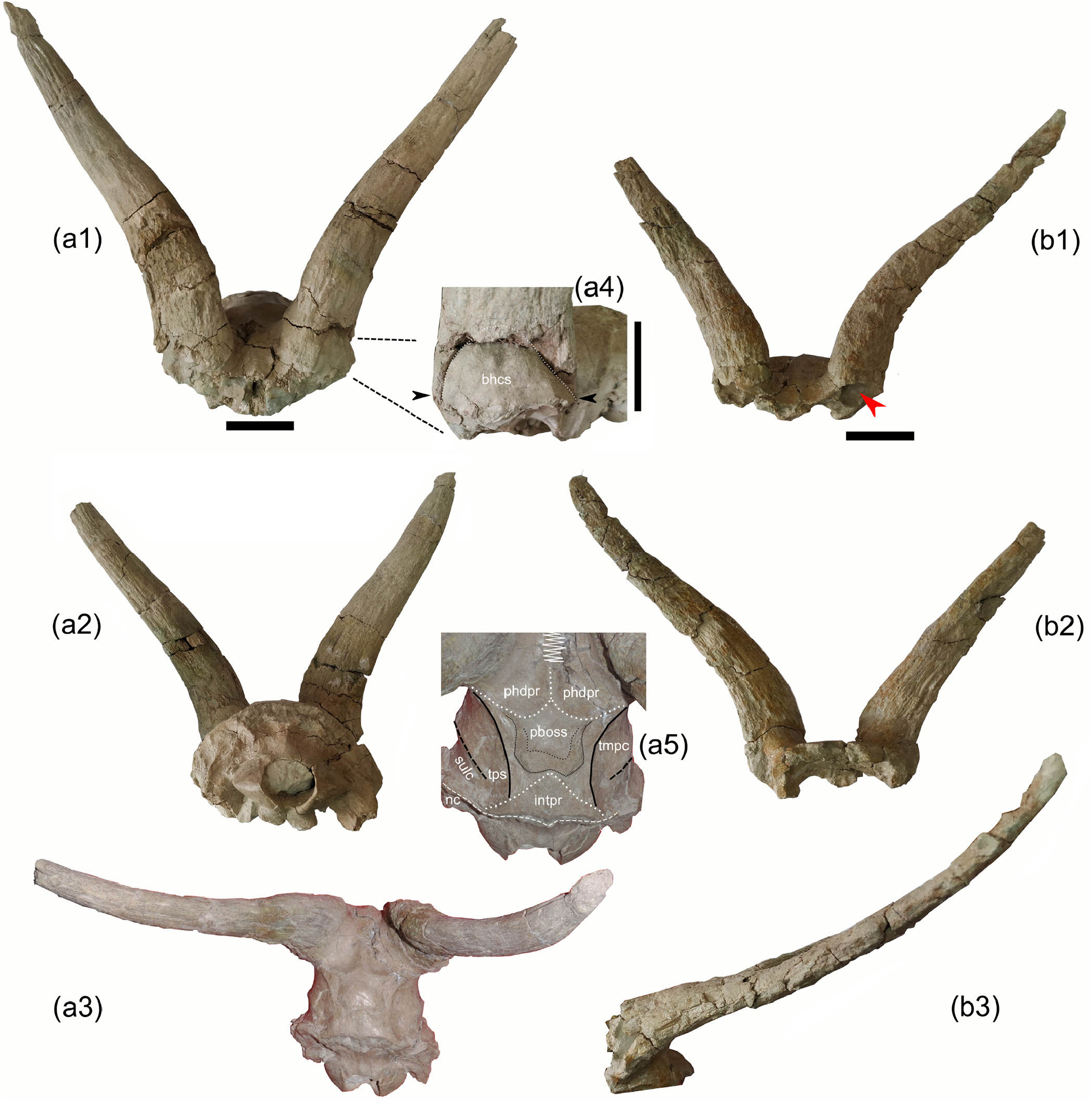
Both ˙O 3187 ( Fig. 1 View Figure 1 ) and ˙O 3476 ( Fig. 2a View Figure 2 ) appear barely affected by taphonomic deformation, which is rather common on the site; hence, the description is based mainly on these two crania. ˙O 3124 ( Fig. 3 View Figure 3 ) is somehow rostrocaudally compressed; as a result, braincase shortening is exaggerated. In some cases (i.e. Table 1 View Table 1 ), one of the two horn cores of the same individual appear more rostrocaudally compressed than the other, especially at its proximal part; we suspect that this is also due to postmortem compression, and we suggest considering the less compressed dimensions as the most reliable.
˙O 3187, ˙O 3476, ˙O 3124, and ˙O 683 form a very homogeneous sample in terms of their morphology, absolute dimensions, and proportions ( Table 1 View Table 1 ; Figs. 1–3 View Figure 1 View Figure 2 View Figure 3 ). On the other hand, the frontlet ˙O 1427 ( Fig. 2b View Figure 2 ) has significantly (20 %) slimmer horn cores and consequently a less broad cranium at the lateral horn core level (∼ 9 %), though the braincase width just behind the horn cores is not much narrower ( Table 1 View Table 1 ). The morphological features of the frontal area in ˙O 1427 are also very similar to those of the holotype, but the interfrontal suture is not pinched but smoothly convex and less complicated. The great length of the horn cores prevents the possibility of a young male individual; we interpret therefore the observed differences between ˙O 1427 and the rest of the crania as representing sexual dimorphism.
The braincase is short and wide ( Figs. 1a View Figure 1 , 2a View Figure 2 . 3, 3a View Figure 3 ) with a length to width ratio (length: from the junction of coronalinterfrontal sutures to inion; width: maximum braincase width) between 59 % and 62 % (n = 2). Placing in a horizontal position the glenoid articular surface, the basioccipital forms an angle of about 70 ◦ with the horizontal plane, inferring a strongly angled braincase compared to the face ( Figs. 1d View Figure 1 , 3c View Figure 3 ). The dorsal braincase plane forms an angle of 115 ◦ (in ˙O 3187) to 125 ◦ (in ˙O 3476) with the occipital plane. The temporal crests are weak but visible ( Figs. 1a, f, View Figure 1
2a.3, a.5); they are wide-apart and run parallel to each other on the dorsal surface of the braincase till the interparietal which is transversally elongated and rather narrow ( Figs. 1f View Figure 1 , 2a View Figure 2 .5). A distinct large parietal boss occurs behind the coronal suture (˙O 3124, 3476; Figs. 1f View Figure 1 , 2a View Figure 2 . 3 View Figure 3 , a.5, 3a).
The occipital condyles with the occipital plane form a ∼ 100 ◦ angle (upper) and are directed caudally. The occipital faces dorso-caudally and has a semi-circular contour defined dorsally by a moderate to strong nuchal crest ( Figs. 1c View Figure 1 , 2a.2 View Figure 2 , 3d View Figure 3 ). The occipital protuberance is weak to moderate, and the mid-occipital crest is slight pinched, surrounded by two well-defined ligament depressions (less deep in ˙O 3187 than in ˙O 3476, in which the occipital relief is much more intense and the occiput projects caudally compared to the main occipital level; Figs. 1c View Figure 1 , 2a.2 View Figure 2 ). The paroccipital processes are compressed, broad at their bases (22.5 mm in ˙O 3187 and 29.3 mm in ˙O 3476), fast tapering and curving caudo-ventrally, and overpassing the ventral condyle level. The auditory bulla (L: 36 mm; TD: 21.4 mm; ˙O 3187) is long, narrow, and slightly curved rostrocaudally, associated with a large stylohyal fossa ( Fig. 1g View Figure 1 ). The basioccipital is weakly angled on the basisphenoid (∼ 160 ◦; ˙O 3476). It is short, quadrangular to square in shape, and grooved with very strong anterior tuberosities, the latter extending significantly rostrally ( Figs. 1b View Figure 1 , 3b View Figure 3 ). The foramen oval is moderately large (12 mm × 6 mm in ˙O 3187), opens at the level of the anterior tuberosities of the basioccipital, and faces mostly ventrally. The mastoids are relatively small and face mostly laterally ( Fig. 1d View Figure 1 ). The temporo-parietal suture is directed obliquely upwards till the interparietal, and it is raised, defining with the nuchal crest a markedly depressed area (sulcus) at the latero-caudal part of the braincase for the temporal musculature (˙O 3187, 3124, 3474; Figs. 1d View Figure 1 , 2a View Figure 2 .5).
The orbital rims protrude significantly laterally ( Fig. 1e View Figure 1 ). The orbits are rather small compared to the cranium size (APD: 35 mm in ˙O 3124) and rounded ( Fig. 3c View Figure 3 ). The frontals are significantly angled in sagittal profile (110– 130 ◦). The frontal area between the orbits is extensively pneumatized: in each half, a medial sinus (TD: 16.8 mm; H: 14.5 mm) is associated with a larger lateral one (TD: 19.0 mm; H: 25.8 mm) ( Fig. 1i View Figure 1 ). This lateral sinus is separated by a thin septum from a single large sinus occupying the rest of the pedicle and the base of the horn core, as is clearly seen in ˙O 3476 and ˙O 1427 ( Fig. 2a View Figure 2 .4, b.1). The interfrontal suture is tightly zigzag in shape (simpler in ˙O 1427); it is slightly raised at the caudal part and in front of the horn cores and appears much more pinched in the specimen ˙O 3476 ( Figs. 1e View Figure 1 , 2a View Figure 2 .5) than in the rest of the examined crania. The fronto-parietal (coronal) suture passes directly behind the pedicles, and it is slightly curved with a medial indentation toward the frontals ( Figs. 1f View Figure 1 , 2a View Figure 2 .5); the two frontal subsectors defined by the coronal and the interfrontal suture appear weakly (˙O 3187, ˙O 683) to strongly (˙O 3476) depressed ( Fig. 2a View Figure 2 .5). The supraorbital foramina are very small, not sunken into pits, and widely apart from each other (∼ 59 mm). They extend rostrally on the frontals by weak furrows. A large (H: 28 mm; TD: 10 mm in ˙O 3476) but very shallow postcornual groove is located below the horn core base (detected also on the right side of ˙O 3124 and ˙O 683; in most other specimens, it is barely or not recognized due to post-burial alterations) ( Fig. 1d View Figure 1 ).
The pedicles are short with sloping sides. Their major basal axis (as that of the horn cores) forms a strong oblique angle compared to the sagittal plane (40–60 ◦). The horn cores are inserted widely apart above the orbits forming a 75–80 ◦ angle with the dorsal braincase plane ( Figs. 1a, c– d View Figure 1 , 2a.1–2 View Figure 2 View Figure 1 , b. 3, 3a, c View Figure 3 ). They are moderately long to long ( Fig. 3c View Figure 3 ) compared to the cranium size; their length along the anterior surface ranges from 275–280 mm (˙O 1427, ˙O 3187, ˙O 3476) to 420 mm (˙O 3124). They are keelless and slightly twisted homonymously. In frontal view, they appear openly lyrated with an internal distance at the apexes reaching 400 mm (˙O 3187, ˙O 3124) and the divergence angle ranging from 55 to 80 ◦. In lateral view, they delineate a very open “S”; their anterior edge is convex at the proximal fifth, then gently concave, leading to straight up or slightly forward re-curved tips ( Figs. 1d, h View Figure 1 , 2a View Figure 2 . 1, b.1 View Figure 1 , 3c View Figure 3 ). They are moderately to strongly compressed rostrocaudally (mean compression index 58 %) with an oval to elliptical cross section throughout their length, flattened posterior, and slightly more convex anterior surface. In ˙O 3187, CO 3476, and ˙O 683, the horn cores taper rather fast upwards but more regularly on ˙O 3124 and ˙O 1427. The horn core surface shows clear developmental transverse ridges all along its length ( Figs. 1h View Figure 1 , 2a View Figure 2 ). Thin irregular longitudinal furrows run along the horn core surface, but more deep and distinct grooves can also be seen, especially in the basal part at the middle of the anterior surface (one to two grooves vanished from the upper and lower third) and at the posterior surface (one main deep groove vanishing towards the two extremes) ( Figs. 1c, e View Figure 1 , 3d View Figure 3 ). Cranial and horn core dimensions are given in Tables 1 View Table 1 and 2 View Table 2 .
No known copyright restrictions apply. See Agosti, D., Egloff, W., 2009. Taxonomic information exchange and copyright: the Plazi approach. BMC Research Notes 2009, 2:53 for further explanation.