
Brachypsectra
|
publication ID |
https://doi.org/ 10.5852/ejt.2017.276 |
|
publication LSID |
lsid:zoobank.org:pub:603DDB0C-081B-4002-8001-A3386D53CAB0 |
|
DOI |
https://doi.org/10.5281/zenodo.3845875 |
|
persistent identifier |
https://treatment.plazi.org/id/03A48798-F75C-FF85-FEA0-F9CAFA87FE37 |
|
treatment provided by |
Carolina |
|
scientific name |
Brachypsectra |
| status |
|
Brachypsectra View in CoL View at ENA sp.
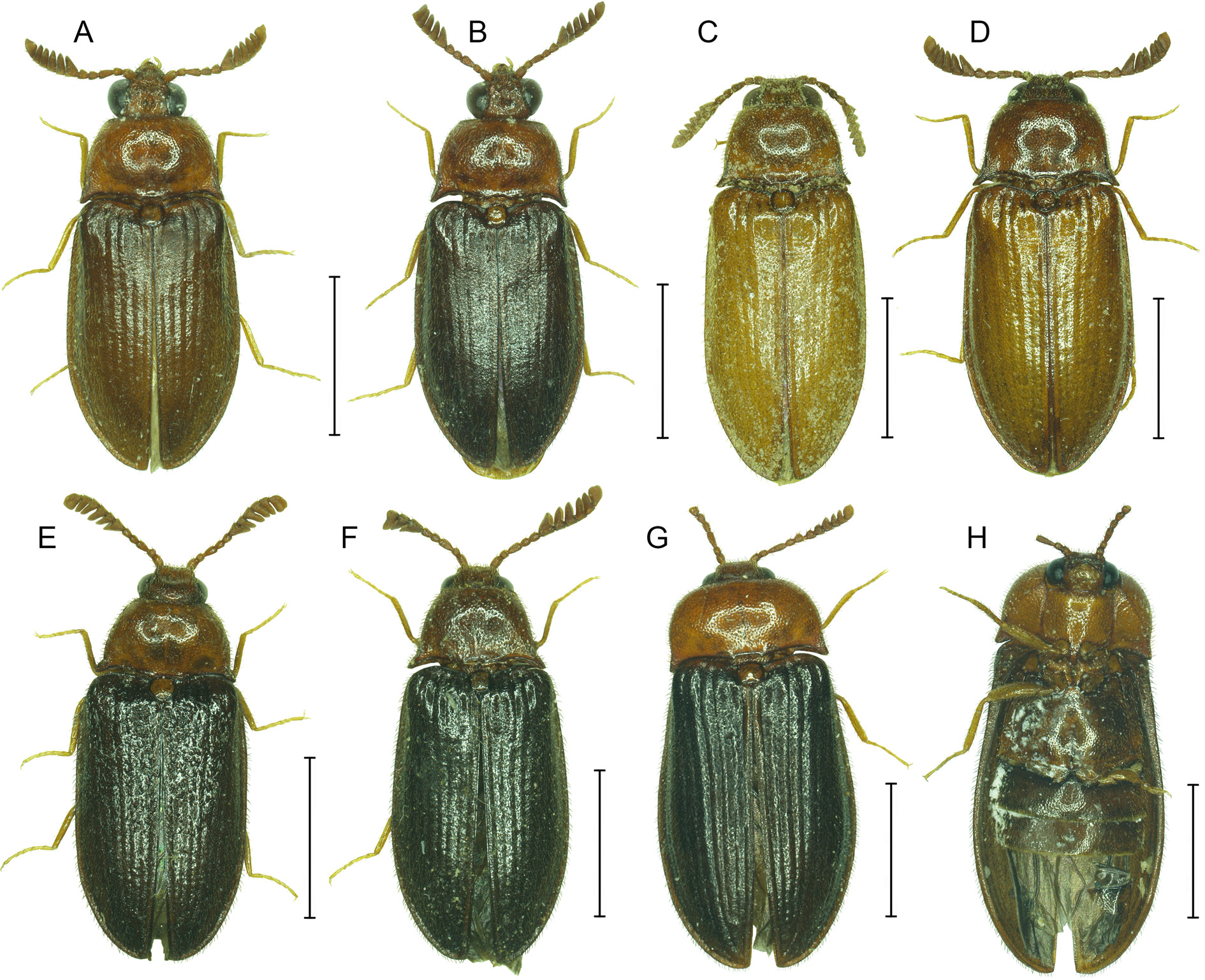
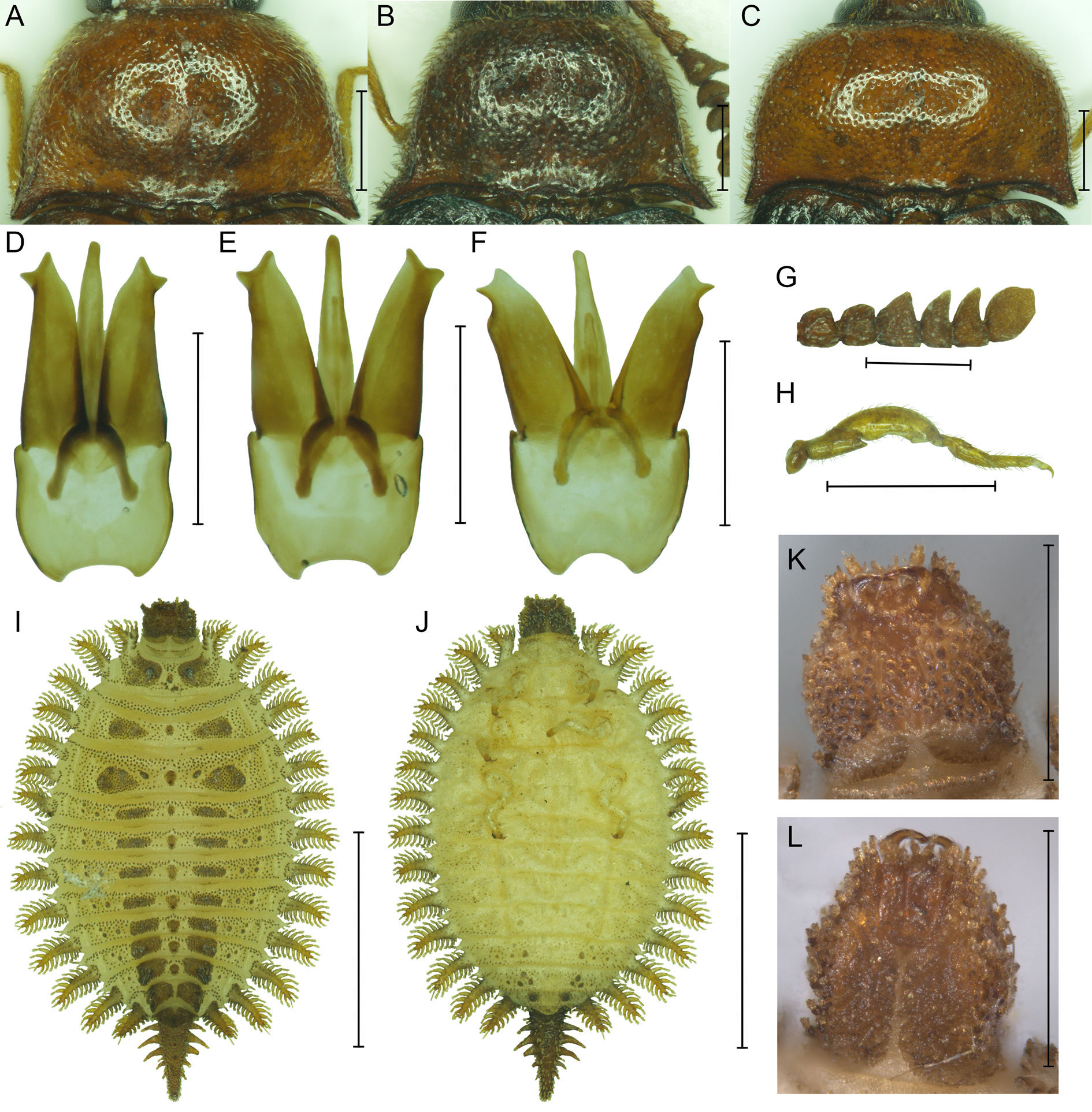
Figs 1 View Fig G–H; 2C, G–L
Material examined
CYPRUS: ♀, Pafos, Vretsia, alt. 600 m, dead under bark of Quercus infectoria , 2 Aug. 2011, Ch. Makris leg. ( PCCM); larva, Pafos, Vretsia, under bark of Quercus infectoria , 3 Jul. 2015, Kundrata and Petrzelkova leg. ( PCRK).
Description
Female
BODY. 6.0 mm long, 2.2 times as long as wide, elongate oblong, dorsoventrally flattened. Body surface testaceous to reddish brown, metaventrite and abdomen darker, elytra dark brown (margins lighter), legs yellowish-testaceous; body covered with yellow pubescence ( Fig. 1 View Fig G–H).
HEAD. Eyes medium-sized; minimum interocular distance 1.39 times maximum eye diameter; frons shallowly depressed medially. Antenna with antennomere 3 elongate, more than 2.5 times as long as wide; antennomeres 4 and 5 simple, subequal, slightly longer than wide; antennomeres 6–7 short, subquadrate, antennomeres 8–9 expanded on one side to form (with apical antennomere) a pectinate club; apical antennomere expanded, with additional trapezoidal broadening apically ( Fig. 2G View Fig ).
PRONOTUM. Transverse, 2.0 times as wide as long, widest posteriorly, width between posterior angles 2.0 times width between anterior angles; lateral sides convex, strongly converging anteriorly and only slightly diverging posteriorly, with distinct lateral carina almost completely concealed in dorsal view. Posterior angles relatively wide, produced posteriorly, apically truncated, with short distinct posterior carina, close to lateral carina in dorsal view ( Fig. 2C View Fig ). Disc strongly convex, posteriorly with shallow longitudinal medial furrow. Prosternum including prosternal process 1.1 times as long as wide, sparsely and coarsely punctured; prosternal process subparallel-sided, considerably curved, converging apically, with apex subacute ( Fig. 1H View Fig ). Scutellum subtrapezoidal, slightly wider than long, apex very slightly emarginate. Elytra gradually widened at posterior third, 4.2 times as long as pronotum; disc clearly striate, covered with moderately fine punctures. Only three legs present; pro- and mesothoracic legs well-developed, moderately long, with tarsomeres 1–4 gradually shortened, tarsomere 4 minute, and apical tarsomere narrow, longest; metathoracic leg minute (anomaly?), only trochanter well-developed, remaining parts reduced, tarsomeres fused together, only one simple claw present ( Figs 1H View Fig , 2H View Fig ).
ABDOMINAL VENTRITES. Finely and moderately densely punctate; only first two ventrites present ( Fig. 1H View Fig ). Genitalia missing.
Larva
BODY. 4.6 mm long, 2.2 mm wide (without lateral lobes). Body broadly ovate, strongly flattened, covered with modified scale-like setae; thoracic segments and abdominal segments I–VIII with lateral lobes bearing elongate setiferous lobules ( Fig. 2 View Fig I–J). Body yellow, except dark brown pigmented head, abdominal segment IX and pigmented areas, median projections and small setiferous tubercles on dorsal surfaces, and apical parts of legs.
HEAD. Prognathous, subtrapezoidal, slightly elevated, slightly wider than long, narrower than prothorax, with large stemma on each side ( Fig. 2 View Fig K–L). Epicranial stem very short, frontal arms slightly lyriform. Antennae less than half of head width; antennomere 1 short, about as long as wide, antennomere 2 enlarged, 2 times as wide as long, widest medially, sparsely covered with modified scale-like setae, shorter in middle, longer and arranged in rows laterally; antennomere 3 highly reduced; sensorium conical.Labrum free, transverse, small, setose apically. Mandibles falcate, moderately stout, considerably curved. Maxillary palpi four-segmented, with segment 2 longest. Labial palpi two-segmented, basal segment about as long as wide, apical segment about as long as basal one, elongate, narrow. Gular sutures present, gula very narrow. Cervical region membranous, dorsally with transverse row of setiferous tubercles, disconnected medially.
THORACIC TERGA. Transverse, sparsely covered with setiferous tubercles, each tergum with pair of laterotergal lobes on each side; lobes equal in length, narrow, lined with narrow, setiferous lobules. Each tergum with median oval projection (largest at metatergum) and pair of irregularly shaped pigment patches (located more posteriorly at meso- and metatergum), consisting of several smaller slightly protruding pigmented areas. Mesotergum transversely divided into anterior very narrow plate covered uniformly with setiferous tubercles and posterior plate bearing also median projection and pigment patches. Thoracic sterna unpigmented, almost smooth, clothed very sparsely with short setae. Legs well-developed, prothoracic leg and mesothoracic spiracle not close together; spiracles located on mesosternal anterolateral transverse sclerites.
ABDOMINAL TERGA. Terga I–VIII transverse, covered with setiferous tubercles and with median projection (missing from tergum VIII) and pair of pigment patches similarly as on thoracic terga (smaller and transverse on anterior segments, larger and plate-like on posterior segments), with pair of laterotergal lobes on each side; posterior lobe similar to thoracic ones, anterior lobe minute, bearing 1–3 setiferous lobules. Lateral parts of abdominal tergites bearing spiracles, with two small, rounded pigment patches behind spiracles on terga I–V, with one patch on tergum VI, remaining terga without such patches. Tergum IX forming long, well-sclerotized, tail-like plate covered with setiferous tubercles, with several lateral branches of different lengths. Abdominal sterna I–V without well-developed pigment patches, sterna VI–VII with four pigment patches of different sizes on each side; sternum VIII with pair of larger patches.
Remarks
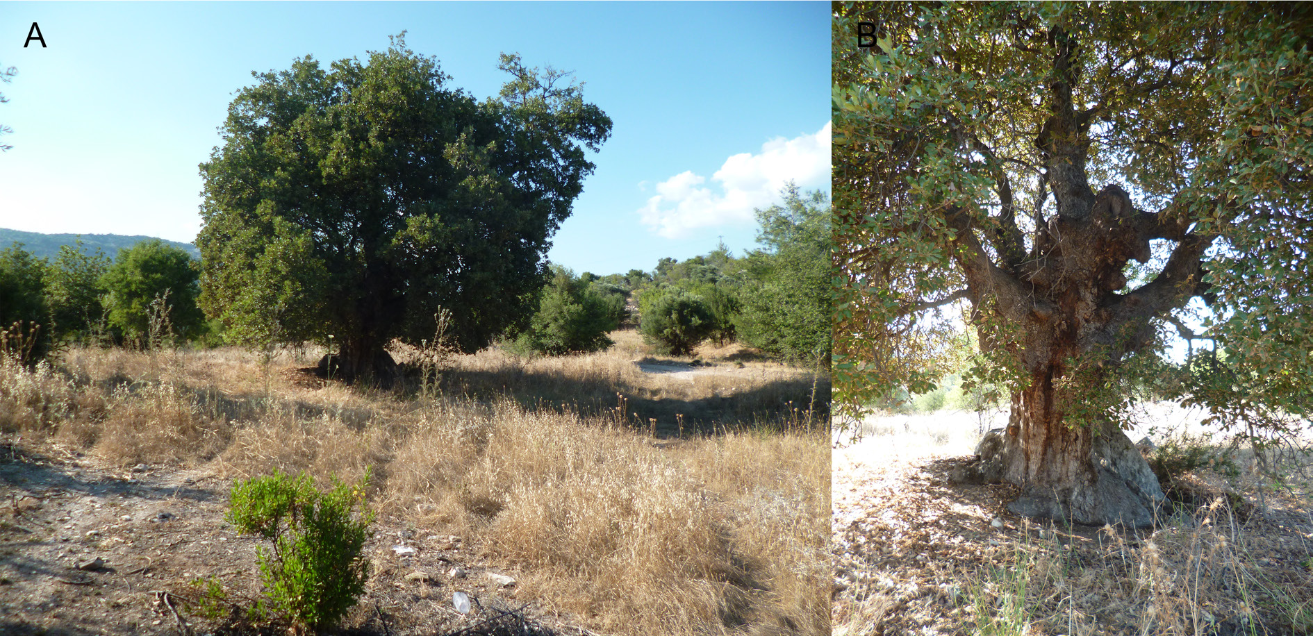
The only adult specimen of Brachypsectra known from Cyprus is a dead, partly damaged female ( Fig. 1 View Fig G–H) found by Ch. Makris in August 2011 under oak bark in Vretsia, Paphos ( Fig. 4 View Fig A–B). Unfortunately, the last three abdominal ventrites are missing in this specimen ( Fig. 1H View Fig ), so we are not able to study the genitalia. In addition, the specimen might be teratological as it has a minute hind leg (the second is missing) with fused tarsomeres and only one claw ( Fig. 2H View Fig ). Subsequent collecting efforts resulted in only finding a single larval specimen.
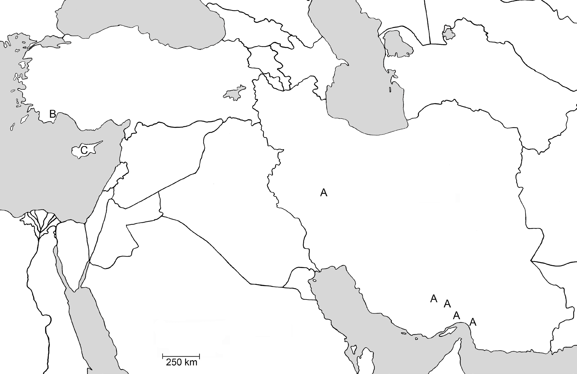
We are not able to assign the Brachypsectra female and larva from Cyprus to any described species since only males are known in B. kadleci and B. jaechi sp. nov. The distribution of Brachypsectra species as well as the larger eyes in the female suggest its close relationship to B. jaechi sp. nov. from Turkey rather than to B. kadleci from Iran. The specimen from Cyprus differs from B. jaechi sp. nov. in having a lighter prosternum and mesoventrite, wider pronotal posterior angles with posterior carina very close to the lateral carina, and a shallower emarginate apex of the scutellum; however, we cannot exclude the possibility that this is only a result of sexual dimorphism. Until more material of both sexes and/or larvae for the Palaearctic species is available for study, we retain the population from Cyprus as Brachypsectra sp.
Key to species of Brachypsectra View in CoL View at ENA (modified from Costa et al. 2006)
Adults (males)
1. Pectinate club with six antennomeres ............................................................................................... 2
– Pectinate club with more than six antennomeres .............................................................................. 5
2. Eyes large; head between eyes less than 2.0 times as wide as eye width in dorsal view; pronotum slightly converging anteriorly ........................................................................................................... 3
– Eyes smaller; head between eyes more than 3.0 times as wide as eye width in dorsal view; pronotum strongly converging anteriorly .......................................................................................................... 4
3. Apical antennomere subtriangular with apical incision; pronotal posterior angles acute; punctation of elytra equal to that of pronotum; aedeagal median lobe with basal struts strongly diverging; paramere subparallel-sided, with lateral preapical acute projection ( Singapore) ............ B. fuscula Blair, 1930
– Apical antennomere trapezoidal, broadening apically; pronotal posterior angles obtuse; punctation of elytra much finer than that of pronotum ( Fig. 1 View Fig A–D); aedeagal median lobe with basal struts U-shaped; paramere constricted apically, with lateral preapical and apical blunt projections ( Fig. 2 View Fig D– E) ( Iran; Fig. 3 View Fig ) .............................................................................................. B. kadleci Hájek, 2010
4. Body unicoloured, yellowish brown; head broad; posterior pronotal carina concealing lateral carina in dorsal view; aedeagal median lobe subparallel-sided, apex broadly rounded; paramere with lateral preapical projection blunt ( India) ............................................................ B. lampyroides Blair, 1930
– Body bicoloured, testaceous, with elytra dark brown; head narrower; posterior pronotal carina separated from lateral carina in dorsal view ( Fig. 1 View Fig E–F); aedeagal median lobe gradually narrowed towards apex, apex subacute; paramere with lateral preapical projection subacute ( Fig. 2F View Fig ) ( Turkey; Fig. 3 View Fig ) ...................................................................................................................... B. jaechi sp. nov.
5. Distal inner angle of antennomeres 6–10 produced in an acute spine (amber, Dominican Republic) .......................................................................... B. moronei Branham in Costa et al., 2006
– Distal inner angle of antennomeres 6–10 not produced in an acute spine ........................................ 6
6. Serration of antennal segments beginning on antennomere 5; antennomere 4, if weakly serrate, much smaller than 5; sides of pronotum distinctly sinuate near base and posterior angles narrowly acute and laterally produced; paramere with lateral apical projection ( Mexico, USA) ............................... ........................................................................................................................ B. fulva LeConte, 1874
– Serration of antennal segments beginning on antennomere 4; antennomeres 4 and 5 equally serrate and similar in size; sides of pronotum more or less straight basally and posterior angles less acute and not laterally produced; paramere with lateral preapical projection ( Dominican Republic) ............................................................................................... B. vivafosile Woodruff, 2004
Larvae
1. Stemmata pedunculate; epicranial suture and gular sutures absent; mandible with retinaculum; anterior lobe of abdominal segment II undivided ( Australia) ............................... Brachypsectra View in CoL sp.
– Stemmata not pedunculate; epicranial suture and gular sutures present; mandible without retinaculum; anterior lobe of abdominal segment II divided into lobules ............................................................. 2
2. Head about as wide as long; mandible slender; first segment of labial palp longer than second ..... 3
– Head wider than long; mandible stouter; first segment of labial palp as long as second ................. 4
3. Head subtrapezoidal; length of antenomere II 0.5 times head width; first segment of labial palp subparallel-sided ( Mexico, USA) .................................................................. B. fulva LeConte, 1874
– Head suboval; length of antenomere II 0.7 times head width; first segment of labial palp expanded distally (amber, Dominican Republic) ............................. B. moronei Branham in Costa et al., 2006
4. Head subrectangular; anterior lobe of abdominal segment II with three or more lobules ( India) ...................................................................................................... B. lampyroides Blair, 1930
– Head subtrapezoidal ( Fig. 2 View Fig K–L); anterior lobe of abdominal segment II with two lobules ( Fig. 2 View Fig I–J) ( Cyprus; Fig. 3 View Fig ) ................................................................................... Brachypsectra View in CoL sp. (this paper)
No known copyright restrictions apply. See Agosti, D., Egloff, W., 2009. Taxonomic information exchange and copyright: the Plazi approach. BMC Research Notes 2009, 2:53 for further explanation.