
Ecdyonurus solus Godunko, Kłonowska-Olejnik and Prokopov, 2007
|
publication ID |
https://doi.org/10.5281/zenodo.179171 |
|
DOI |
https://doi.org/10.5281/zenodo.6240749 |
|
persistent identifier |
https://treatment.plazi.org/id/03A3C701-1E69-FF8A-54C2-FCB8BB6956EE |
|
treatment provided by |
Plazi |
|
scientific name |
Ecdyonurus solus Godunko, Kłonowska-Olejnik and Prokopov |
| status |
sp. nov. |
Ecdyonurus solus Godunko, Kłonowska-Olejnik and Prokopov View in CoL , sp. nov.
( Figs 1–25 View FIGURES 1 – 3 View FIGURES 4 – 6 View FIGURES 7 – 14 View FIGURES 15 – 19 View FIGURES 20 – 25 )
Ecdyonurus venosus: Kiseleva and Ezernitskii 1985: 112 View in CoL ; Kiseleva 1993: 163; Godunko 2001: 81 Ecdyonurus fluminum: Kiseleva and Ezernitsikii 1985: 112 ; Kiseleva 1992: 116; Kiseleva 1997: 39
Description. Male imago (all adults were reared from larvae). Size: body 10.0– 10.4 mm; fore wing 11.0– 11.2 mm; hind wings 3.9–4.3 mm; fore legs 10.2–10.9 mm (femur 2.6–2.7 mm; tibia 2.5–2.7 mm; tarsal segments: T1 = 0.8–1.0 mm; T2 = 1.4–1.5 mm; T3 = 1.3–1.4 mm; T4 = 1.1 mm; T5 = 0.5 mm; gradation of tarsal segments: 2>=3>4>1>5); cerci 29.6–30.5 mm. General color of body yellow to brown.
Head yellowish with brownish and brownish-gray smudges on short facial keel. Antennae brown. Ocelli black basally with subapical yellowish-brown ring and whitish or white-gray apically. Eyes are separated by a distinct gap comparable in size with the central ocellus, gray-blackish, with light gray ring around of margin. Lateral portion of eyes with two light gray rings ( Figs 1, 2 View FIGURES 1 – 3 ).
Thorax yellow to brown, dorsally with dark brown maculation on metanotum and hind part of mesonotum. Pronotum light yellowish-brown. Lateral part of thorax generally whitish to yellowish-brown. Ventral part of thorax distinctly brown to brownish-black. Wing hyaline, transparent, unicolorous. Pterostigmatic area clearly yellow, costal area yellowish-white. Venation brown to blackish, lighter basally. Fore legs distinctly darker than middle and hind legs: femora dark brown; tibiae and tarsi brown, with slightly dark last tarsal segment. Middle and hind legs with yellow femora and tibiae; tarsi yellowish-brown basally with brownish last segment.
Abdominal terga yellow to yellowish-brown. Tergum 1 brown, with blackish-brown strips near hind margin of segment; tergum 2 with central longitudinal light band and two brownish paramedian bands; terga 3–10 yellowish, with two longitudinal narrow paramedia bands (terga 7–10 slightly darker that others ones). Laterally terga 2–8 with distinct blackish-brown stripes connected dorsally in posterior part of segments ( Fig. 3 View FIGURES 1 – 3 ). Sterna 1 and 9 brown, sterna 2–8 uniformly yellowish with two unclear brownish spots. Violet or blackish nerve ganglia well visible on the sterna 2–8. Cerci brownish, lighter apically. Joints of segments blackish.
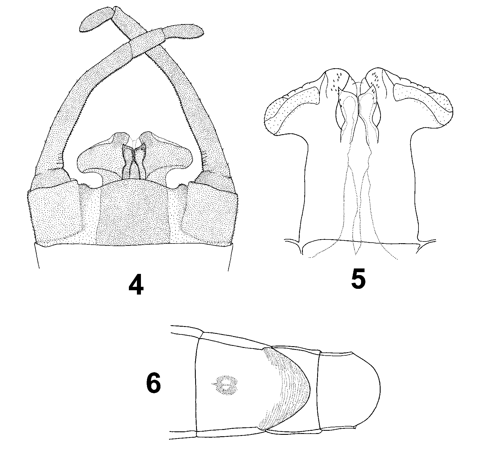
Styliger plate light brown, with two small and rounded apically protuberance near forceps base ( Fig. 4 View FIGURES 4 – 6 ). Forceps brown. Penis lobes light brown, slightly stretched laterally and convergent toward outer margins, with rounded distal part. Penis light brown. Basal sclerite large, partially cover the basal part of lateral sclerite. Lateral sclerite narrow and curved, distinctly convergent distally. Apical sclerite relatively wide, not convergent apically. Titilators wide, brown ( Fig. 5 View FIGURES 4 – 6 ).
Female imago. Size: body 12.2 mm; fore wing 12.0 mm; hind wings 4.0 mm; cerci 19.5 mm. Head yellow with blackish smudges on facial keel. Eyes and apical part of ocelli grayish-black. Central part of ocelli light yellow; apical part whitish. Antennae light brown.
Thorax yellow to dark brown. Pronotum yellowish-gray with two brownish spots centrally. Mesonotum yellowish-brown with brown to dark brown distal part. Metanotum dark brown. Lateral part of thorax yellowish-brown. Sterna of thorax brown to dark brown. Wings transparent, hyaline slightly yellowish color. Pterostigmatic area yellowish, opaque. Venation yellow to dark brown. Fore legs with light brown femora, and brown to dark brown tibiae and tarsi. Middle and hind legs with unicolorous yellow femora and yellowish-brown tibiae. Tarsi brown to dark-brown.
Abdominal terga with central longitudinal bands and two short reddish strokes on the segments. Two distinct triangular reddish or brownish spots are present on central part of tergum 1; lateral part of terga 2–8 with blackish stripes. Abdominal sterna yellow to reddish-brown, with uniformly pattern on the segments 1–7 (generally present two central light short strokes). Sterna 2-8 with visible violet or blackish nerve ganglia. Subgenital and subanal plates as in Fig. 6 View FIGURES 4 – 6 .
Male subimago. Size: body 9.5 mm; fore wing 12.0 mm; hind wings 4.0 mm; cerci 19.5 mm. General color of body yellow and yellowish-gray to brown and dark brown.
Head grayish brown, antennae brownish. Eyes grayish-black with one hard visible grayish-white ring laterally. Ocelli blackish basally, with grayish-white tip.
Pronotum brownish-gray. Mesonotum and metanotum yellowish-white with distinct dark brown bands. Wings unicolorous yellowish, relatively transparent. Venation whitish-gray to brownish. Transversal veins darker than longitudinal with grayish-brown smudges around veins. Fore legs dark: femora brown slightly darker distally; tibiae dark brown; tarsi unicolorous brown. Middle and hind legs: femora yellow with elongate brownish spot apically; tibiae yellowish-brown; tarsi yellowish.
Abdominal terga yellow to yellowish-brown. Sterna 1–8 slightly darker than terga with two light short strokes and two light spots centrally. Sternum 8 with violet nerve ganglia. Lateral part of terga 1–8 with distinct blackish-brown stripes connected distally in posterior part of segments. Styliger plate yellowish-brown, forceps slightly darker. Cerci light brown. Joints of segments blackish.
Female subimago. Size: body 12.7–13.8 mm; fore wing 13.0– 14.2 mm; hind wings 4.2–5.0 mm; cerci 12.2–13.7 mm. General color of body very similar to those of female imago except wings and legs.
Thorax yellow to dark brown. Wings distinctly yellowish to light brown, slightly opaque, especially in pterostigmatic area. Veins dark brown to black. Brownish smudges around veins are presented. Fore legs unicolorous dark brown with black tarsi. Middle and hind legs light brown. Tarsi dark brown to black.
Abdominal terga with distinct drawing: tergum 1 with diffuse central brownish spots; terga 2–3 with central diffuse brownish and two yellow spots; terga 4–5 with longitudinal brownish spot; terga 6–7 with two pair of small brownish spots near anterior part of segment; terga 8–10 uniformly yellowish with central brownish smudges. Lateral side of terga 1–8 with distinct dark brown strips. Sternum 8 with violet nerve ganglia. Cerci dark brown.
Mature larva. Body length 11.2–13.5 mm; caudal filaments 7.4–10.0 mm. General color yellow to light brown. Abdominal segments of mature larvae with well visible pattern similar to adults.
Head yellow, oval, relatively large, the part at the eyes level not widest. Antennae yellow to light brown. Mouthparts: Labrum not large, slightly stretched laterally, dorsally with one row of 7–9 median strong bristles ( Fig. 7 View FIGURES 7 – 14 ), typical for the E. venosus species-group ( Belfiore & Buffagni 1994). Hypopharynx without specific features, generally with long hair on the outer margin and distal part, typical for the E. venosus species-group ( Fig. 8 View FIGURES 7 – 14 ). Mandibles (n = 20) with 6–9 prosthecal bristles. Maxillae (n = 20) (characters listed by Haybach (1999)): number of comb-shaped bristles (N_CBS) = 17–22; number of teeth on 5th comb-shaped bristle (N_TCB5) = 14–17; number of hairs on dorsal upper side (N_DOR) = 14–18; outer margin of maxillae without hairs (N_OUT = 0); number of hairs on ventral basal part of maxillae (N_VEN) = 21–24; number of hairs at the base of maxillary palps (N_PLBas) = 16–20; outer base of the first segment of maxillary palps without hairs (N_PLH = 0); number of setae on the outer margin of the first segment of maxillary palps (N_PLS)>50; number of setae on the inner side of the first segment of maxillary palps (N_PLP)>40. Labial palps with numerous hairs of the group on the dorsal side of its first segment (N_LPH = 28–36) arranged on 2–3 rows.
Pronotum yellow with some light central smudges. Lateral projection of pronotum short, distinctly asymmetrical, with apical part curved towards the body ( Fig. 10 View FIGURES 7 – 14 ). The width/length ratio of semipronotum to caudal projection (see Bauernfeind & Humpesch 2001) is 3.8–4.2. Mesonotum yellow to yellowish-brown, with whitish spots laterally. Legs yellow to light brown. Femora relatively slender and long. The length/width ratio of metafemora on average 2.67. Dorsal surface of femora mainly with distinct pattern (four yellowish lateral spots on a dark field) ( Fig. 11 View FIGURES 7 – 14 ). Femoral scales of various types ( Fig. 12 View FIGURES 7 – 14 ). Outer margin of femora with row of long slender bristles and small pointed spines; subapical spines is pointed or bluntly pointed ( Fig. 13 View FIGURES 7 – 14 ). Tibiae yellow. Tarsi yellow to light brown basally with brown or dark brown apical ring. Tarsal claw brown to dark brown with 2–3 teeth ( Fig. 14 View FIGURES 7 – 14 ).
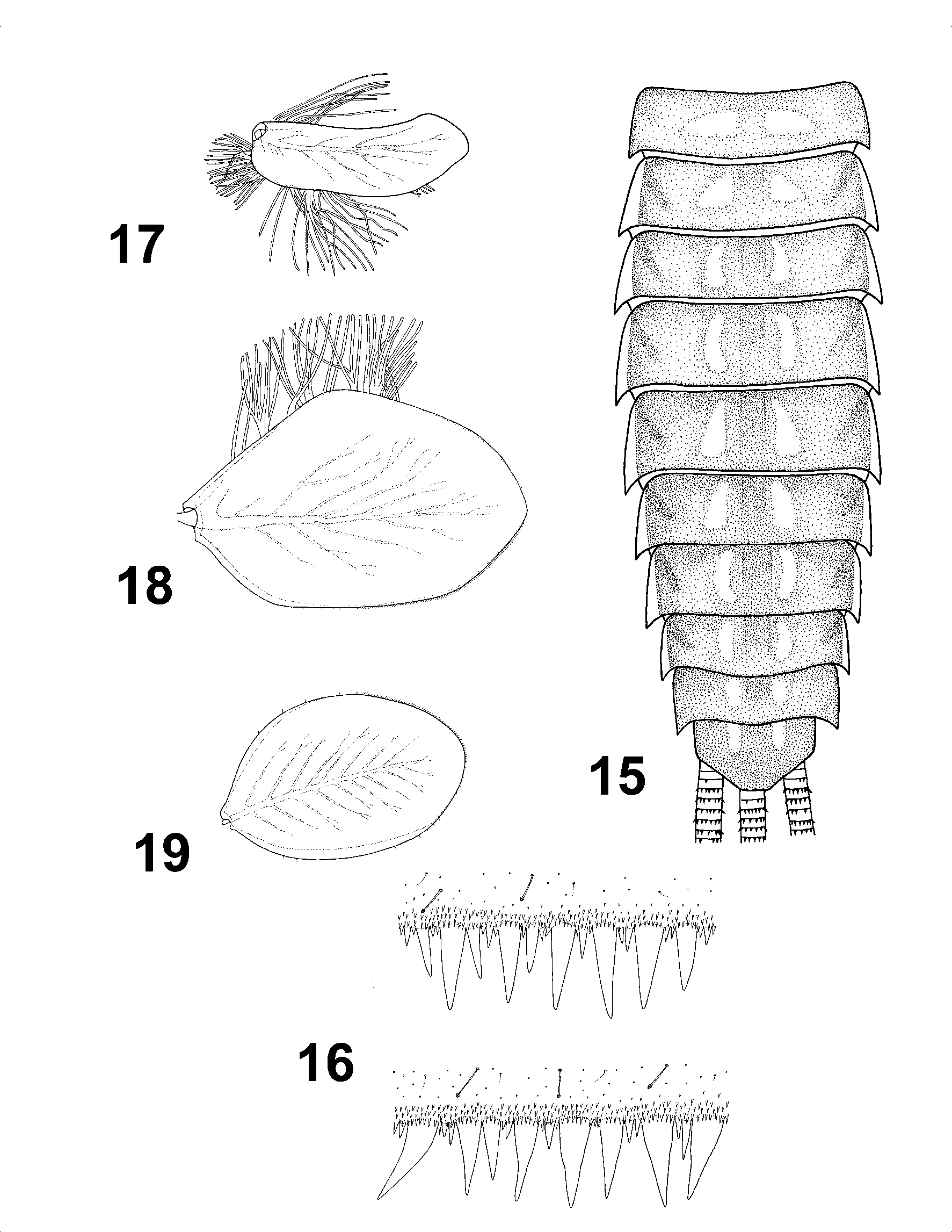
Abdominal terga yellow to light brown. Tergum 1 uniformly light; terga 2–7 and 10 (sometime 8) with two light central longitudinal spots (sometimes, with two pair of light spots); Tergum 9 with wide central light spot ( Fig. 15 View FIGURES 15 – 19 ). Posterior margins of terga with large pointed marginal teeth ( Fig. 16 View FIGURES 15 – 19 ). Sterna unicolorous yellowish-brown with well visible violet nerve ganglia on the segments 2–8. Gills yellowish to light brown. Gill 1 relatively long with broadly rounded apical part ( Fig. 17 View FIGURES 15 – 19 ). Gill 4 distinctly wide and asymmetrical ( Fig. 18 View FIGURES 15 – 19 ). Gill 7 as in Fig. 19 View FIGURES 15 – 19 , without tuft. Cerci yellow to yellowish brown.
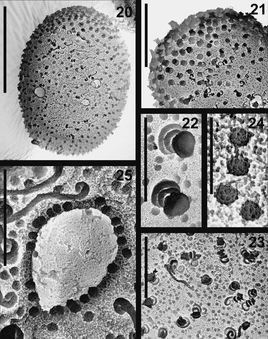
Egg. Size: length: 164–171 µm; width 108–116 µm. Egg oval. The eggs are characterized by attachment structures, tubercles, and very small granules ( Fig. 20 View FIGURES 20 – 25 ). The attachment structures are represented by knob-terminated coiled threads (KCTs) ( Koss & Edmunds 1974) and are of two different kinds. The first are larger (diameter 3.9–5.3 µm) and concentrated (aggregated) at one egg pole, covering a densely chorionic surface (spaced at a distance of 2.6–3.9 µm) ( Fig. 21 View FIGURES 20 – 25 ). This structure is similar to that of E. venosus KCTs ( Gaino & Rebora 2003) ( Fig. 22 View FIGURES 20 – 25 ). The second attachment structure is slightly smaller (diameter 2.7–3.3 µm) and is regularly distributed over the chorion surface (spaced at a distance of 6.7–12.0 µm) ( Fig. 23 View FIGURES 20 – 25 ). The chorionic surface has many small rounded tubercles (0.7–0.9 µm), spaced at a distance of 0.4–1.3 µm. The chorionic surface and tubercles are covered by very small granules (granular ground matrix) 0.09–0.17 µm in diameter ( Fig. 24 View FIGURES 20 – 25 ). Five to eight micropyles are visible in the subequatorial area. The sperm guide ovoidal is 11.1–11.8 µm in length and 7.9–9.0 µm in width (micropylar opening situated at the side). The micropylar rim is narrow with tubercles similar to those found on the chorionic surface ( Fig. 25 View FIGURES 20 – 25 ).
Etymology. The species name originates from the Latin noun solus , i.e. single, solitary. It is the only one species of the genus Ecdyonurus living in the Crimea.
Specimens examined. HOLOTYPE: male imago, UKRAINE, Autonomous Republic of the Crimea, Chorna River, Chornorichens’kyi gap, 8.06. 2003, 300 m. a.s.l., Long. 33o46’19’’ E, Lat. 44o30’23’’ N, leg. G.A. Prokopov. PARATYPES: 3 larvae (two on slides), ibid, Stilia River, 7.07. 1989, 150 m. a.s.l., Long. 34o02’50’’ E, Lat. 44o38’11’’ N, leg. G.A. Kiseleva; 2 larvae, ibid, Upper Chorna River, Chornorichens’kyi gap, 15.06. 1998, 300 m. a.s.l., Long. 33o46’19’’ E, Lat. 44o30’23’’ N, leg. G.A. Prokopov; 5 larvae, ibid, Upper Chorna River, Chornorichens’kyi gap, 3.01. 2001, 250 m. a.s.l., Long. 33o46’19’’ E, Lat. 44o30’23’’ N, leg. G.A. Prokopov; 6 larvae, ibid, Upper Chorna River near Chornorichens’kyi gap, 4.01. 2001, 300 m. a.s.l., Long. 33o46’19’’ E, Lat. 44o30’23’’ N, leg. G.A. Prokopov; 9 larvae, ibid, Al’ma River, downstream of “Asport” Boundary, 29.06. 2001, 450 m. a.s.l., Long. 34o14’21’’ E, Lat. 44o43’14’’ N, leg. G.A. Prokopov; 29 larvae, ibid, Al’ma River downstream of “Tar’er” Boundary, 30.06. 2001, 400 m. a.s.l., Long. 34o10’43’’ E, Lat. 44o43’11’’ N, leg. G.A. Prokopov; 4 larvae, ibid, Al’ma River downstream of “Sosnovyi” Boundary, 1.07.2001, Long. 34o08’04’’ E, Lat. 44o45’33’’ N, 350 m. a.s.l., leg. G. A. Prokopov; 1 larva, ibid, Al’ma River near Partyzans’ke Reservoir, 1.07. 2001, 300 m. a.s.l., Long. 34o05’43’’ E, Lat. 44o47’30’’ N, leg. G.A. Prokopov; 1 larva, ibid, Upper Chorna River, Chornorichens’kyi gap, 16.08. 2001, 300 m. a.s.l., Long. 33o46’19’’ E, Lat. 44o30’23’’ N, leg. G.A. Prokopov; 6 larvae, ibid, Chorna River near Chornorichyns’ke village, 17.08. 2001, 200 m. a.s.l., Long. 33o41’23’’ E, Lat. 44o32’12’’ N, leg. G.A. Prokopov; 1 male subimago, 1 female imago, 3 female subimagines, 25 larvae, Ukraine, Autonomous Republic of the Crimea, Chorna River, Chornorichens’kyi gap, 8.06. 2003, 300 m. a.s.l., Long. 33o46’19’’ E, Lat. 44o30’23’’ N, leg. G.A. Prokopov.
All specimens were preserved in 75% alcohol. All adults were reared from larvae. The holotype and part of the paratypes are housed in the collection of the State Museum of Natural History, National Academy of Sciences of Ukraine (Lviv, Ukraine). The other paratypes are stored in the Biology Centre of the Academy of Science of the Czech Republic, Institute of Entomology (Č eské Budĕjovice, Czech Republic) and in the first and second author’s collections.
Affinities. E. solus sp. nov. belongs to the E. venosus species-group by the presence of following characteristics: in male imago: (1) apical sclerite of penis subparalel to the body axis, straight, relatively short and blunt; (2) abdominal terga with mainly visible darker pattern; in larva: (3) superlinguae of hypopharynx with long fine setae on the anterior margin (up to the lateral tips); (4) labrum with one row of setae ventrally; (5) lateral prolongation of sterna are markedly visible. The new species can be easily distinguished from all other representatives of this species-group by the following features: in male imago: (1) eyes gray-blackish, with two light gray rings laterally, divided by a small gap; (2) wing hyaline, transparent and unicolorous; (3) venation brown to blackish, lighter basally, without maculation around veins; (4) color of abdominal terga and sterna; (5) styliger plate with two small rounded apically protuberance; (6) penis lobes slightly stretched laterally and convergent toward outer margins, with rounded distal part; (7) peculiarities of the penis structure; in larva: (8) number of some groups of setae, bristles and hairs on maxillae and labial palps (9) lateral projection of pronotum short, distinctly asymmetrical, with curved towards the body apical part; (10) femora relatively slender and long; (11) outer margin of femora with row of long slender bristles and small pointed spines; (12) coloration of abdominal terga and sterna; (13) coloration of tarsi; (14) denticulation of posterior margins of terga; (15) shape of scales of dorsal surface of femora and (16) shape of gills (especially gills 1, 4 and 7).
The penis structure discriminates the new species from the all other European species of the E. venosus species-group (except for E. ornatipennis , see below). Some aspects of the penis shape of E. solus sp. nov., are similar to E. ruffii Grandi, 1953 ( Bauernfeind & Humpesch 2001: 139, Fig. 474), but the new species can be distinguished by its body coloration and especially by the pattern on the terga.
The male imago of E. solus sp. nov. can be easily distinguished from the two Caucasian species, viz. E. autumnalis and E. ornatipennis . By laterally stretched penis lobes the new species is similar in appearance to E. ornatipennis . The combination of following features markedly differs E. ornatipennis from E. solus sp. nov.: (1) unicolorous eyes without any rings, not close to each other; (2) penis lobes relatively square-shape distally; (3) abdominal sterna with brown longitudinal central band ( Tshernova 1938, 57–58, Fig. 4 View FIGURES 4 – 6 , Kluge pers. com.). E. autumnalis differs from E. solus sp. nov. by (1) the distinctly darker color of body; (2) the presence one light ring on eyes; (3) the presence of brown longitudinal central band on abdominal sterna; (4) the shape of penis, with large and not stretched laterally lobes; (5) the distinctly convergent towards the tip apical sclerites; (6) the shape of lateral sclerites ( Braasch 1980b, 103–104, Fig. 1, 1 View FIGURES 1 – 3 a, 2, Kluge pers. com.).
The larvae of E. solus sp. nov., partly resembles to E. aurantiacus (Burmeister, 1839) , E. belfiorei Haybach and Thomas, 2002 and E. dispar (Curtis, 1834) by the structure of the pronotal projection and gills (especially gill 1). However, larval characteristics 8, 11, 14 and 16 easily differs new species from E. aurantiacus ; characters 8, 10, 12, 13 and 14 from E. dispar ; characters 8, 12 and 16 from E. belfiorei (see Haybach and Thomas 2002). The larvae of E. solus sp. nov. can be separated from larvae described as E. ornatipennis (see Braasch 1980a), by the shape of the pronotum expansion, femoral scales, gills and by the coloration of the abdominal terga.
Distribution and biology. The larvae of E. solus sp. nov. are found in metarithral and hyporithral rivers on the northern slopes of the Crimean mountains at altitudes of 150– 450 m. a.s.l. In summer, the average water temperature is 12 0C and the water is of hydrogen carbonate type. The larvae inhabit the waters with current velocities of about 1 m /s. Width of river bed width is around 3–6 m, depth 0.3–0.5 m, with cobble and gravel bottoms. The banks of beds are partly shaded by Fagus orientalis Lipsky , Carpinus betulus Linnaeus and Quercus pubescens Willd. E. solus sp. nov. has a univoltine winter cycle (Uw), with one generation per year ( Clifford 1982). The population overwinters in the nymphal stage and larval growth is very slow. The flying period extends from May to July, with most of the population emerging by the end of June or the beginning of July. Other insects groups found in these rivers with E. solus sp. nov. include: stoneflies Nemoura cinerea (Retzius, 1783) ; dragonflies Calopteryx taurica Selys, 1853 , Gomphus vulgatissima (Linnaeus, 1758) , Onychogomphus forcipata (Linnaeus, 1758) ; mayflies Heptagenia samochai (Demoulin, 1973) and Baetis rhodani tauricus Godunko and Prokopov, 2003 , caddisflies Halesus tessellatus (Rambur, 1842) , Limnephilus lunatus Curtis, 1834 , Hydropsyche acuta Martynov, 1909 , Wilhelmia balcanica (Enderlein, 1924) , W. paraequinum Puri, 1925 and blackflies Simuluim acutiphallus (Rubtsov, 1956), S. ponticum (Rubtsov, 1956).


No known copyright restrictions apply. See Agosti, D., Egloff, W., 2009. Taxonomic information exchange and copyright: the Plazi approach. BMC Research Notes 2009, 2:53 for further explanation.
|
Kingdom |
|
|
Phylum |
|
|
Class |
|
|
Order |
|
|
Family |
|
|
Genus |
Ecdyonurus solus Godunko, Kłonowska-Olejnik and Prokopov
| Kłonowska-Olejnik, Małgorzata, Prokopov, Grigorii A. & Godunko, Roman J. 2007 |
Ecdyonurus venosus:
| Godunko 2001: 81 |
| Kiseleva 1993: 163 |
| Kiseleva 1992: 116 |
| Kiseleva 1985: 112 |
| Kiseleva 1985: 112 |