
Lepidocyrtus mariani Traser & Dányi, 2008
|
publication ID |
https://doi.org/ 10.11646/zootaxa.4375.3.6 |
|
publication LSID |
lsid:zoobank.org:pub:1E331F1B-1216-4190-B088-F585B7A008B1 |
|
DOI |
https://doi.org/10.5281/zenodo.5946390 |
|
persistent identifier |
https://treatment.plazi.org/id/03A38468-2D09-FFAE-FF55-5D49EFC4E1E6 |
|
treatment provided by |
Plazi |
|
scientific name |
Lepidocyrtus mariani Traser & Dányi, 2008 |
| status |
|
Lepidocyrtus mariani Traser & Dányi, 2008 View in CoL
Figs 24–29 View FIGURE 24 View FIGURE 25 View FIGURE 26 View FIGURES 27–28 View FIGURE29 , Tab 2
Material examined. Holotype, dissected on three slides (codes B–11, B–12, B–13), Porva, Bakony Mts., Hungary, 390 m above sea level, N47°18'42"; E17°47'30", from moss along the stream Hódos-ér, 31.v.2008, leg. Gy. Traser GoogleMaps .
Other material examined. Three specimens from the type locality (LmPOR), hand collecting, 24.ix.2014, leg. Gy. Traser, Á. Erdő & D . Winkler; twelve specimens from Nagykapornak (LmNK), Mid-West Hungary, 214 m above sea level, N46°50'35"; E16°58'26", from beech forest soil and moss, hand collecting, 15.vii.2015, leg. Á. Erdő & D GoogleMaps . Winkler; three specimens from Farkasgyepű (LmFGY), Bakony Mts. , 417 m above sea level, N47°11'30"; E17°38'18", from beech forest soil, hand collecting, 15.vii.2016, leg. Á. Csiszár & D. Winkler GoogleMaps ; two specimens from Zákányfalu (LmGYE), South-West Hungary, 210 m above sea level, 46°17'8"; E16°57'24", from soil of Illyrian oak-hornbeam forest, hand collecting, 29.viii.2016, leg. Á. Erdő & D. Winkler. All specimens preserved in 96% alcohol and deposited in the first author’s collection in the Faculty of Forestry, University of Sopron .
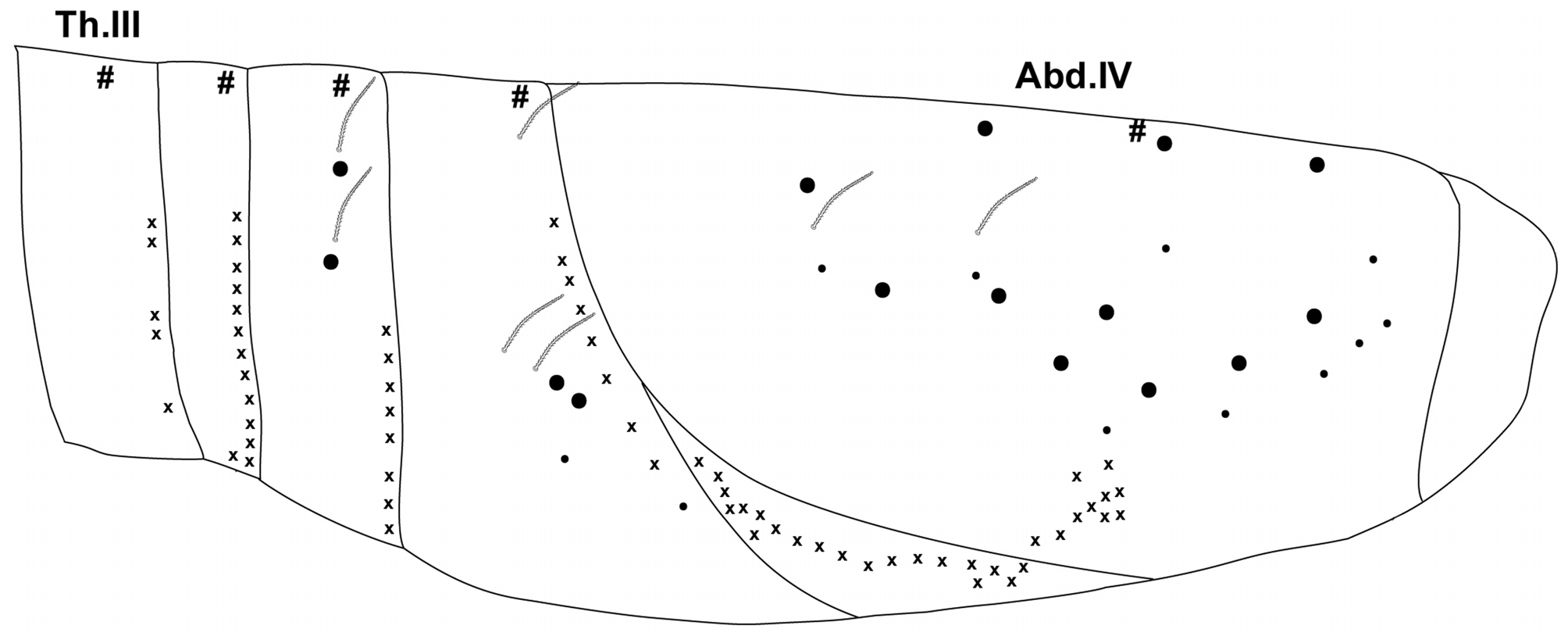
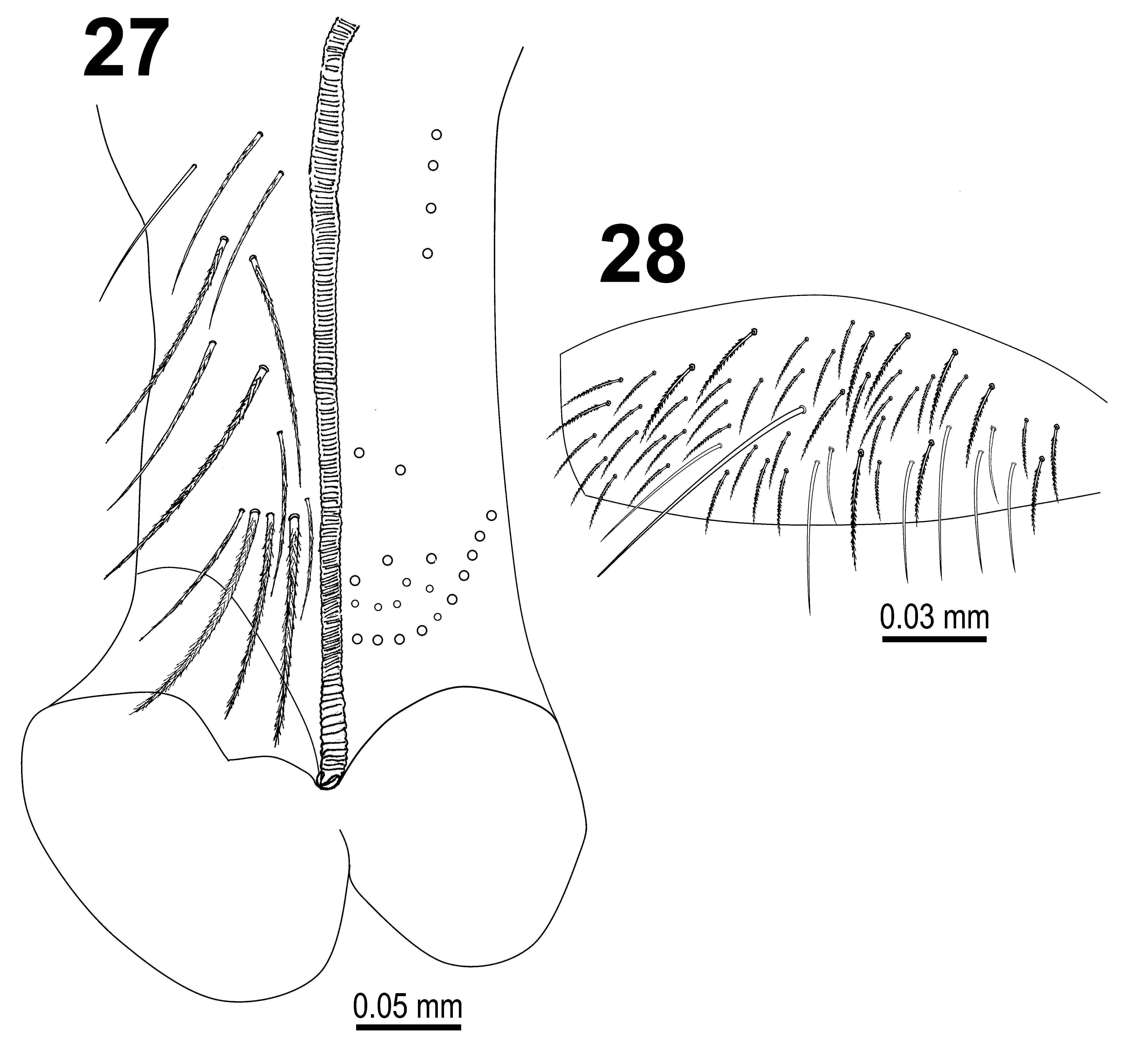
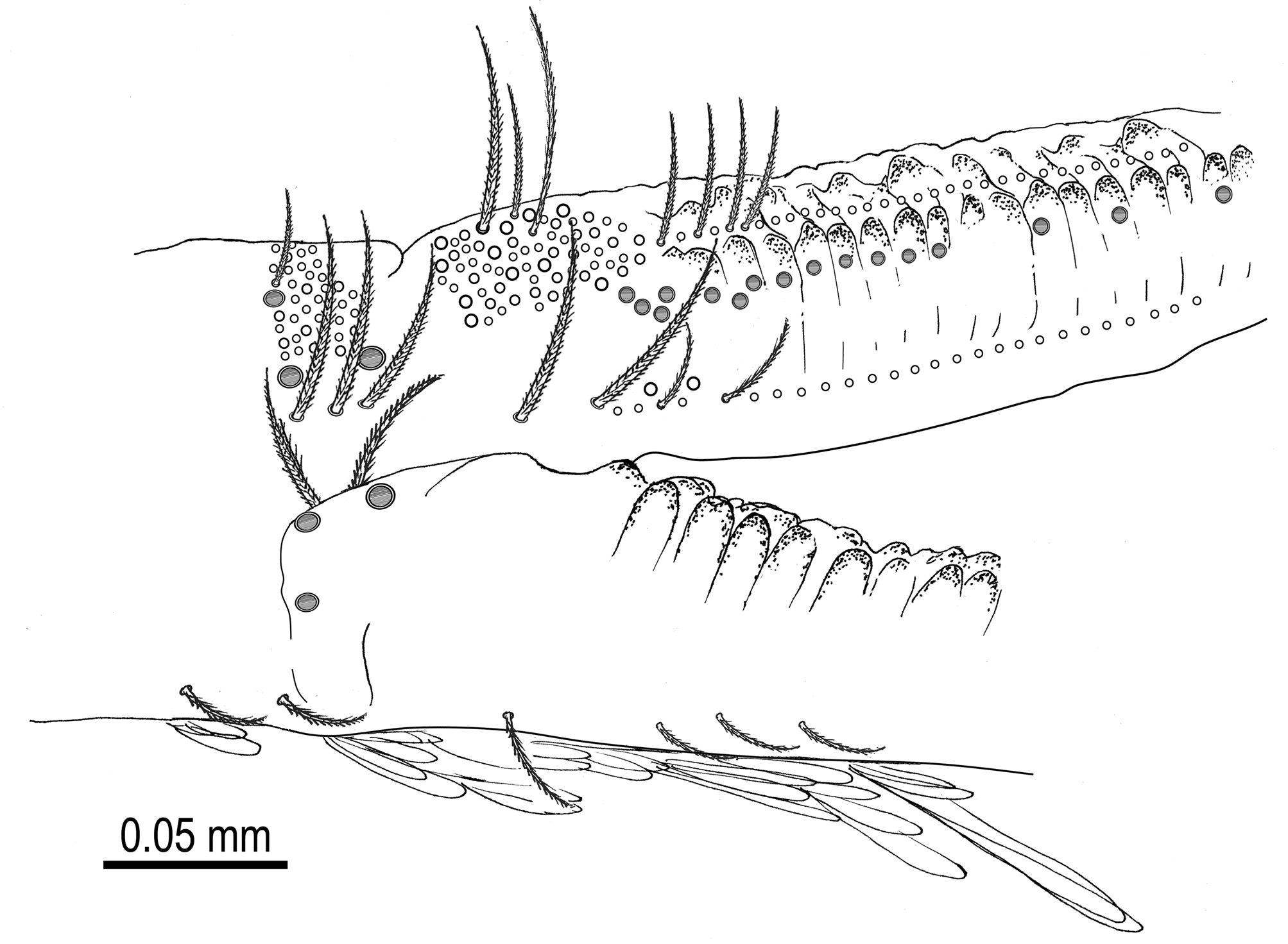
Diagnosis. Large-sized Lepidocyrtus (3.8 mm of maximum trunk length), with mesothorax strongly projecting over the head ( Fig. 24 View FIGURE 24 ) and white-yellowish main body color. Blue pigmented areas include ant. II–IV (seldom also ant. I), cx. I–II, with occasional blue shade also on anterior region of the head, legs, manubrium, ventral tube, anterior part of th. II, posterior parts of abd. III–IV and abd. V–VI. Ocular area dark bluish-black. Ant. I–III and ant. IV base, legs, manubrium and ventral tube scaled. Ant. IV without apical bulb. Small pseudopori (5–7) on apical membranous part of antennal segments present. Labial chaetotaxy M1*M2R*EL1L2 (chaetae M1 and R shorter than others, marked with *) with occasional symmetrical or asymmetrical variations (see Table 2). Labrum 4/554, prelabral and labral chaetae smooth, apical intrusion inverted V-shaped. Lateral labral papillae rounded and bigger in size, medial labral papillae with conical spine expansion. External maxillary palp with two smooth chaetae. Lateral process on labial papilla E slightly curved, tip not reaching apex of papilla. Ventral cephalic groove with 4+4 ciliated chaetae. Head with 16–22 macrochaetae in row A on each side. Interocular chaetotaxy with ciliated chaetae s, t, p, q and 3–4 scales. Dorsal macrochaetae formula R0R1S o/00/0101+3, with small supplementary macrochaetae R1S between R0 and R1. A number of small lateral pseudopori present on th. III (4–6), abd. I (9–13), abd. II (4–8), abd. III (4–8) and abd. IV (21–34) ( Fig 25 View FIGURE 25 ). Chaetae associated with the dorsal trichobothria on abd. II–III–IV as fan-shaped mesochaetae except for abd. II chaeta a2 (smooth mesochaeta). All specimens with five long S-chaetae on abd. IV ( Fig. 26 View FIGURE 26 ). Abd. V with three S-chaetae typical for Lepidocyrtus . Rectangular shaped trochanteral organ formed by 32–74 smooth chaetae. Ventral tube with 12+12 ciliated and 1+1 smooth chaetae on anterior side and 24+24 ciliated chaetae on posterior side ( Fig. 27 View FIGURES 27–28 ). Lateral flap ( Fig. 28 View FIGURES 27–28 ) with a maximum of 54 laterodistal chaetae (45 ciliated and 9 smooth). Dorsal surface of dens with a number of small pseudopori ( Fig. 29 View FIGURE29 ).
Comparative remarks. The morphological and chaetotaxic characters of the studied specimens are in agreement with the original description of Traser & Dányi (2008). In the original description abd. III chaeta d2 (which is a ms chaeta sensu Szeptycki, 1972) is omitted, while it is present in all specimens examined in the present paper, and also present in all European Lepidocyrtus species. New characters added in this paper include the presence and pattern of small pseudopori on the apical membranous area of antennal segments, the dorsal surface of dens and on the lateral region of th. III–abd. IV; presence and distribution of long S-chaetae on abd. IV; and chaetotaxy of the ventral tube. Since the original description was made based on a single specimen only, the newly collected specimens from both the type locality and other locations gave us the opportunity to describe the variability of certain characters ( Table 2). Labial chaetotaxy shows intraspecific variability and even asymmetrical differences on the same specimen. Apart from the characteristic chaetotaxy M1*M2R*EL1L2 with all chaetae ciliated, chaetae m1, r, l1 and l2 might appear as smooth chaetae as well. Bilateral asymmetry of abd. IV chaeta D1 was observed on one specimen (fan-shaped mesochaeta on the right side and smooth mesochaeta on the left side). Small dorsolateral pseudopori shows variability regarding their number, depending on specimen development stage. In one case, bilateral asymmetry on abd. I has been observed, with a small pseudoporus present in the place of the normal-sized dorsomedial pseudoporus. It should be noted that the presence of lateral small pseudopori from th. III to abd. IV, on the apical membranous area of antennal segments and dorsal surface of dens are characters not previously described for any Lepidocyrtus species. Only lateral pseudopori on abd. IV have been described for several species: species of subgenus Setogaster have two lateral pseudopori located externally to chaetae r3–r4–r5, while in L. fimetarius Gisin, 1964 eight lateral pseudopori are present in this position (Mateos & Greenslade 2015). Another species possessing abd. IV pseudopori is L. curvicollis ( Bourlet, 1839) , but they are not in a lateral position but located dorsally within the rectangle formed by macrochaetae B5, B6, T6 and D3 (Mateos 2008).
L. mariani is only known from Hungary, but probably is more widely distributed. Due to a possible confusion, re-investigations of earlier L. curvicollis records are therefore recommended.


No known copyright restrictions apply. See Agosti, D., Egloff, W., 2009. Taxonomic information exchange and copyright: the Plazi approach. BMC Research Notes 2009, 2:53 for further explanation.
|
Kingdom |
|
|
Phylum |
|
|
Class |
|
|
Order |
|
|
Family |
|
|
Genus |