
Lepidocyrtus peisonis Traser & Christian, 1992
|
publication ID |
https://doi.org/10.11646/zootaxa.4375.3.6 |
|
publication LSID |
lsid:zoobank.org:pub:1E331F1B-1216-4190-B088-F585B7A008B1 |
|
DOI |
https://doi.org/10.5281/zenodo.5946386 |
|
persistent identifier |
https://treatment.plazi.org/id/03A38468-2D02-FFA9-FF55-5886EDC3E6DE |
|
treatment provided by |
Plazi |
|
scientific name |
Lepidocyrtus peisonis Traser & Christian, 1992 |
| status |
|
Lepidocyrtus peisonis Traser & Christian, 1992 View in CoL
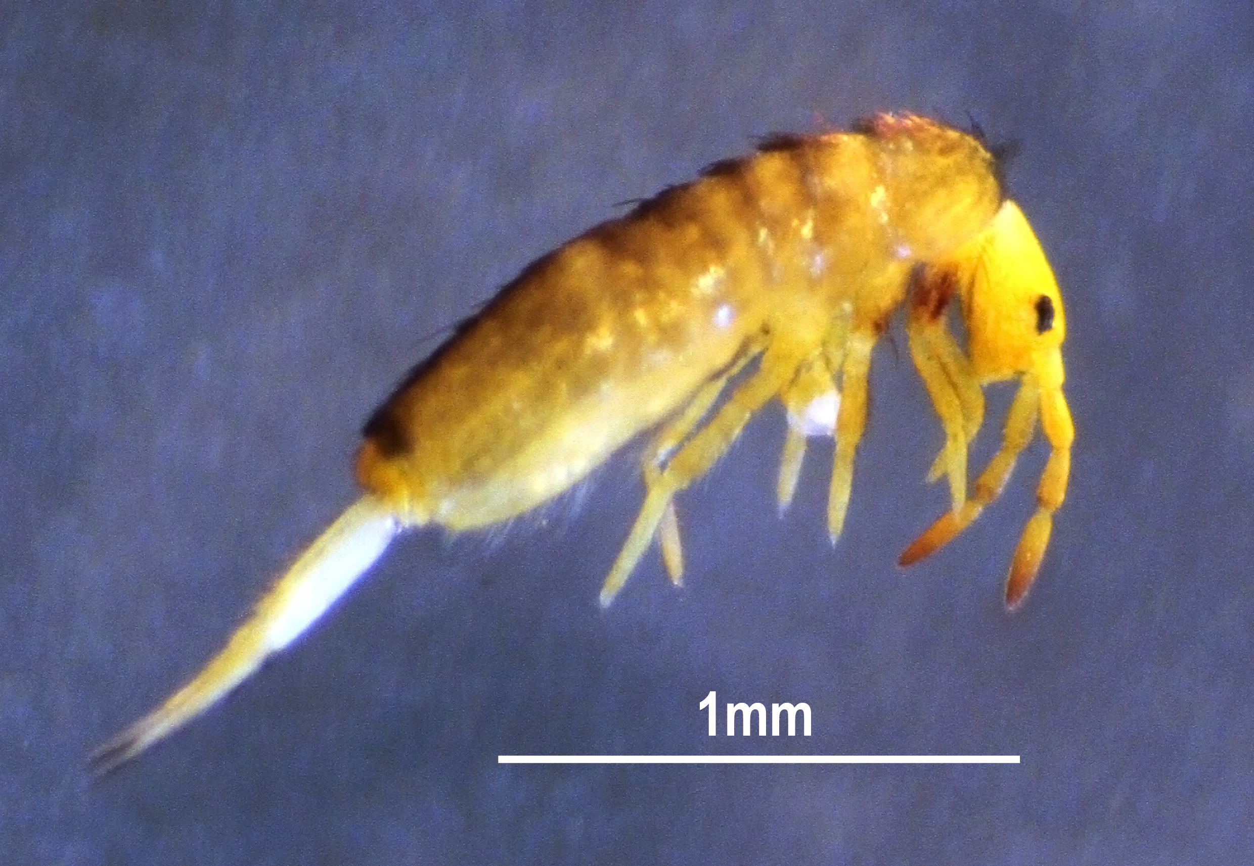
Figs 1–23 View FIGURE 1 View FIGURES 2–7 View FIGURES 8–9 View FIGURES 10–12 View FIGURES 13–15 View FIGURES 16–17 View FIGURES 18–23 , Tab 1
Material examined. Holotype and six paratypes on slide (code HNHM collpr-367), Fertőrákos, Hungary, 116 m above sea level, N47°42'54"; E16°40'14", from Carex riparia and Phragmites australis vegetation, 5.vi.1987, leg. Gy. Traser.
Other material examined. Four specimens on slides (slide code, EFE-LP), same data as type material, deposited at the University of West-Hungary. Ten specimens from type locality (code LpFER), hand collecting, 22.ii.2017 and 25.iii.2017, leg. D. Winkler; five specimens from Sárosfő (code LpSAR), Mid-West Hungary , 164 m above sea level, N47°3'14"; E17°23'38", alder forest soil, hand collecting, 16.xi.2015, leg. D. Winkler & Á. Erdő; five specimens from Bakonygyepes (code LpBGY), Mid-West Hungary GoogleMaps , 225 m above sea level, N47°8'32"; E17°33'39", from soil of non-tussock tall-sedge bed, hand collecting, 3.xii.2015, leg. D. Winkler & Á. Erdő; preserved in 96% alcohol deposited in the first author’s collection in the Faculty of Forestry , University of Sopron GoogleMaps .
Redescription. Medium sized Lepidocyrtus , adult trunk length 0.9–1.4 mm (without head nor furca). Holotype trunk length 1.3 mm. Mesothorax not or slightly projecting over head. Main body color white-yellowish to orange ( Fig. 1 View FIGURE 1 ). Purple pigmentation on ant. II–IV with increasing intensity from base to distal part of each antennal segment, on cx. I–III and occasionally on whole legs, ventral tube and manubrium. Ocular area dark-bluish-black. Dark spot between antennae.
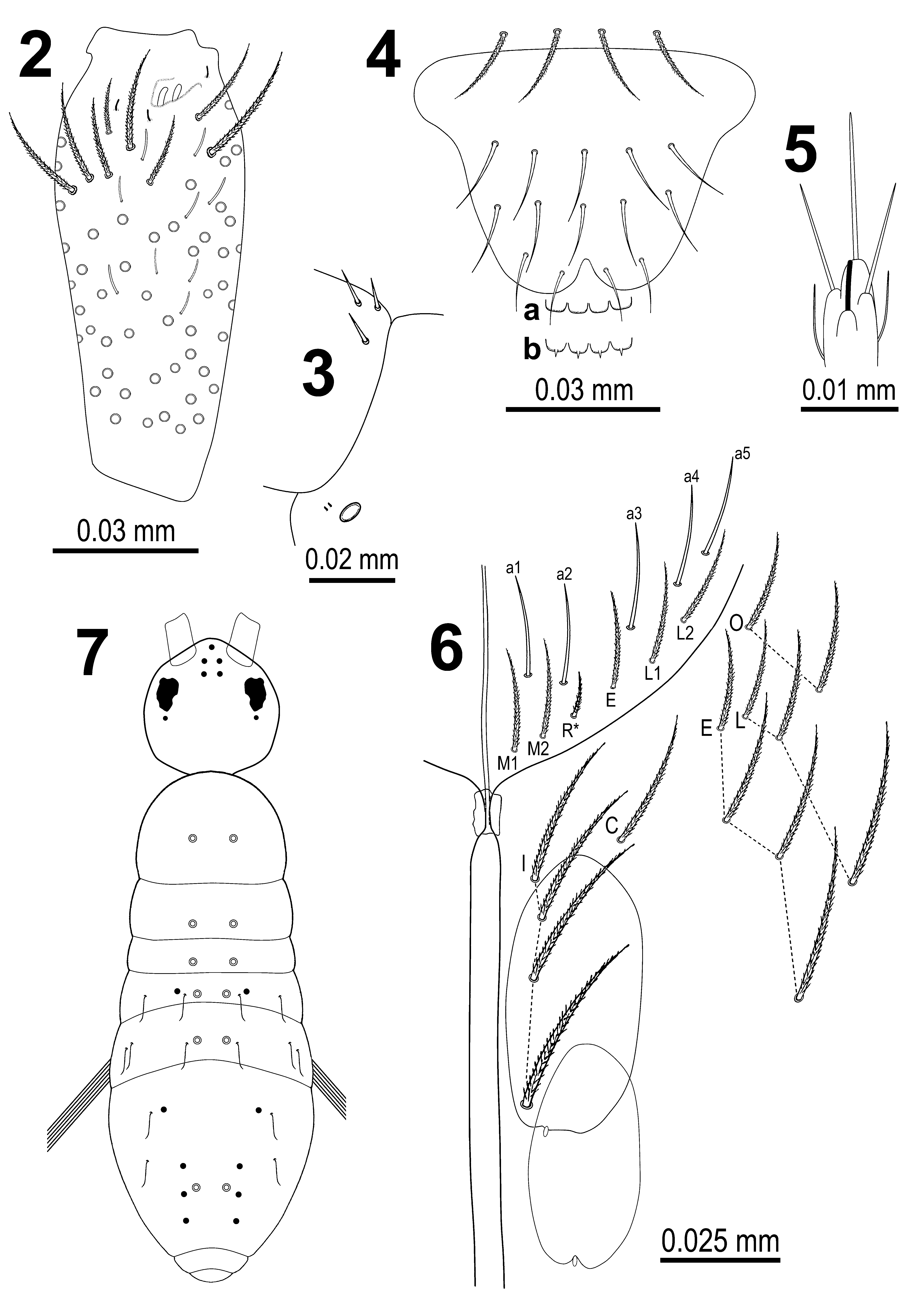
Light brown colored scales cover whole body including ant. I–II (III), entire legs without foot complexes, ventral tube, dorsal and ventral surface of manubrium and ventral side of dentes. Antennae rather short, antennal length to head diagonal length ratio 1.3–1.7 (head diagonal measured from the cervical edge to the apex of the mouth parts). Relation of the antennal joints I–IV as 1: 2.2: 2.1: 2.9 (holotype). Apical bulb on ant. IV absent. Ant. III sensillary organ composed of two bent sensory rods partially hidden by a cuticular fold plus three guard sensilla ( Fig. 2 View FIGURES 2–7 ). Antennal-I-organ (sensu Hüther 1986) composed of three acute smooth chaetae forming a triangle ( Fig. 3 View FIGURES 2–7 ). Ventrolateral side of antennal base with antennobasal-organ (sensu Hüther 1986) guarded with two short microsensilla ( Fig 3 View FIGURES 2–7 .). Arrangement of chaetae on labrum 4/554, prelabral chaetae ciliated, first and second rows of labral chaetae smooth, third (apical) row of labral chaetae simple (pointed) ( Fig. 4 View FIGURES 2–7 ). Labrum intrusion inverted Ushaped, rounded labral papillae simple ( Fig. 4a View FIGURES 2–7 ) or each armed with a conical spine ( Fig. 4b View FIGURES 2–7 ). Outer maxillary palp with two smooth chaetae and three smooth sublobal chaetae. Lateral process (sensu Fjellberg 1999) of outer labial papilla E slightly curved ( Fig. 5 View FIGURES 2–7 ), tip barely reaching top of papilla. Labium chaetotaxy formed by 5 smooth "a" chaetae and in the basal row by ciliated chaetae M1M2R*EL1L2 ( Fig. 6 View FIGURES 2–7 ) with R smaller than other chaetae (marked with*, ratio of R/M ~0.6). Ventral cephalic groove associated with 4+4 ciliated chaetae and 2+2 scales ( Fig. 6 View FIGURES 2–7 ). All other postlabial chaetae ciliated and of the same shape and length ( Fig 6 View FIGURES 2–7 ).
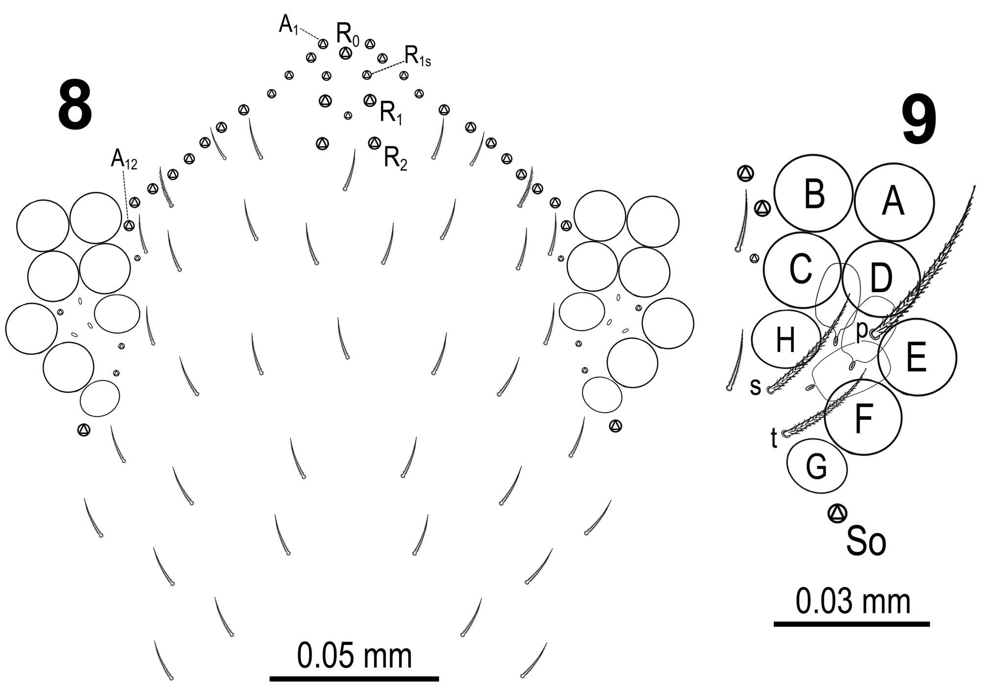
Dorsal cephalic macrochaetae chaetotaxy R0R1R2S o ( Fig. 7 View FIGURES 2–7 ), with a pair of smaller supplementary macrochaetae R1s between R0 and R1 ( Fig. 8 View FIGURES 8–9 ). Head with 11–12 macrochaetae in row A on each side ( Fig. 8 View FIGURES 8–9 ). Eye patches dark blue, eyes well visible. Diameters of ocelli A–F about the same. Ocelli G and H somewhat smaller (A:G; A:H ≈ 1.4), and more oval than circle. Interocular chaetotaxy ( Fig. 9 View FIGURES 8–9 ) with s, t, p chaetae and 2–3 intraocular scales. Head and body dorsal mesochaetae finely ciliated.
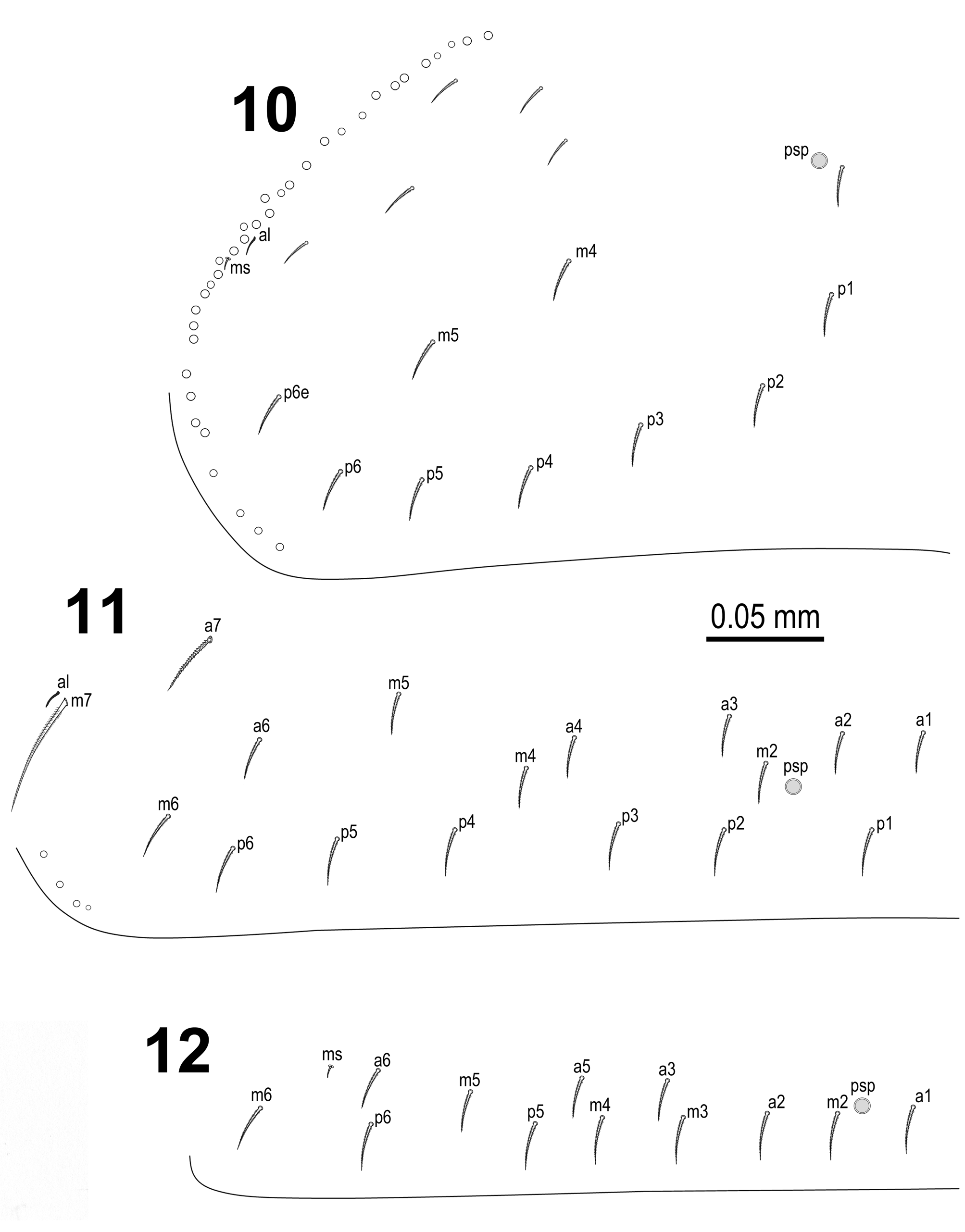
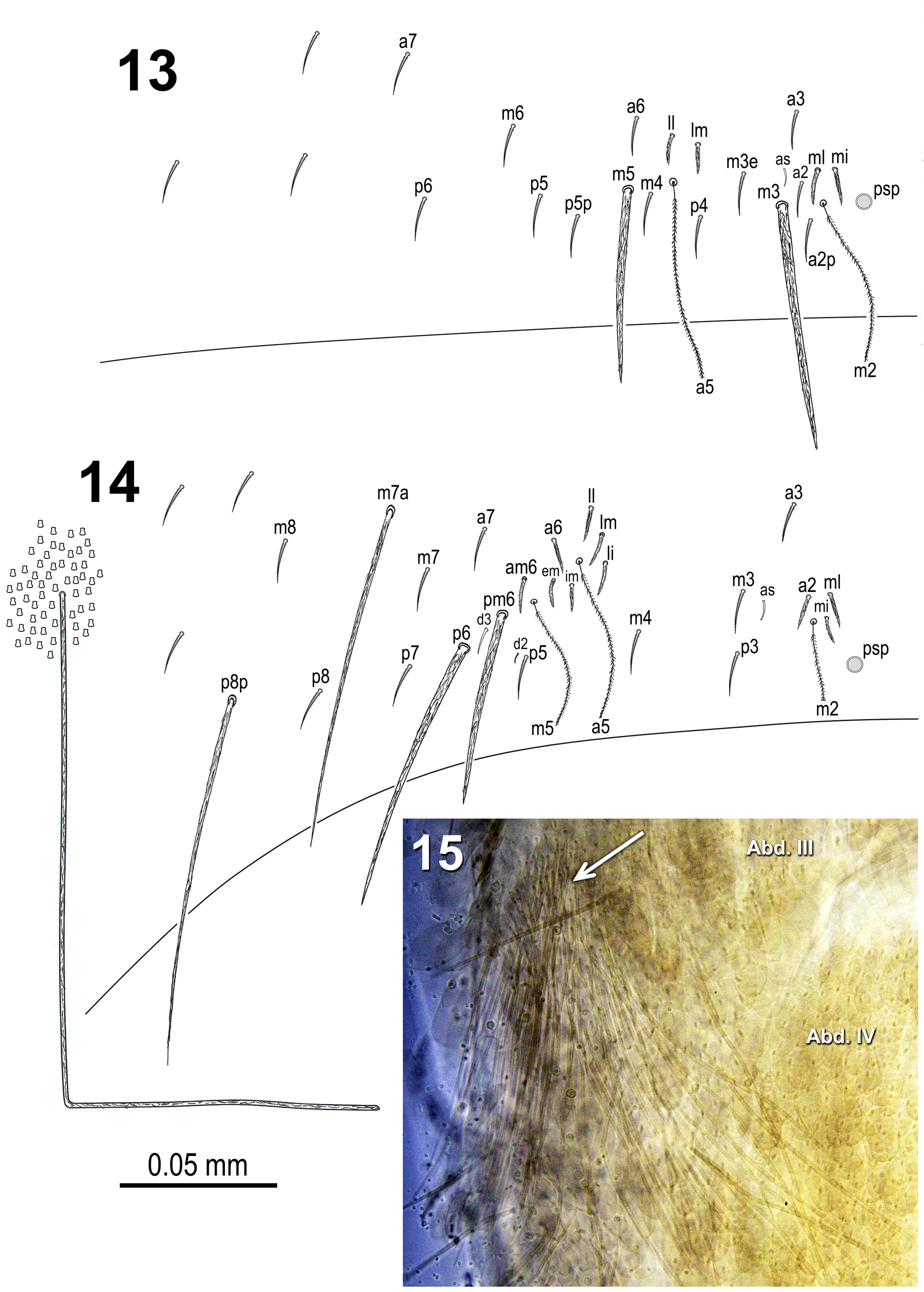
Body macrochaetae 00/0101+3 ( Fig. 7 View FIGURES 2–7 ). Dorsal chaetotaxy of th. II–III and abd. I as on Figs 10–12 View FIGURES 10–12 . Mesothorax with 2 anterolateral s-chaetae (al and ms). Th. III with sensillum (al) next to chaeta m7. Abd. I with a lateral S-microchaeta (ms) external to a6. Chaetotaxy of abd. II–III as in Figs 13–14 View FIGURES 13–15 . Abd. II chaetotaxy between two dorso-medial trichobothria a2p, a2, as, a3, m3, m3e, p4. Abd. II chaeta ml present on 15 and absent on 16 specimens. Abd. III chaeta mi present on 25 and absent on 6 specimens. Chaetae associated with trichobothria on abd. II–III acuminate and ciliate (not fan-shaped). Abd. III chaeta d3 present. Abd. III with lateral tuft of 10–60 (depending on specimen) long ciliated filiform chaetae ( Figs 14–15 View FIGURES 13–15 ).
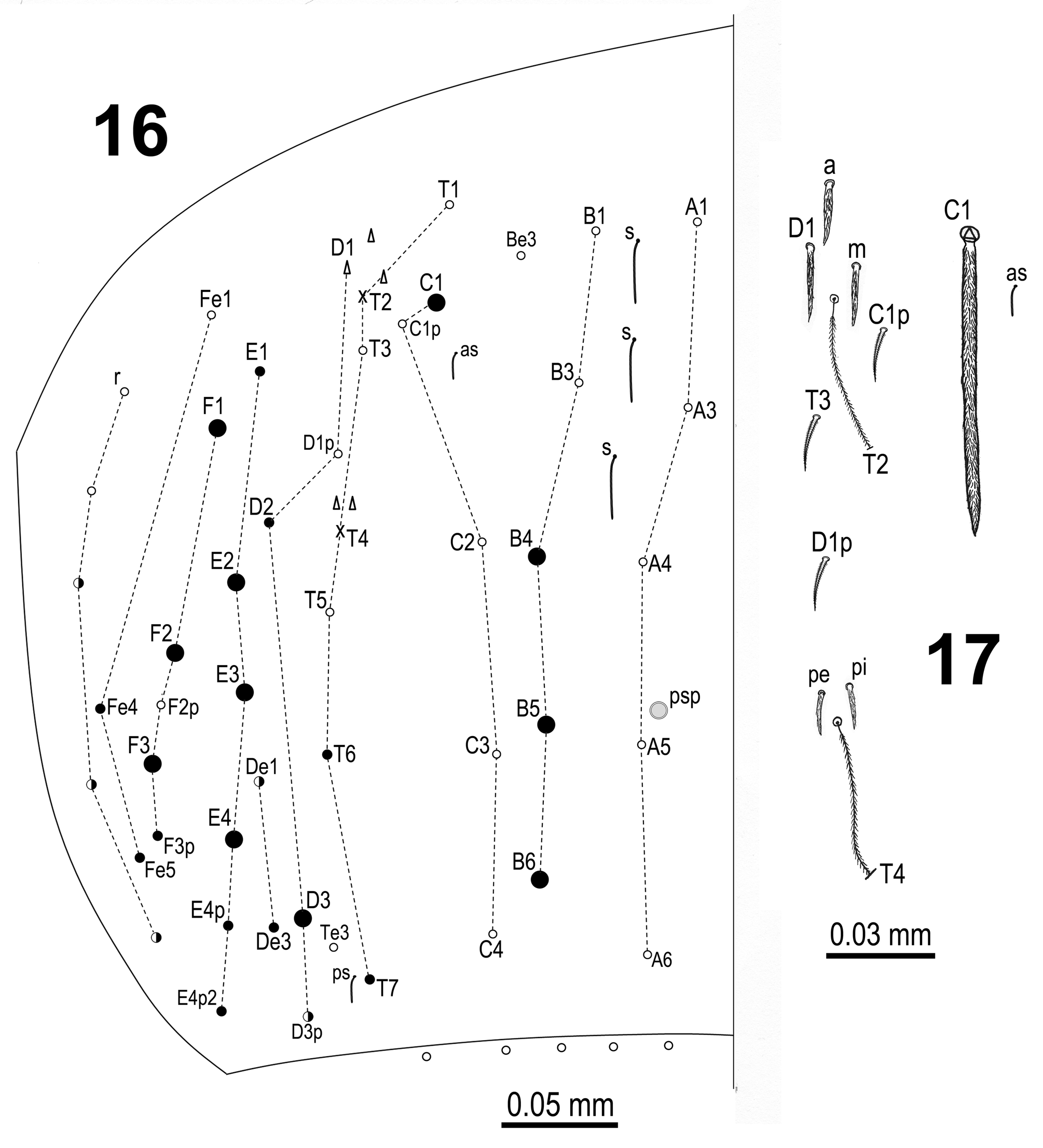
Chaetotaxy and trichobothrial complexes on abd. IV as on Figs 16–17 View FIGURES 16–17 . Macrochaetae B4, B5, B6, C1, D3, E2, E3, E4, F1, F2 and F3 broader with broad socket, while D2, De3, E1, E4p, E4p2, Fe4, Fe5, T6 and T7 thinner with smaller socket. Chaetae De1 and D3p thin ciliated macrochaetae or mesochaetae depending on specimens. Lateral r chaetae r3, r4 and r5 irregularly and asymmetrically thin ciliated macrochaetae on several specimens. Macrochaeta F2 above macrochaeta E3. The ratio of distances between macrochaetae C1–B4 / B4–B6 on abd. IV 0.6–0.9. Accessory chaeta s associated with trichobothrium T2 absent. All chaetae associated with trichobothria on abd. IV (D1, a, m, pe, pi) acuminate and ciliate. All specimens with three long S-chaetae on anterior dorsomedial part of abd. IV. Abd. V with three S-chaetae typical for Lepidocyrtus .
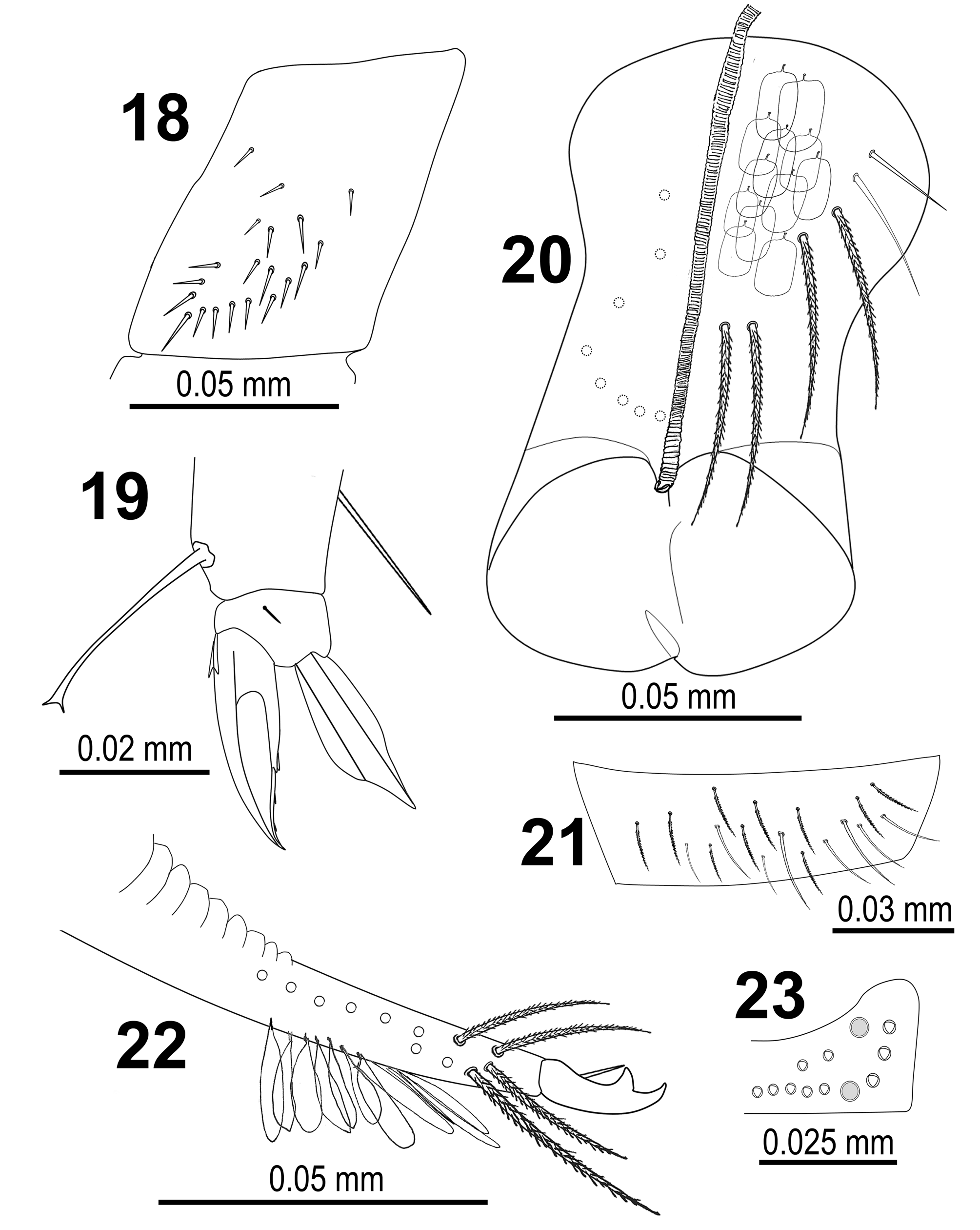
Legs with scales except on empodial complexes. Trochanteral organ with 14–25 smooth spiny chaetae forming a +/- V-shaped pattern ( Fig. 18 View FIGURES 18–23 ). Unguis and unguiculus of claw III as on Fig. 19 View FIGURES 18–23 . Unguis with sub-equal paired basal teeth distinct at 54% from inner edge, and also with two more small unpaired teeth at 75% and 87%, respectively. Apart from two small lateral teeth an outer tooth also present. Unguiculus truncate, without denticles along the outer lamella. Tibiotarsal tenent hair smooth and spatulate, as long as claw. Ratio of supraempodial chaeta (smooth chaeta on tibiotarsus III opposite to the tenent hair) / unguiculus around 0.9.
Ventral tube with scales on anterior side; 4+4 ciliated and 2+2 smooth chaetae on anterior side ( Fig. 20 View FIGURES 18–23 ); 8+8 weakly ciliated chaetae on posterior side; lateral flap with a maximum of 8 smooth and 10 ciliated chaetae ( Fig. 21 View FIGURES 18–23 ).
Manubrium with scales on dorsal and ventral surface, dens with scales on ventral surface. Dental tubercle absent. Mucro as on Fig. 22 View FIGURES 18–23 . Ratio manubrium/dens/mucro as 15:16:1. Manubrial plate with 2(3) inner chaetae and 4–7 chaetae outer the 2 psp ( Fig. 23 View FIGURES 18–23 ).
Variability. Selected characters of the studied specimens are summarized in Table 1. Other variabilities found on single specimens include the presence of a third labial M chaeta (on both sides); and the absence of abd. IV macrochaeta F1 (on left side only).
Comparative remarks. The original description indicates the morphology of labral papillae as rounded (Traser & Christian 1992: Fig. 2 View FIGURES 2–7 ), while re-examination of the type material as well as newly collected specimens from the type locality revealed variability in the morphology; apart from the simple rounded form, labral papillae can also have pointed expansions, occurred more frequently in the early-spring-collected specimens. This kind of variability has not been observed in specimens from the other two localities (Sárosfő, Bakonygyepes).
The lateral tuft of filiform chaetae on abdomen III has been described for several species of subgenera Setogaster Salmon, 1951 and Cinctocyrtus Yoshii & Suhardjono, 1989, but never for European species of subgenera Lanocyrtus Yoshii & Suhardjono, 1989 and Lepidocyrtus s.str. As Mateos & Greenslade (2015) noted this tuft could be present or absent depending on the specimen within the same species, and according to Yoshii & Suhardjono (1992) this tuft is not present in all development stages. Notably, all specimens studied in the present paper have this tuft on abd. III.
| HNHM |
Hungarian Natural History Museum (Termeszettudomanyi Muzeum) |
No known copyright restrictions apply. See Agosti, D., Egloff, W., 2009. Taxonomic information exchange and copyright: the Plazi approach. BMC Research Notes 2009, 2:53 for further explanation.
|
Kingdom |
|
|
Phylum |
|
|
Class |
|
|
Order |
|
|
Family |
|
|
Genus |