
Gephyromantis ambohitra ( Vences and Glaw, 2001 )
|
publication ID |
https://doi.org/10.5281/zenodo.175444 |
|
DOI |
https://doi.org/10.5281/zenodo.6236715 |
|
persistent identifier |
https://treatment.plazi.org/id/03A32236-FFD6-E754-F1A0-FB77FAC6B581 |
|
treatment provided by |
Plazi |
|
scientific name |
Gephyromantis ambohitra ( Vences and Glaw, 2001 ) |
| status |
|
Gephyromantis ambohitra ( Vences and Glaw, 2001) View in CoL
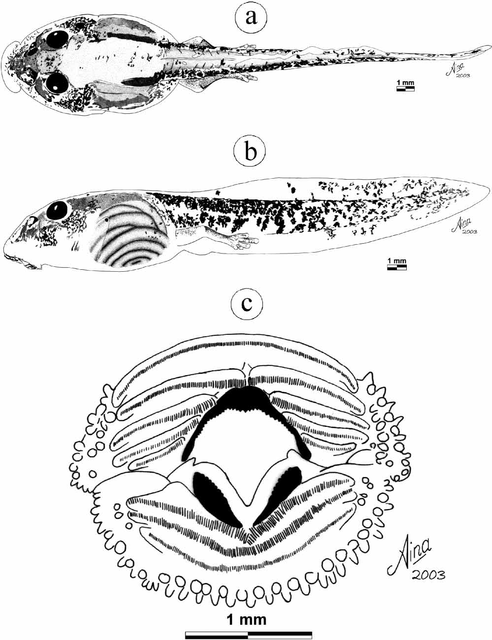
The following description refers to one tadpole in developmental stage 37 (field number FG/MV 2002.1963, catalogued as ZSM 790/2004, TL 26.6 mm, BL 9.5 mm) ( Fig. 1 View FIGURE 1 ), from Montagne d’Ambre National Park. Generalized tadpole of type IV of Orton (1953). In dorsal view, body ovoid with small constrictions of the body wall at the plane of spiracle. Snout large, edge of anterior labium in front of the snout. In lateral view, body depressed, maximum width attained behind midbody, BW 122 % of BH. Eyes of moderate size, ED 12 % of BL, not visible in ventral view, positioned dorsally and directed dorsolaterally situated at about 1/3 of body length. Nares elliptical, moderately sized, with dark spots, positioned dorsally, oriented anterolaterally and equidistant between snout tip and eye. NN 64 % of PP. Spiracle sinistral with its inner wall free and formed such that the aperture opens laterally instead of posteriorly, visible in dorsal view. Spiracular opening round, positioned laterally, directed dorsolaterally, situated at the height of the longitudinal axis of caudal musculature and much closer to the end of the body than to the snout, SS 55 % of BL. Medial vent tube tubular, medial, with both walls attached directly to ventral fin. Caudal musculature moderately developed, myosepta visible in dorsal view in the proximal 3/5 of the tail. TMH 42 % of BH and 49 % of MTH, TMW 36 % of BW; its height 2/5 of total height at midlength of tail. Dorsal fin inserts at the tail muscle posterior to the dorsal tailbody junction, increases quickly to attain the maximum height, then diminishes gradually to the tail tip. Ventral fin begins at the ventral terminus of the body, is constant until midtail, diminishes quickly towards tail tip. Tail tip pointed. MTH 86 % of BH, lateral line visible on the proximal 2/5 of the tail. Oral disc relatively large, ODW 29 % of BL and 51 % of BW, positioned anteroventrally, directed anteroventrally, emarginated. Two rows of marginal papillae, large medial gap anteriorly (DG 72 % of ODW), no medial gap on the posterior labium; total number of marginal papillae 79, submarginal papillae positioned in the lateral parts of the anterior and posterior labia (9 on the right and 13 on the left side). Papillae of moderate size, round or conical with rounded tip. No denticulate papillae. Labial tooth row formula (LTRF) after McDiarmid and Altig (1999) 5(25)/3(1), after Dubois (1995) 1:4+4/1+1:2. The density of keratodonts on A1 is about 57 per millimetre (a total of about 100). The length of interrupted anterior keratodont rows (A2, A3 and A4) decreases gradually towards the centre of the disc. Rows A1 and P3 have small keratodonts, in the other rows, keratodont size declines from the centre to the periphery. Both jaw sheaths coarsely serrated, with three parts having different colorations: edge, totally keratinised (black); medial part, partially keratinised (brown); base, not keratinised (white). Upper jaw sheath medially with pointed serrations, each side of the sheath smoothly curved, inversed Ushaped opening with medial convexity. Lower jaw sheath Vshaped, rounded serration on each edge, medial smoothly curved arc. General coloration in life brownish. Coloration in preservative: Dorsally body with two different pigmentation patterns: first, speckled with differently sized and irregularly formed brown spots; second, irregular pigmentation that forms a network of patches in some areas; intestinal coils and spiracle well visible; tail musculature speckled with brown spots that are irregularly distributed on each side, forming a network of spots; laterally body speckled brownish with network of brown pigments below the eyes. Caudal musculature whitish, with large networks of irregularly distributed brown spots, size and density of spots diminishing towards the tail tip. Fins translucent, with brown variable sized spots that are irregularly distributed on the fins, density of spots higher in the upper fin. Ventrally translucent over the whole surface. Variation: TL and BL of 90 tadpoles at stages 3044, all from Montagne d’Ambre National Park (field numbers: FG/MV 2002.1926, 1940, 19461949, 19541956, and 19631964, catalogued as ZSM 717, 743 746, 756761, 769773, 790795/2004) are 18.730.8 mm and 5.49.7 mm, respectively. The ratios vary in the following proportions: BW 106134 % of BH; ED 1124 % of BL; SS 1472 % of BL; TMH 4596 % of MTH; TMW 3472 % of BW; MTH 70114 % of BH; ODW 1733 % of BL; ODW 3360 % of BW.
No known copyright restrictions apply. See Agosti, D., Egloff, W., 2009. Taxonomic information exchange and copyright: the Plazi approach. BMC Research Notes 2009, 2:53 for further explanation.
|
Kingdom |
|
|
Phylum |
|
|
Class |
|
|
Order |
|
|
Family |
|
|
Genus |