
Pavoniocotes, Gustafsson & Grossi & Ren & Zou, 2023
|
publication ID |
https://doi.org/10.1080/00222933.2023.2226375 |
|
publication LSID |
lsid:zoobank.org:pub:FFDF1435-92D6-4C19-9B51-3AB61E1BD7DA |
|
DOI |
https://doi.org/10.5281/zenodo.8270857 |
|
persistent identifier |
https://treatment.plazi.org/id/03A25902-FFAE-CF06-3D32-FA3DFDF0FF45 |
|
treatment provided by |
Plazi (2023-08-21 09:28:02, last updated 2023-11-10 05:36:35) |
|
scientific name |
Pavoniocotes |
| status |
gen. nov. |
Pavoniocotes gen. nov.
Goniodes Nitzsch, 1818: 293 View in CoL in partim.
Goniocotes Burmeister, 1838: 431 View in CoL in partim.
Type species
Goniodes parviceps Piaget, 1880: 277 View in CoL .
Diagnosis
It is unclear what group of lice within Goniodidae is most closely related to Pavoniocotes gen. nov., partly because there are no useful characters to delimit Goniodes and Goniocotes as currently circumscribed [following Price et al. (2003)]. The great morphological variation within Goniodes also makes direct comparisons difficult. We therefore compare this genus to typical members of Goniocotes , here represented by G. rectangulatus and G. gallinae , and with the type species Goniodes pavonis . Note that species belonging to the Dictyocotes group, currently placed within Goniocotes , do not exhibit all characters listed for Goniocotes below; von Kéler (1940) included G. parviceps in the genus Gonotyles , and a comparison between Pavoniocotes and members of this proposed genus is given below as well.
Pavoniocotes can be separated from Goniocotes by the following characters: preantennal head shortened in Pavoniocotes ( Figure 26 View Figures 26–30 ) compared to Goniocotes ( Figure 20 View Figures 20–22 ); antennae sexually dimorphic in Pavoniocotes ( Figure 26 View Figures 26–30 ), but not in Goniocotes ( Figure 20 View Figures 20–22 ); mts4–5 situated close together and at least mts5 thorn-like in Pavoniocotes ( Figure 26 View Figures 26–30 ), but mts4–5 widely separated and both microsetae in Goniocotes ( Figure 20 View Figures 20–22 ); sternal setae absent on metathorax in Pavoniocotes ( Figures 24–25 View Figure 24 View Figure 25 ), but present in Goniocotes ( Figures 18–19 View Figure 18 View Figure 19 ); at least some tps present as microsetae on at least tergopleurites II–VI in both sexes in Pavoniocotes ( Figures 24–25 View Figure 24 View Figure 25 ), but absent on all tergopleurites or, if present, then not microsetae in Goniocotes ( Figures 18–19 View Figure 18 View Figure 19 ); male genitalia stout, either asymmetrical ( Figures 28–29 View Figures 26–30 ) or symmetrical ( Figures 42–43 View Figures 40–44 ), but in either case with clearly defined mesosome and dorsally displaced parameres, all contained in a partially rugose or spiculate genital sac in Pavoniocotes , but much reduced, without a mesosome and with parameres fused entirely to basal apodeme and with genital sac either absent or, if present, not rugose or spiculate in Goniocotes ( Figure 21 View Figures 20–22 ); vulval margin with dense, continuous row of mesosetae in Pavoniocotes ( Figure 30 View Figures 26–30 ), but with spare row of microsetae widely separated medianly in Goniocotes ( Figure 22 View Figures 20–22 ); post-vulval area with distinct sublateral lobes and areas with dense minute, seemingly hyaline, microsetae in Pavoniocotes ( Figure 30 View Figures 26–30 ), but without such structures in Goniocotes ( Figure 22 View Figures 20–22 ).
Pavoniocotes can be separated from Goniodes s. str. by the following characters: male scape much widened distally, not rugose or squamous, with spike- or thorn-like seta apically on posterior extension in Pavoniocotes ( Figure 26 View Figures 26–30 ), but narrowing distally, with singe protruding squamous thumb at about mid-length and thorn-like seta near posterodistal end in Goniodes s. str. ( Figure 3 View Figures 3–7 ); male os short or mesoseta (in some species spike-like) in Pavoniocotes ( Figure 26 View Figures 26–30 ), but macroseta in Goniodes s. str. ( Figure 3 View Figures 3–7 ); at least some of pos, mts2 and mts4–5 much thickened, thorn-like in Pavoniocotes ( Figure 26 View Figures 26–30 ), but slender in Goniodes s. str. ( Figure 3 View Figures 3–7 ); temples with distinct posterior extension at base of mts 4–5 in Goniodes s. str. ( Figure 3 View Figures 3–7 ), but without such extension in Pavoniocotes ( Figure 26 View Figures 26–30 ; in Pavoniocotes mayuri Lakshminarayana and Emerson, 1971 , distal bulge present but not shaped as in Goniodes ; Figure 40 View Figures 40–44 ); lpts and ipts widely separated and ipts clearly on posterior margin of pteronotum in Pavoniocotes ( Figures 24–25 View Figure 24 View Figure 25 ), but lpts and ipts closely associated, both sets more or less lateral in Goniodes s. str. ( Figures 1–2 View Figure 1 View Figure 2 ); ventral chaetotaxy of legs II–III of Goniocotes type, dominated by large, spike-like setae in Pavoniocotes ( Figure 24 View Figure 24 ), but without large, spike-like setae in Goniodes s. str. ( Figure 1 View Figure 1 ); tergopleurite II lengthened anteriorly, pushing pterothorax anteriorly, in Pavoniocotes ( Figure 24 View Figure 24 ), but lengthened distally to increase dorsal and lateral overlap with tergopleurite III in Goniodes s. str. ( Figure 1 View Figure 1 ); male tergopleurite X extended antero-laterally to reach tergopleurite VIII, and genital opening hidden underneath tergopleurite X in Goniodes s. str. ( Figures 1 View Figure 1 , 8–9 View Figures 8–9 ), but male tergopleurite X not extended and genital opening not underneath tergopleurite X in Pavoniocotes ( Figure 24 View Figure 24 ); basal apodeme fused with mesosome and parameres, and parameres displaced dorsally in Pavoniocotes ( Figures 28–29 View Figures 26–30 , 42–43 View Figures 40–44 ), but basal apodeme distinct from mesosome and parameres, and parameres lateral to mesosome in Goniodes s. str. ( Figures 5–6 View Figures 3–7 ); inner sclerite present in female genitalia of Goniodes s. str. ( Figure 7 View Figures 3–7 ), but absent in Pavoniocotes ( Figure 30 View Figures 26–30 ); vulval margin with dense marginal row of short setae and submarginal row of thorn-like setae in Pavoniocotes ( Figure 30 View Figures 26–30 ), but with dense brushes of mesosetae laterally and without thorn-like submarginal setae in Goniodes s. str. ( Figure 7 View Figures 3–7 ); post-vulval area without subvulval plates but with distinct sublateral lobes and areas with dense minute, seemingly hyaline, microsetae in Pavoniocotes ( Figure 30 View Figures 26–30 ), but with subvulval plates and without sublateral lobes or areas of microsetae in Goniodes s. str. ( Figure 7 View Figures 3–7 ).
Pavoniocotes can be separated from members of the proposed genus Gonotyles , presently considered a synonym of Goniodes , by the following characters, based on comparisons with Goniodes cervinicornis Giebel, 1874 : male scape much widened distally, not rugose or squamous, with spike- or thorn-like seta apically on posterior extension in Pavoniocotes ( Figure 26 View Figures 26–30 ), but narrowed distally, with a single squamous, bifid thumb on posterior margin in inner half and only thorn-like seta of scape positioned distal to thumb in ̍ Gonotyles ̾; male pas, os, and pns short setae or mesosetae in Pavoniocotes ( Figure 26 View Figures 26–30 ), but macrosetae in ̍ Gonotyles ̾; temples more or less parallel (with posterior modification in P. mayuri ; Figure 40 View Figures 40–44 ) and at least mts5 thorn-like and not situated on posterior extension removed from mts 4 in Pavoniocotes ( Figure 26 View Figures 26–30 ), but temples flaring laterally, mts5 situated on distinct posterior extension and well separated from mts 4 in ̍ Gonotyles ̾; pteronotum with postero-lateral corner rounded, and lpts situated at corner in Pavoniocotes ( Figures 24–25 View Figure 24 View Figure 25 ), but flattened laterally, with lpts clearly lateral, situated anterior to corner in ̍ Gonotyles ̾; basal apodeme fused to mesosome and parameres in Pavoniocotes ( Figures 28–29 View Figures 26–30 ), but basal apodeme separate from fused mesosome and parameres in ̍ Gonotyles ̾; parameres small, simple, displaced dorsally in Pavoniocotes ( Figure 28 View Figures 26–30 ), but longer than mesosome, lateral, and with extensive ventral scaly combs and two triangular extensions (one median, one lateral) on each paramere in ̍ Gonotyles ̾; female vulval margin more or less straight, with dense more or less continuous row of thin marginal setae in Pavoniocotes ( Figure 30 View Figures 26–30 ), but strongly convex medianly, without marginal setae for most of convex part, and with dense lateral brushes of mesosetae in ̍ Gonotyles ̾; post-vulval area without subvulval plates but with distinct sublateral lobes and areas with dense minute, seemingly hyaline, microsetae, and without lateral fleshy lobes of the abdomen in Pavoniocotes ( Figure 30 View Figures 26–30 ), but in ̍ Gonotyles ̾ with lateral fleshy, setae-bearing lobes but no sublateral lobes, and no minute hyaline setae.
Description
Both sexes. Head shape sexually dimorphic ( Figures 24–25 View Figure 24 View Figure 25 ); frons rounded to flattened, marginal carina uninterrupted. Preantennal nodi not elongated, but may be longer in females than in males. Head chaetotaxy sexually dimorphic; os short; mts1 and mts3 only temporal macrosetae; at least mts5 thornlike, but pos, mts2, and mts4 may also be thorn-like. Antennae sexually dimorphic. Prothorax may flare posteriorly, with a single ppss macrosetae on postero-lateral corner ( Figures 24–25 View Figure 24 View Figure 25 ). Anterior margin indented, rhombic sclerite fused to pronotum medianly. Proepimera not fused medianly. Pteronotum roughly triangular, with lpts and ipts widely separated; smns present, but mpts absent. Pterothorax not fused to tergopleurite II. Sternal setae present on mesothorax but absent on metathorax. Abdominal plates and chaetotaxy as in Figures 24–25 View Figure 24 View Figure 25 ; tergopleurites II– VIII extended medianly. Central sternal plates absent, accessory lateral sternal plates present on segments II–VI ( Figures 24–25 View Figure 24 View Figure 25 ) or absent ( Figures 38–39 View Figure 38 View Figure 39 ). Abdominal chaetotaxy: ss present on segments II–VIII; tps microsetae present on at least segments II–VII, more numerous in anterior segments and there forming rows between ss and post-spiracular setae; psps present on segments II–VII in males and at least IV–VII in females; more than one post-spiracular seta present on anterior segments in male; single sts on each side of segments II–VIII, but accessory sts present on segment VI. Tergopleurites IX– XI sexually dimorphic. Measurements as in Table 1 View Table 1 .
Male. Frons shortened, temples more or less parallel, not flaring much lateral to eye ( Figure 26 View Figures 26–30 ). Head setae as1, as3, pcs clearly dorsal; head sensilla s1–4, s6–9 present; pos, mts2, mts4 may be thorn-like ( Figure 26 View Figures 26–30 ) or slender ( Figure 40 View Figures 40–44 ). Antennae modified: scape much expanded posteriorly into broad, bluntly triangular process with single apical spike-like ( Figure 26 View Figures 26–30 ) or thorn-like ( Figure 40 View Figures 40–44 ) seta; pedicel broadened on posterior margin, slightly arched; flagellomere I with large thumb-like process on posterior margin; flagellomeres II–III roughly as in female. Tergopleurites II–IV with tps and post-spiracular setae more numerous than in female. Tergopleurite IX small, largely rounded or drop-shaped, not fused to central tergopleurite X, which is roughly quadratic with indented anterior margin. Subgenital plate roughly square-shaped with anterior margin indented, but poorly sclerotised and exact outline not clearly visible. Basal apodeme fused to mesosome and parameres ( Figures 28–29 View Figures 26–30 ). Distal genitalia asymmetrical ( Figures 28–29 View Figures 26–30 ) or symmetrical ( Figures 42–43 View Figures 40–44 ) and described under each species.
Female. Frons gently rounded, temples extended lateral to eye but not flaring ( Figure 25 View Figure 25 ). Head setae as1, as3, pcs clearly ventral; head setae s1–4, s8–9 present; pos, mts2, mts4 slender. Antennae not modified. Tergopleurites II–IV with fewer tps and post-spiracular setae than males; post-spiracular setae absent on tergopleurite II. Tergopleurites IX–XI fused, medianly continuous, with median indentation of posterior margin. Subgenital plate absent, but small accessory lateral sternal plates present on segment VIII ( Figure 30 View Figures 26–30 ). Vulval margin flat to slightly convex, differing between specimens, with dense, medianly continuous row of short, slender marginal setae and submarginal set of thorn-like setae. Subvulval plates absent, but post-vulval area with fleshy sublateral lobes; lobes and other areas of post-vulval surface covered in small, seemingly hyaline and soft, setae.
Geographical range
Indo-Malayan region, but presumably spread more widely due to domestication of host.
Host range
Peafowls of the genus Pavo Linnaeus, 1758 ( Galliformes : Phasianidae ).
Etymology
Pavoniocotes is a portmanteau of the generic names Pavo Linnaeus, 1758 , and Goniocotes Nitzsch, 1818 . Gender: masculine.
Burmeister H. 1838. Mallophaga Nitzsch. In: Handbuch der Entomologie. Zweite Abteilung. Besondere Entomologie. Zweite Abteilunf. Lauskerfe. Gymnognatha. (Zweiter Halfte; vulgo Neuroptera), Vol. 2. Theod. Chr. Fried Enslin, Berlin; p. 418 - 443.
Giebel CG. 1874. Insecta epizoa. Die auf Saugetieren und Vogeln schmarotzenden Insecten nach Chr. L. Nitzsch ̾ s Nachlass bearbeitet. Leipzig: Otto Wigand; xiv + 308 pp., 20 plates.
Lakshminarayana KV, Emerson KC. 1971. Mallophaga Indica. VI. Notes on Goniocotes (Mallophaga: Philopteridae) found on Pavo cristatus, with description of a new species. Orient Insects. 5: 95 - 102. doi: 10.1080 / 00305316.1971.10433994.
Linnaeus CV. 1758. Systema naturae per Regna Tria Naturae, Secundum Classes, Ordines, Genera, Species, cum Characteribus, Differentiis, Synonymis, Locis. 10 th ed. Stockholm (Sweden): Salvius; p. iv + 824.
Linnaeus CV. 1766. Systema naturae per Regna Tria Naturae, Secundum Classes, Ordines, Genera, Species, cum Characteribus, Differentiis, Synonymis, Locis. 12 th ed. Stockholm (Sweden): Salvius; p. iv + 824.
Nitzsch CL. 1818. Die Familien und Gattungen der Thierinsekten (insecta epizoica); als Prodromus einer Naturgeschechte derselben. Magazin der Entomologie. 3: 261 - 316.
Piaget E. 1880. Les Pediculines. Essai monographique. Leiden (The Netherlands): E. J. Brill; p. xxxix + 714.
Price RD, Hellenthal RA, Palma RL. 2003. World checklist of chewing lice with host associations and keys to families and genera. In: Price RD, Hellenthal RA, Palma RL, Johnson KP, Clayton DH, editors. The chewing lice: world checklist and biological overview. Illinois Natural History Survey Special Publications 24, Illinois Natural History Survey. Champaign (IL USA); p. x + 501.
von Keler S. 1940. Baustoffe zu einer Monographie der mallophagen. II. Teil: Uberfamilie der Nirmoidea. Nova Acta Leopoldiana Abhandlungen der Kaiserlich Leopoldinisch-Carolinisch Deutschen Akademie der Naturforscher Neue Folge. 8: 1 - 354 + 4 plates.
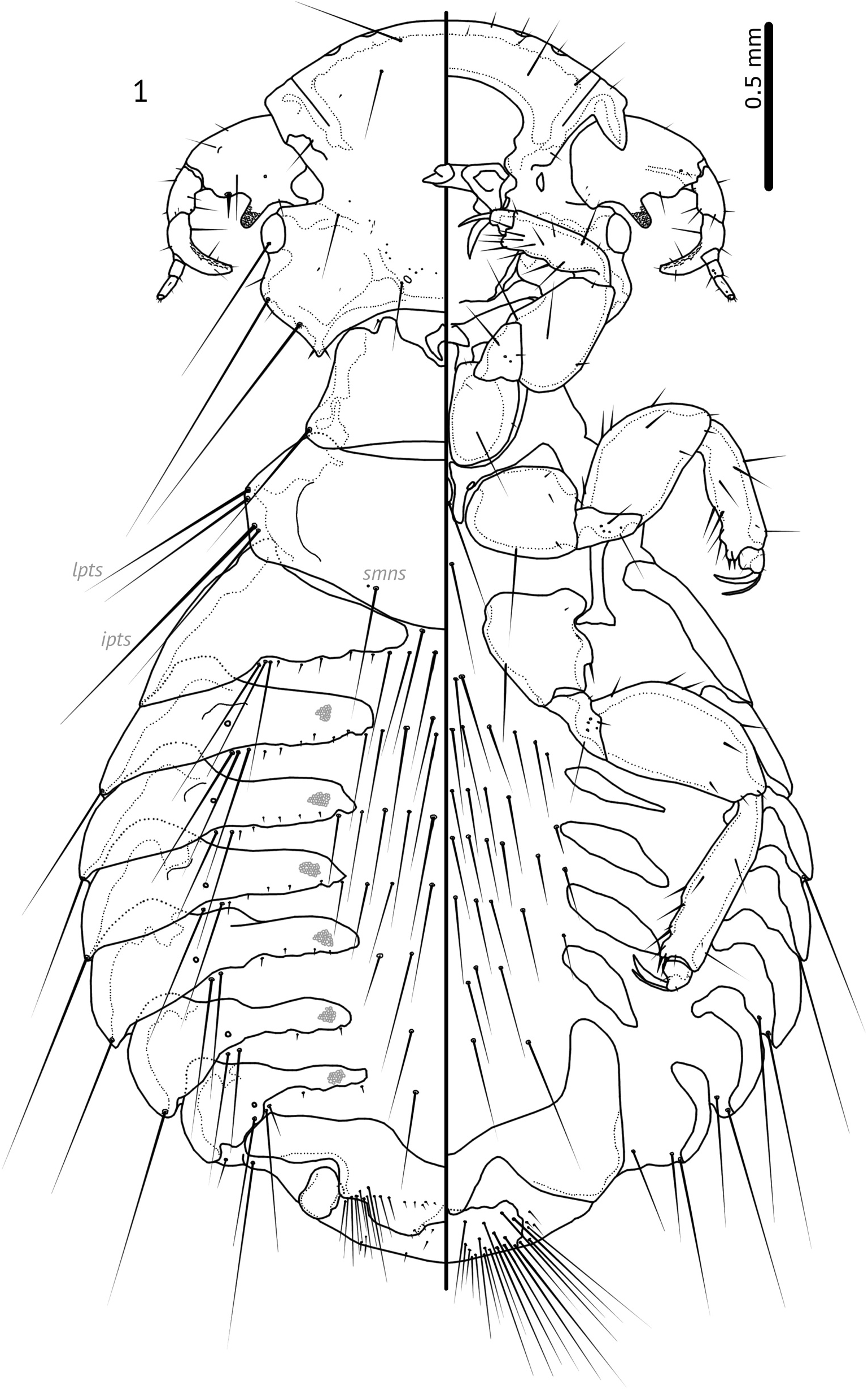
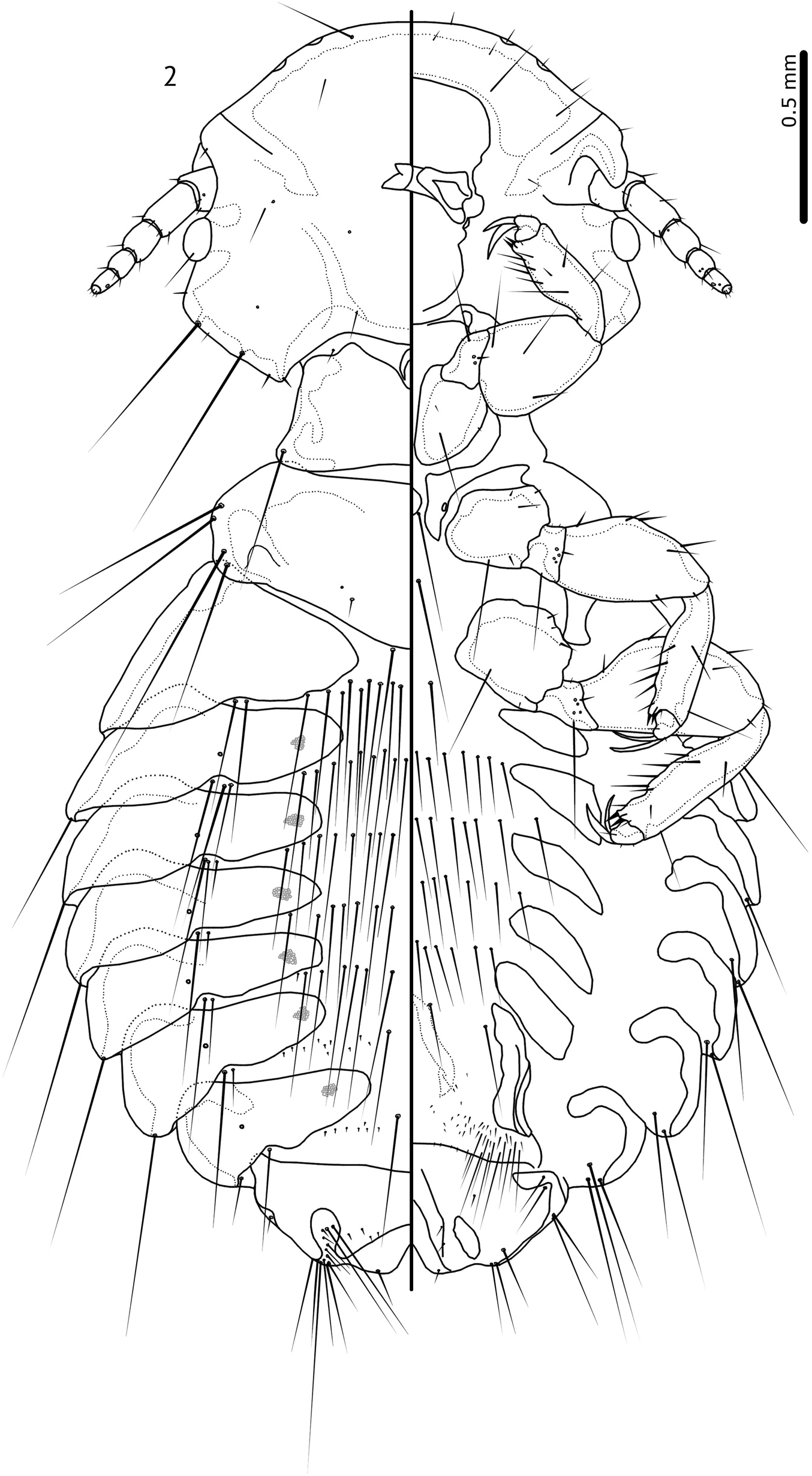
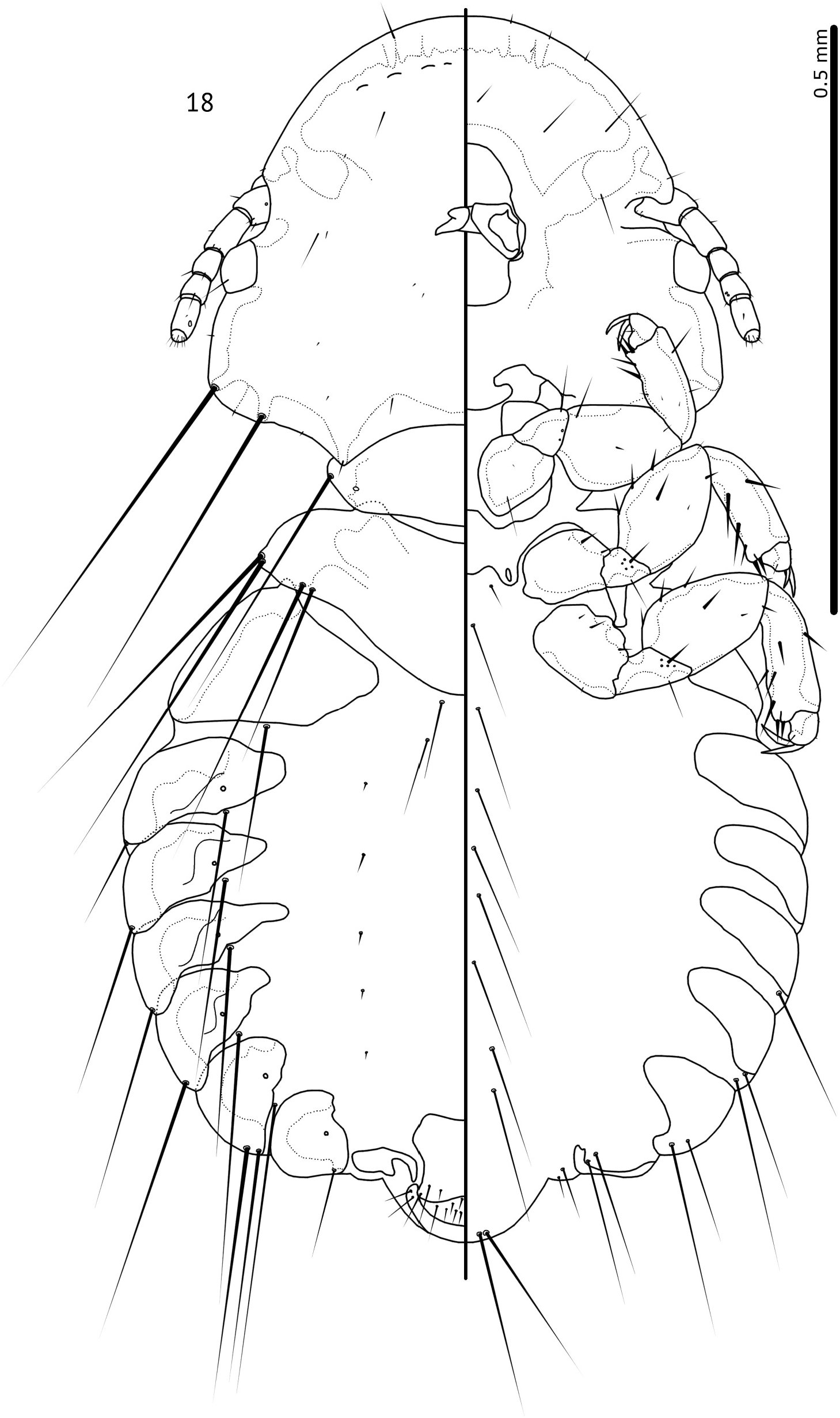
Figure 18. Goniocotes rectangulatus Nitzsch [in Giebel], 1866, male habitus, dorsal and ventral views. Paratergal plates not clearly demarcated in specimen, and illustrated approximately.
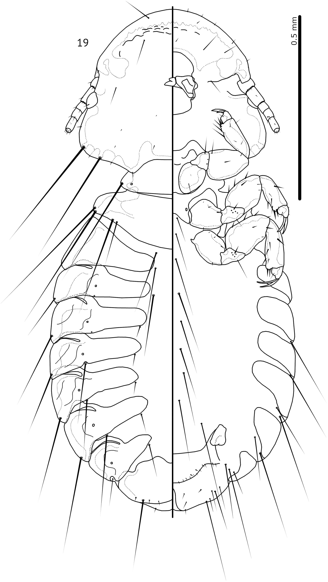
Figure 19. Goniocotes rectangulatus Nitzsch [in Giebel], 1866, female habitus, dorsal and ventral views. Paratergal plates not clearly demarcated in specimen, and illustrated approximately.
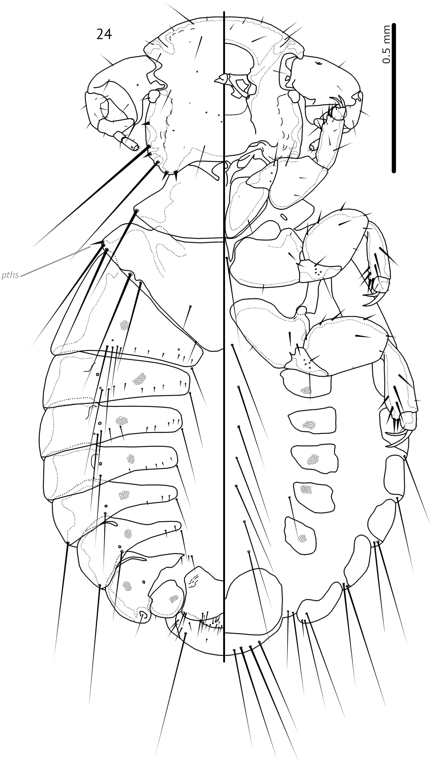
Figure 24. Pavoniocotes parviceps (Piaget, 1880) ex Pavo cristatus Linnaeus, 1758, male habitus, dorsal and ventral views.
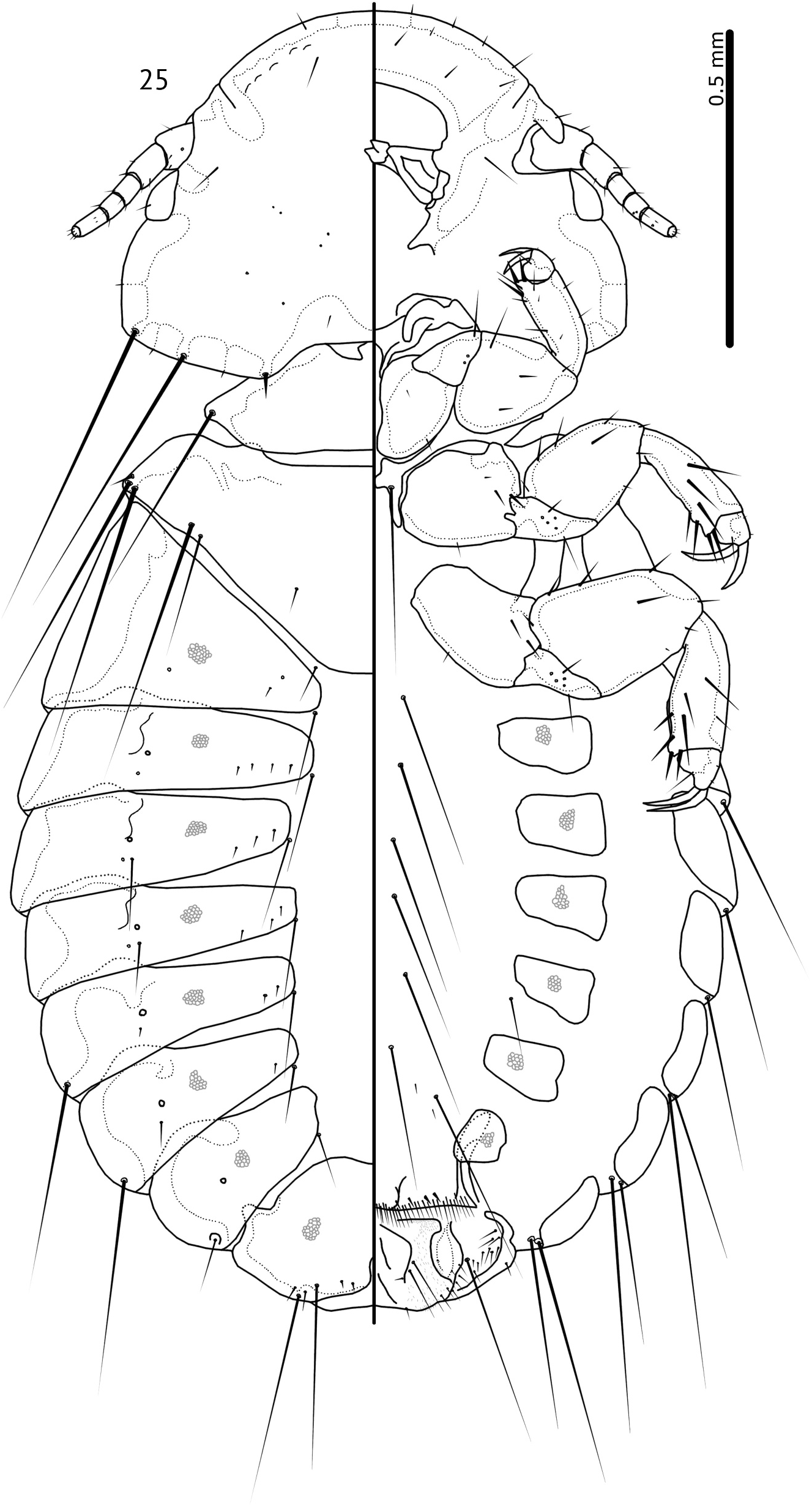
Figure 25. Pavoniocotes parviceps (Piaget, 1880) ex Pavo cristatus Linnaeus, 1758, female habitus, dorsal and ventral views.
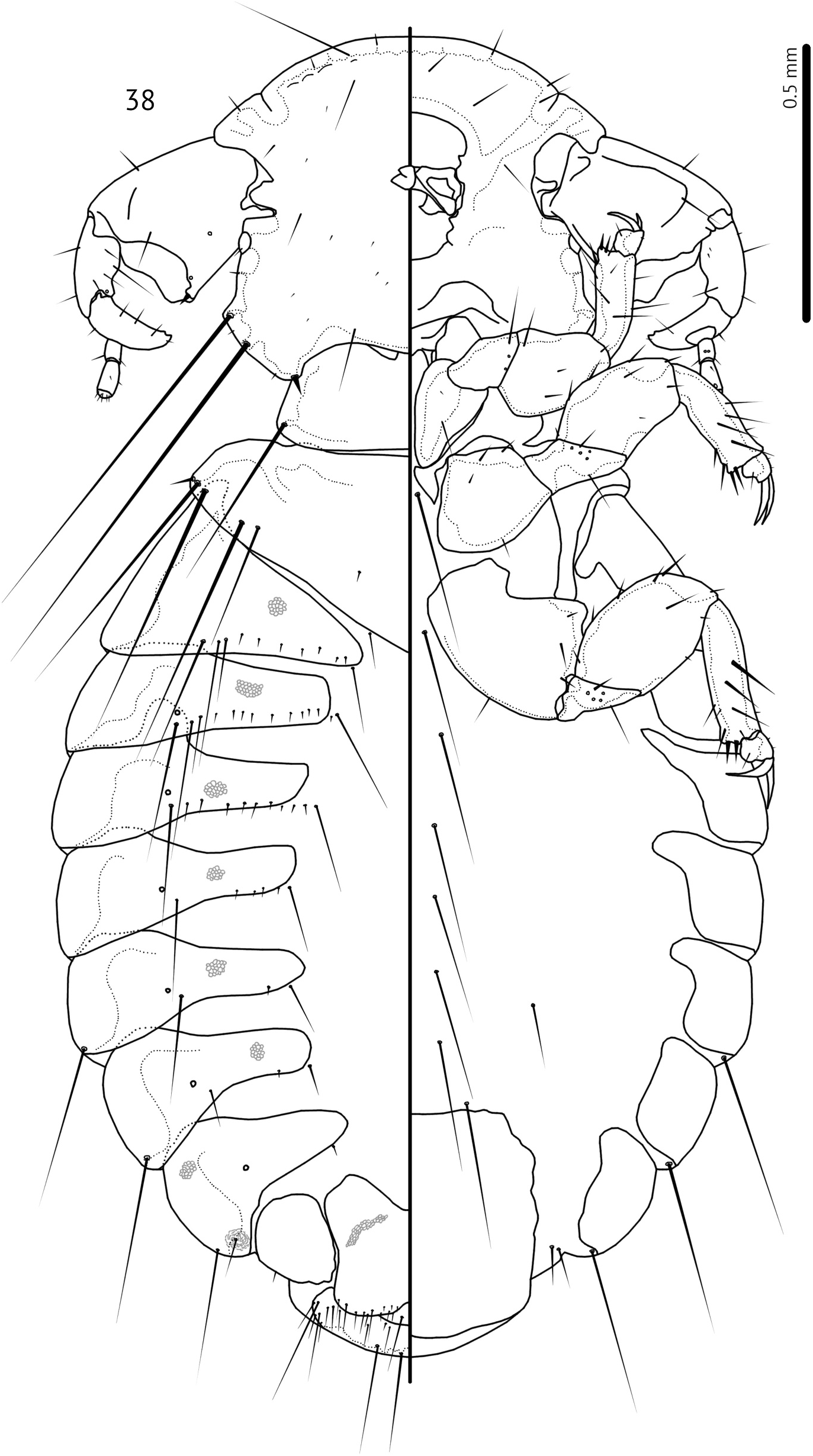
Figure 38. Pavoniocotes mayuri (Lakshminarayana and Emerson, 1971), male habitus, dorsal and ventral views.
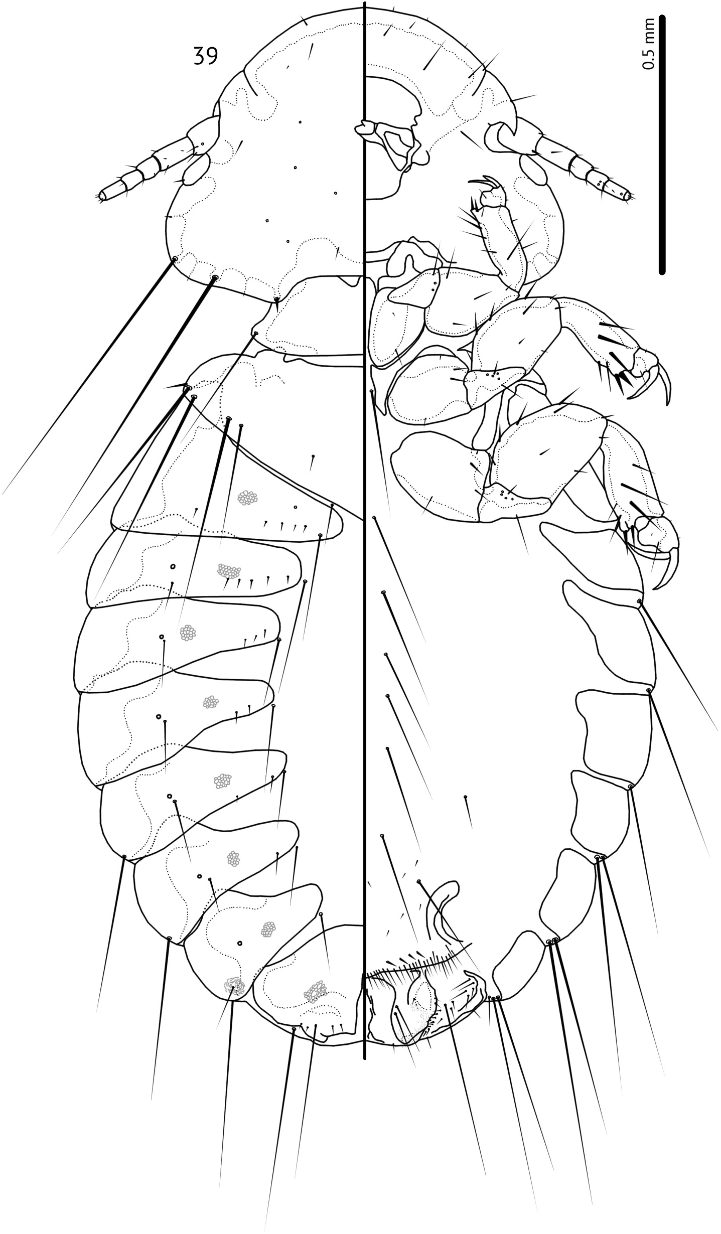
Figure 39. Pavoniocotes mayuri (Lakshminarayana and Emerson, 1971), female habitus, dorsal and ventral views.
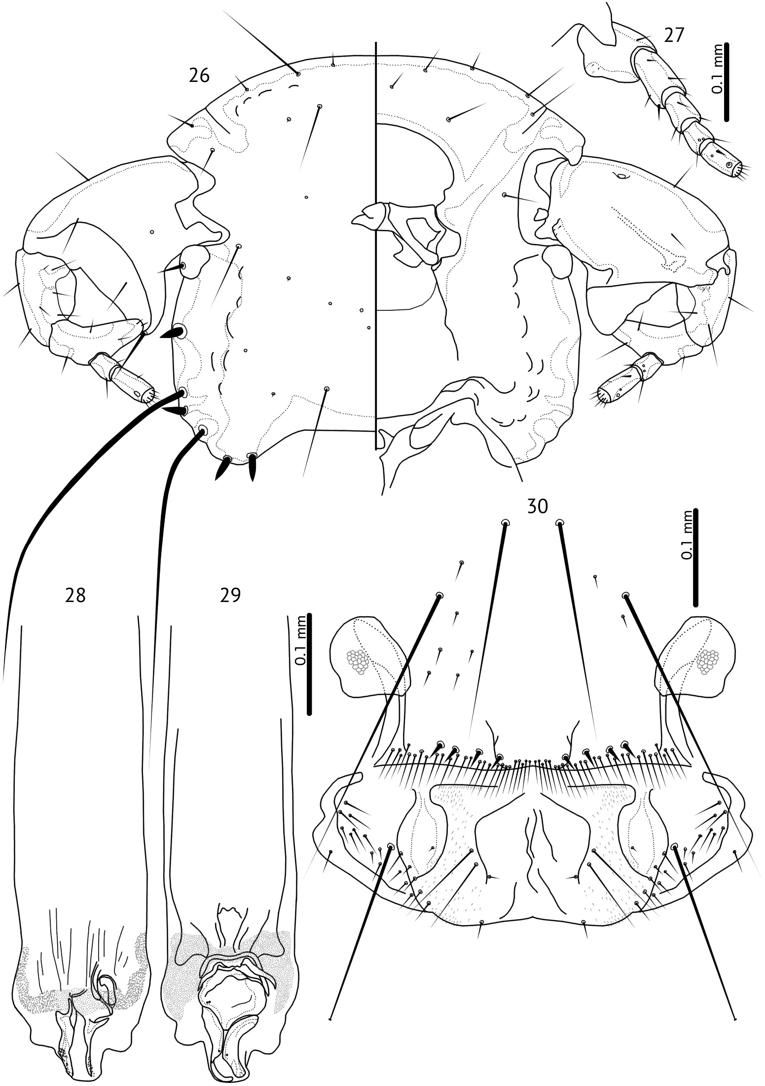
Figures 26–30. Pavoniocotes parviceps (Piaget, 1880) ex Pavo cristatus Linnaeus, 1758. 26, male head, dorsal and ventral views. 27, female antenna and conus, ventral view. 28, male genitalia, dorsal view. 29, male genitalia, ventral view. 30, female genitalia and abdominal segments VII–XI, ventral view.
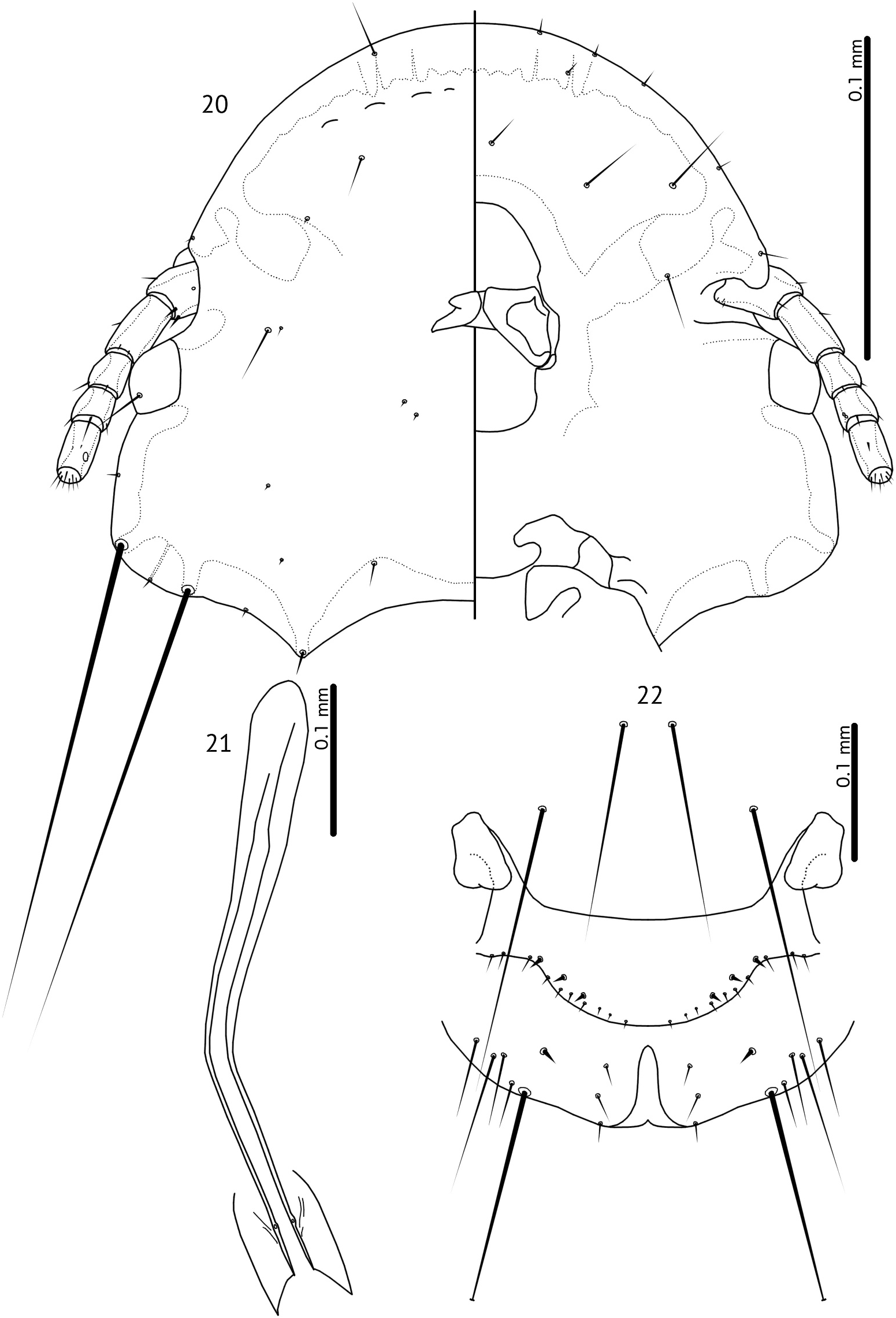
Figures 20–22. Goniocotes rectangulatus Nitzsch [in Giebel], 1866. 20, male head, dorsal and ventral views. 21, male genitalia, dorsal view. 22, female genitalia and abdominal segments VIII–XI, ventral view.
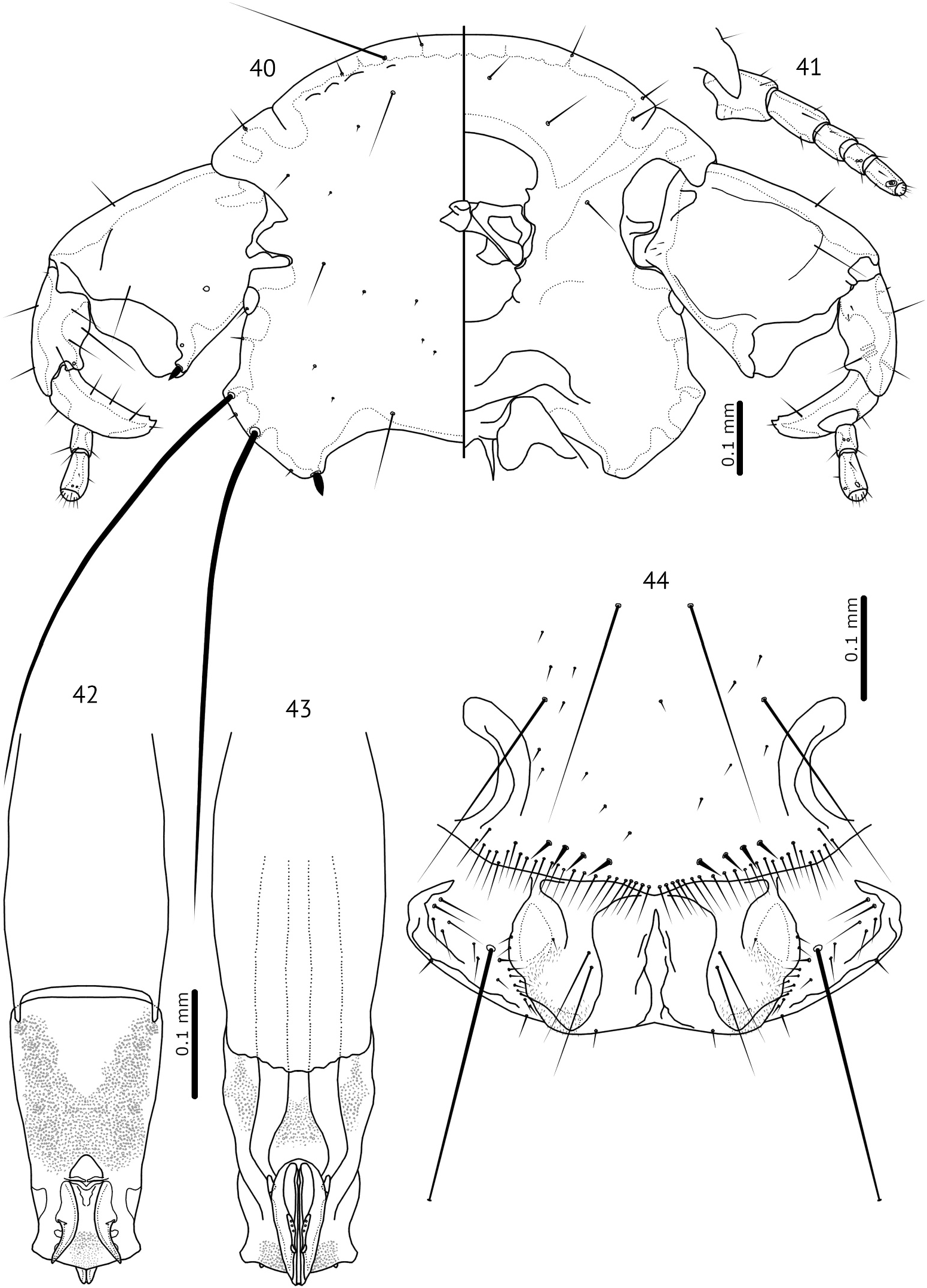
Figures 40–44. Pavoniocotes mayuri (Lakshminarayana and Emerson, 1971). 40, male head, dorsal and ventral views. 41, female antenna and conus, ventral view. 42, male genitalia, dorsal view. 43, male genitalia, ventral view. 44, female genitalia and abdominal segments VII–XI, ventral view.
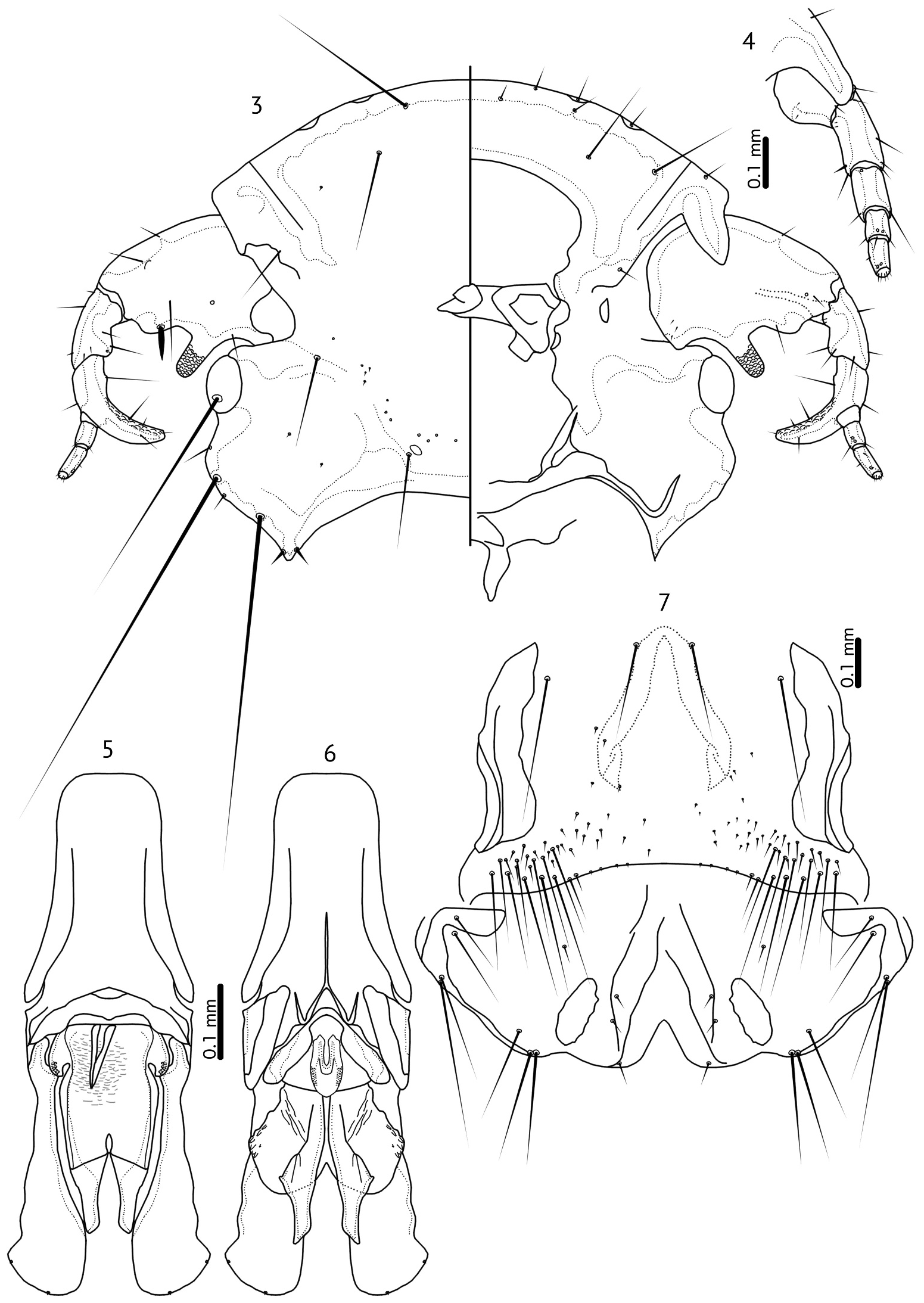
Figures 3–7. Goniodes pavonis (Linnaeus, 1758). 3, male head, dorsal and ventral views. 4, female antenna and conus, ventral view. 5, male genitalia, dorsal view. 6, male genitalia, ventral view. 7, female genitalia and abdominal segments VII–XI, ventral view.
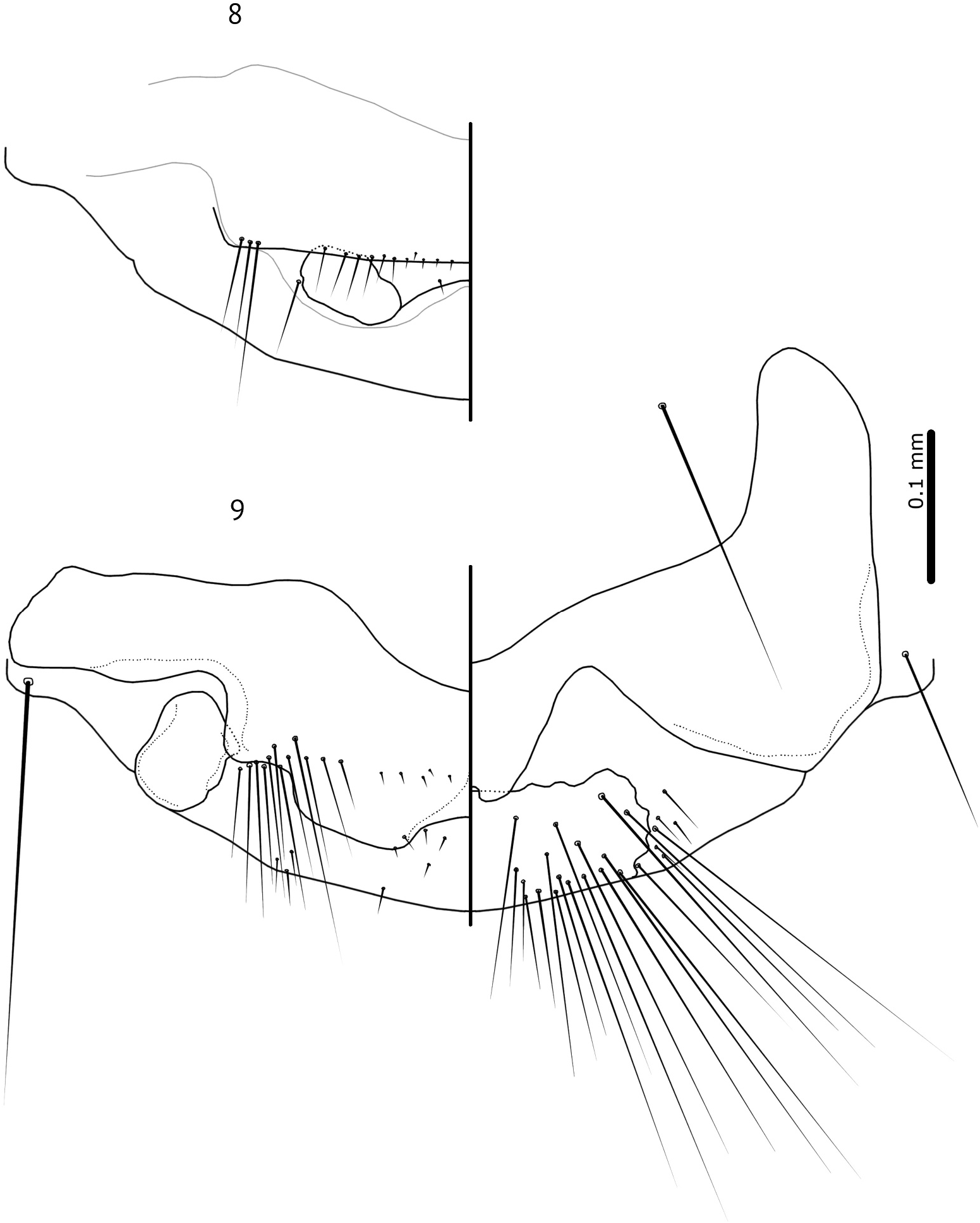
Figures 8–9. Goniodes pavonis (Linnaeus, 1758). 8, male abdominal segments IX–XI, dorsal view, showing the inner margin of the ano-genital opening; grey line indicates the approximate outline of the central dorsal plate of abdominal segment IX+X. 9, male abdominal segments IX–XI, dorsal and ventral views, showing the outer surface. Plates and setae shown in Figure 8 are located underneath the central plate of segment IX+X.
No known copyright restrictions apply. See Agosti, D., Egloff, W., 2009. Taxonomic information exchange and copyright: the Plazi approach. BMC Research Notes 2009, 2:53 for further explanation.
|
Kingdom |
|
|
Phylum |
|
|
Class |
|
|
Order |
|
|
Family |
Pavoniocotes
| Gustafsson, Daniel R., Grossi, Alexandra A., Ren, Mengjiao & Zou, Fasheng 2023 |
Goniodes parviceps
| Piaget E 1880: 277 |
Goniocotes
| Burmeister H 1838: 431 |
Goniodes
| Nitzsch CL 1818: 293 |