
Zorotypus hainanensis Yin and Li
|
publication ID |
https://doi.org/10.11646/zootaxa.4007.4.6 |
|
publication LSID |
lsid:zoobank.org:pub:0EB2FE9A-749C-4555-9CF4-E202F74331F9 |
|
DOI |
https://doi.org/10.5281/zenodo.6096896 |
|
persistent identifier |
https://treatment.plazi.org/id/03A17F3B-FF96-FFA1-05F6-77A79D48FC01 |
|
treatment provided by |
Plazi |
|
scientific name |
Zorotypus hainanensis Yin and Li |
| status |
sp. nov. |
Zorotypus hainanensis Yin and Li , new species
( Figs 1–5 View FIGURE 1 View FIGURE 2 View FIGURE 3 View FIGURE 4 View FIGURE 5 )
Type material (2 ♂♂, 3 ♀♀). Holotype: CHINA: apteron ♂, labeled ‘ China: Hainan, Ledong Hsien [ffi÷θ], Jianfengling N.R. [*RΚ], Mingfenggu [d¼☺], 18°44’30’’N, 108°50’29’’E, rainforest, decaying log, 995 m, 25.i.2015, Xiao-Bin Song ( SNUC). Paratypes: CHINA: 1 apteron ♂, same label data as holotype; 1 apteron ♀, also at Jianfengling, ‘District V [Ϫ∬U], 18°43’57’’N, 108°52’41’’E, broad-leaved forest, decaying log, 975 m, 24.i.2015, Zhong Peng’; 1 apteron ♀, labeled ‘ China: Hainan, Qiongzhong Hsien [¼φθ], Limu Mt. [ϔDzM], nr. reservoir, 19°09’01’’N, 109°45’08’’E, mixed forest, decaying log, 580 m, 30.i.2015, De-Yao Zhou’; 1 apteron ♀, also at Limu Mt., ‘path to peak, 19°10’27’’N, 109°45’29’’E, mixed forest, decaying log, 1000 m, 31.i.2015, Zhong Peng’. (all SNUC)
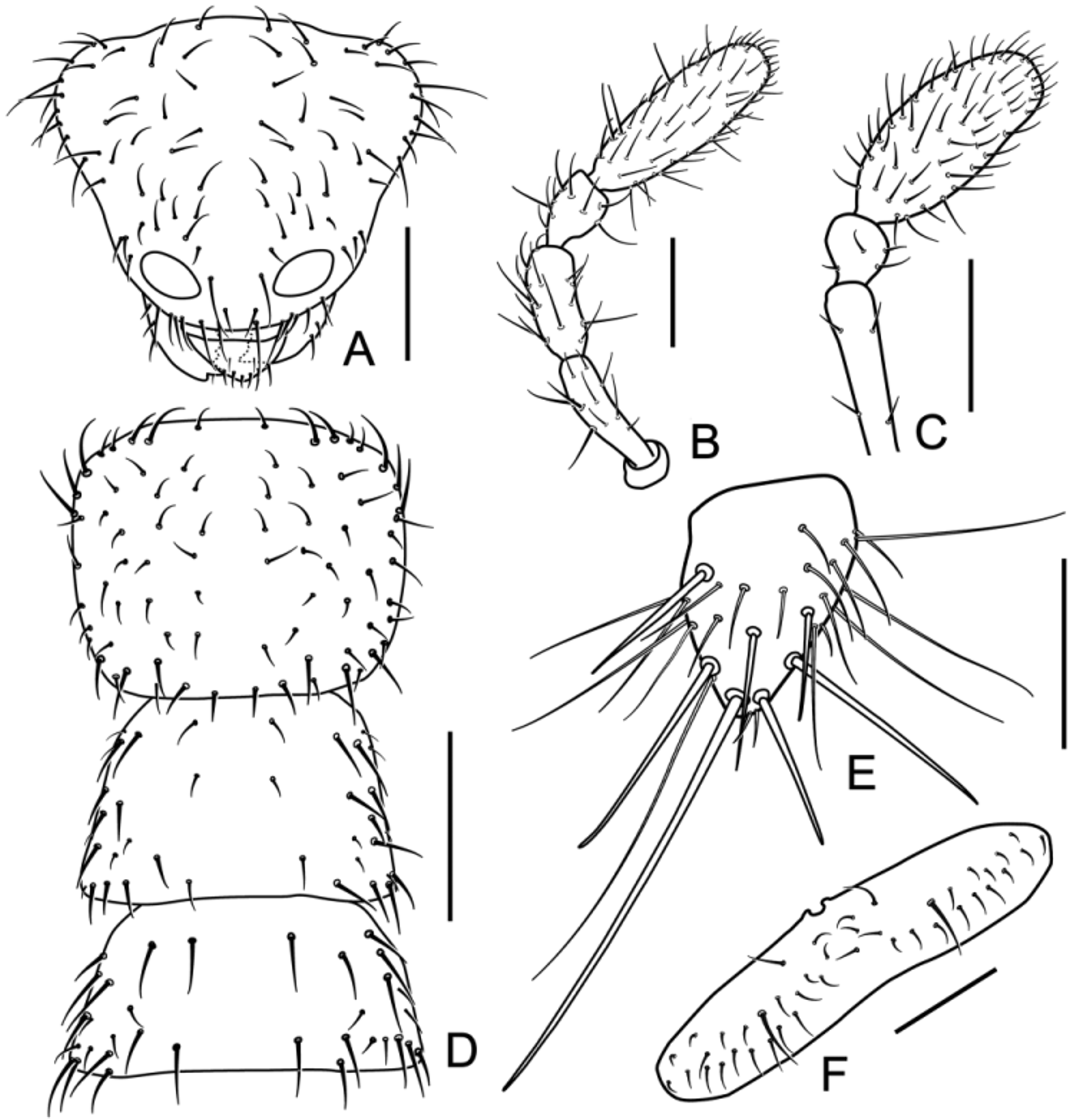
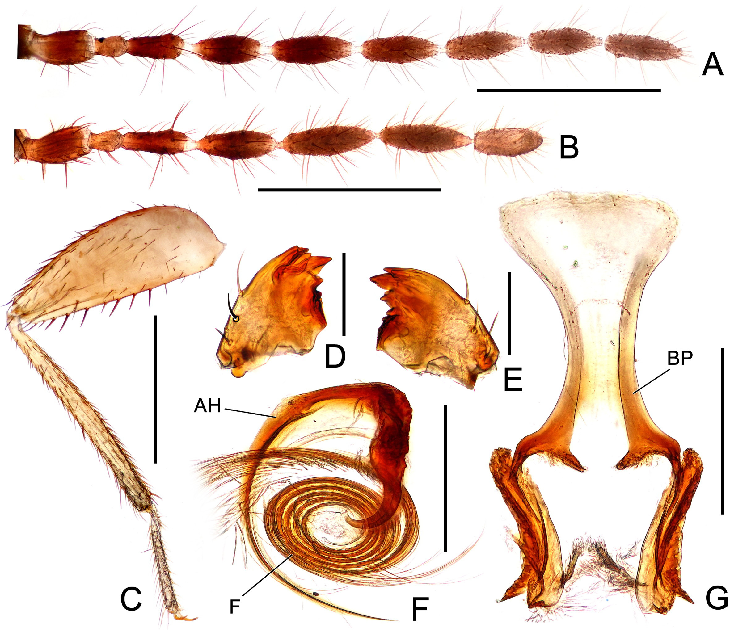
Description. Apteron male ( Fig. 1 View FIGURE 1 ). Body length 2.54–2.60 mm (exclusive of antennae), color glossy brown except membranous regions and yellowish white cercus; head ( Fig. 2 View FIGURE 2 A) sub-triangular, slightly wider than pronotum, with whitish area in posterolateral corner; cephalic chaetotaxy as depicted in Fig. 2 View FIGURE 2 A; compound eyes and ocelli absent; maxillary palpus ( Fig. 2 View FIGURE 2 B) five-segmented; labial palpus ( Fig. 2 View FIGURE 2 C) three-segmented; antennae nine-segmented, antennomeres VII paler than preceding ones (antennomeres VIII–IX missing) ( Fig. 4 View FIGURE 4 B), antennomere I slightly curved outward, antennomere II faintly curved, short, about one-half length of antennomere III, antennomeres III–VII longer than wide, length of each subequal to that of antennomere I ( Fig. 4 View FIGURE 4 B); both mandibles with five apical teeth and well-developed molar region ( Fig. 4 View FIGURE 4 D–E). Pronotum ( Fig. 2 View FIGURE 2 D) subrectangular, slightly narrowed posteriorly; mesonotum ( Fig. 2 View FIGURE 2 D) trapezoidal, slightly shorter than pronotum; metanotum ( Fig. 2 View FIGURE 2 D) trapezoidal, distinctly wider than long, shorter than mesonotum; thorax sparsely setose as depicted in Fig. 2 View FIGURE 2 D. Legs with setae of moderate length; tibiae and tarsi of all legs paler in color; posterior surface of profemur broadly setose, anterior and dorsal surfaces covered with setae of moderate length only distally; protibia covered with setae of moderate length, bristles arranged like comb in distal half along ventral surface, with two apical spurs; mesofemur about as broad as profemur, anterior surface broadly setose, posterior and dorsal surfaces covered with setae of moderate length on distal half and several short setae on proximal half; mesotibia covered with setae of moderate length, with two apical spurs; metafemur broader than profemur, more swollen proximally than distally ( Fig. 4 View FIGURE 4 C), anterior surface broadly setose, posterior and dorsal surfaces covered with setae of moderate length on distal half and several short setae on proximal half, ventral surface with eight to nine long stout bristles ( Fig. 4 View FIGURE 4 C); metatibia covered with setae of moderate length, with two apical spurs.
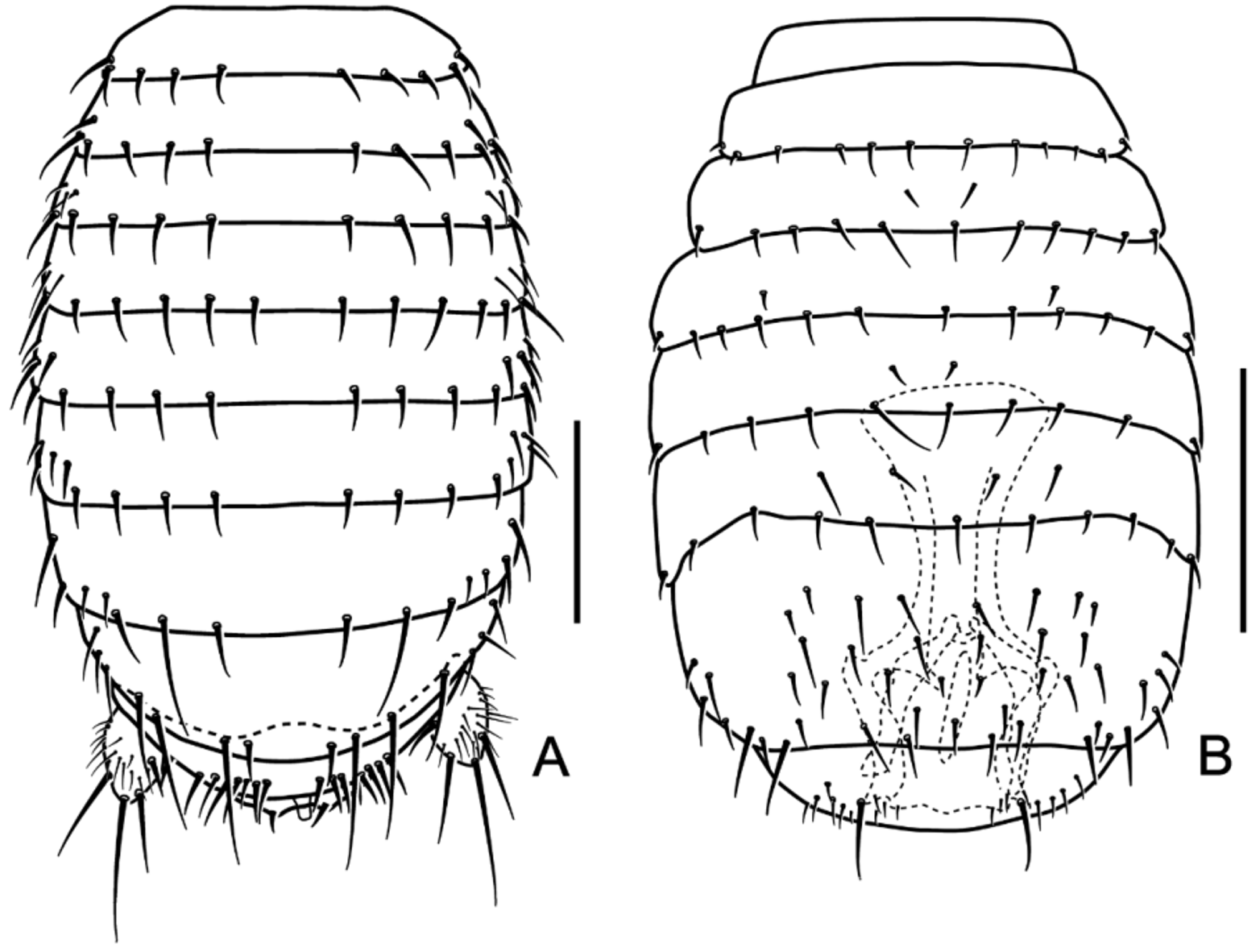
Abdominal tergites I–VI ( Fig. 3 View FIGURE 3 A) with single transverse row of four to six setae of moderate length and a few lateral, short setae on each side of midline; tergite VII ( Fig. 3 View FIGURE 3 A) with single transverse row of one long seta, two moderate-length setae, and a few short setae laterally on each side of midline; tergite VIII ( Fig. 3 View FIGURE 3 A) with single transverse row of one short seta, three long erect setae, and three moderate-length setae on each side of midline; tergite IX ( Fig. 3 View FIGURE 3 A) short, anterior trapezoidal expansion extending beneath tergite VIII, with single row of seven to eight short, thick setae on each side of midline; tergite X ( Fig. 3 View FIGURE 3 A) posteromedially incised, with several moderate-length setae on each side of midline, one pair of stout setae near incision; tergite XI with small median upcurved projection (= male mating hook) and two lateral, subtriangular sclerites, several moderate-length setae on each sclerite; epiproct and paraproct unsclerotized; cercus ( Fig. 2 View FIGURE 2 E) unsegmented, oval, with one long apical seta, three or four preapical moderate-length setae, several short setae, and very long, fine setae; surface covered with numerous minute spicules except at base and apex; sternite I scarcely sclerotized; sternite II ( Fig. 3 View FIGURE 3 B) weakly sclerotized; sternite III ( Fig. 3 View FIGURE 3 B) with single transverse row of several moderate-length; sternites IV–V ( Fig. 3 View FIGURE 3 B) with single transverse row of five to six moderate-length setae on each side of midline; sternites VI–VII ( Fig. 3 View FIGURE 3 B) with two transverse rows of moderate-length setae, anterior row with several setae, posterior row with five or six setae on each side of midline; sternite VIII ( Fig. 3 View FIGURE 3 B) wider than long, with evenly scattered fine setae, one longer setae on posterior margin on each side of midline; sternite IX trapezoidal, with small, fine setae; posterior margin with two moderate-length setae on each side of midline; sternite X invaginated beneath sternite IX, not visible externally; sternite XI with two lateral sclerites, each with fine setae. Genitalia symmetrical; basal plate ( Fig. 4 View FIGURE 4 G) well sclerotized, posteriorly bifurcate, with long, anterior tongue-like process; flagellum ( Fig. 4 View FIGURE 4 F) long, sclerotized, coiled; aedeagus with hook ( Fig. 4 View FIGURE 4 F).
Apteron female. Body length 2.48–2.51 mm (exclusive of antennae). General features corresponding to those of males. Antennomere III ( Fig. 4 View FIGURE 4 A) slightly shorter than that of male; tergite X not posteromedially incised, with three or four setae of moderate length on each side of midline; tergite XI evenly sclerotized, not divided into two halves (hemitergites), with small scattered setae and a pair of paramedian setae of moderate length apically; sternite IX ( Fig. 2 View FIGURE 2 F) transverse, short, anterior margin with bifurcate projection extending beneath sternite VIII, several fine setae scattered and one pair of setae as depicted in Fig. 2 View FIGURE 2 F.
Alate form. Unknown.
Comparative notes. This new species is similar to Z. sinensis and Z. medoensis in general appearance, but can be readily separated from both by a different placement of the bristles on the ventral margin of the metafemur ( Fig. 4 View FIGURE 4 C; Hwang 1974: Fig. 3 View FIGURE 3 ), relatively shorter cerci ( Fig. 1 View FIGURE 1 ; Hwang 1974: Fig. 1 View FIGURE 1 ), setation on the posterior margin of male sternum VIII ( Fig. 3 View FIGURE 3 B; Hwang 1976: Figs 1 View FIGURE 1 , 2 View FIGURE 2 ), and form of the male genitalia ( Fig. 4 View FIGURE 4 F–G; Hwang 1976: Figs 3–4 View FIGURE 3 View FIGURE 4 ). The Taiwanese Z. newi has a unique combination of elongate first tarsomere and cercus ( Chao & Chen 2000: Figs 1 View FIGURE 1 , 3 View FIGURE 3 , 6 View FIGURE 6 ), which leads to a quick separation of this species from all other zorapterans ( Engel & Grimaldi 2000: 155). Zorotypus hainanensis seems closely allied to the Malaysian Z. caudelli Karny , Z. magnicaudelli Mashimo, Engel, Dallai, Beutel & Machida , and Z. cervicornis Mashimo, Yoshizawa & Engel , and possibly to the central American Z. cramptoni Gurney , Z. gurneyi Choe , Z. hamiltoni New , and Z. snyderi Caudell by sharing a strongly elongate and coiled flagellum. As discussed in Mashimo et al. (2013), this character possibly ‘represents a complex synapomorphy of these species’, and ‘it appears plausible that these species comprise a clade within Zorotypus .’ On the other hand, Zorotypus hainanensis can be separated from all these congeners by the unique forms of the male genital basal plate and aedeagal hooks.
Biology. The two male and one female adults from Jianfengling were collected from several fallen decomposing logs ( Fig. 5 View FIGURE 5 B) in rainforests at an elevation of ca. 1000 m ( Fig. 5 View FIGURE 5 A, C–D). The other two females from Limu Mountain were also collected from decaying logs in broad-leaved forests, one at an elevation of ca. 1000 m ( Fig. 5 View FIGURE 5 E), and the other at ca. 580 m.
Distribution. Zorotypus hainanensis is currently known only from the Jianfengling Nature Reserve and Limu Mountain ( Fig. 5 View FIGURE 5 F), but it seems reasonable to expect the occurrence of this species in other well-preserved forested areas of the Hainan Island.
Etymology. The specific epithet refers to the region in which specimens were collected, i.e., Hainan Island.


No known copyright restrictions apply. See Agosti, D., Egloff, W., 2009. Taxonomic information exchange and copyright: the Plazi approach. BMC Research Notes 2009, 2:53 for further explanation.
|
Kingdom |
|
|
Phylum |
|
|
Class |
|
|
Order |
|
|
Family |
|
|
Genus |