
Rosalydia, Gonçalves & Viegas, 2022
|
publication ID |
https://doi.org/ 10.11646/zootaxa.5093.5.5 |
|
publication LSID |
lsid:zoobank.org:pub:1350EC4B-A9A7-49FB-A823-6E55F803B41A |
|
DOI |
https://doi.org/10.5281/zenodo.6302226 |
|
persistent identifier |
https://treatment.plazi.org/id/039FFF13-EB6A-AC4A-FF0D-A9E4FA153D8F |
|
treatment provided by |
Plazi |
|
scientific name |
Rosalydia |
| status |
gen. nov. |
Rosalydia View in CoL gen. nov.
Figs 1–36 View FIGURES 1–6 View FIGURES 7–15 View FIGURES 16–25 View FIGURES 26–36
Type-species. Rosalydia xavieri View in CoL gen. et sp. nov., by present designation.
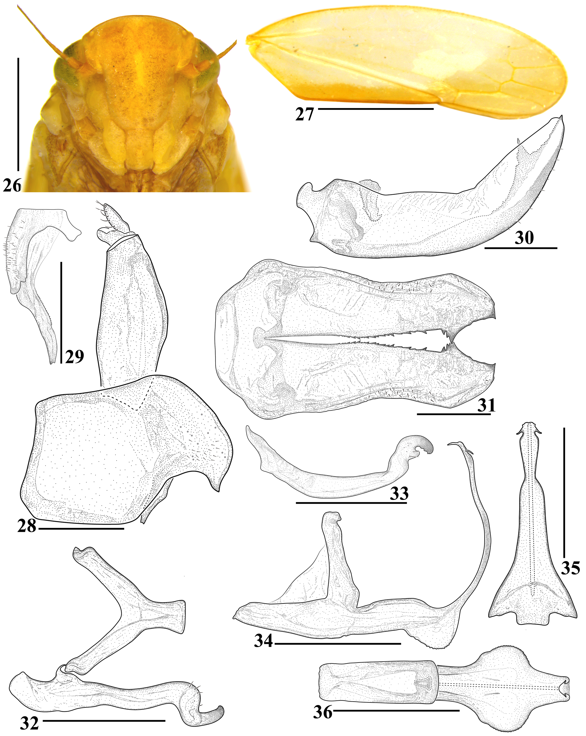
Diagnosis. Medium-sized cylindrical leafhoppers; head ( Figs 1–6 View FIGURES 1–6 ), moderately produced anteriorly, crownface transition angled, with marginal carina; crown ( Figs 1, 3, 5 View FIGURES 1–6 ) subpentagonal, with four orange stripes, three longitudinal and one transverse forming E-shaped macula; lateral margins of crown ( Figs 2, 4, 6 View FIGURES 1–6 ), adjacent to eyes, raised and not carinate; ocellus ( Figs 2, 4, 6 View FIGURES 1–6 ) on anterior margin of head, distant from eye margin; pronotum ( Figs 1, 3, 5 View FIGURES 1–6 ) acutely emarginate; forewing ( Figs 17 View FIGURES 16–25 , 27 View FIGURES 26–36 ) with venation indistinct, except in apical portion, with four apical and three anteapical cells; male pygofer ( Figs 8 View FIGURES 7–15 , 18 View FIGURES 16–25 , 28 View FIGURES 26–36 ) with dorsoapical margin expanded and produced dorsally or posteriorly, caudal margin with process on inner surface or contiguous to posterior margin; subgenital plates ( Figs 10–11 View FIGURES 7–15 , 20–21 View FIGURES 16–25 , 30–31 View FIGURES 26–36 ) fused together only at base, inner margin with several sclerotized teeth; connective ( Figs 12 View FIGURES 7–15 , 22 View FIGURES 16–25 , 32 View FIGURES 26–36 ) Y-shaped; style ( Figs 13 View FIGURES 7–15 , 23 View FIGURES 16–25 , 33 View FIGURES 26–36 ) with tooth below hooked apex; aedeagus ( Figs 14–15 View FIGURES 7–15 , 24–25 View FIGURES 16–25 , 34–36 View FIGURES 26–36 ) with shaft long and slender, with pair of short preapical processes, gonopore apical.
Etymology. The generic name Rosalydia (feminine noun), is a tribute to the Dipterologist Profa. Dra. Rosaly Ale-Rocha for their friendship and importance for the scientific development of the authors of this paper. We are grateful to Dra. Rosaly for being our advisor in the Graduate Program in Entomology at Instituto Nacional de Pesquisas da Amazônia.
Description. Head, in dorsal view ( Figs 1, 3, 5 View FIGURES 1–6 ), moderately produced anteriorly, median length of crown approximately equal to or slightly less than interocular width; transocular width about six-sevenths of humeral width of pronotum; crown subpentagonal, anterior margin bluntly angulate, surface flat and smooth; ocellus on anterior margin of head, distant from eye margin, not visible in dorsal view; coronal maculae and coronal suture indistinct. Head, in frontal view ( Figs 7 View FIGURES 7–15 , 16 View FIGURES 16–25 , 26 View FIGURES 26–36 ), with face approximately as high as wide; frontogenal suture reaching to ocelli; antennal ledge oblique and carinate; frons approximately 1.3 times longer than wide; muscle impressions indistinct; epistomal suture distinct, complete and straight; clypeus approximately 1.6 times longer than maximum width, lateral margins parallel, apex straight; maxillary plate produced ventrally, slightly surpassing the clypeus apex; lorum ellipse-shaped, apical margin not reaching apex of clypeus; gena incompletely covering episternum. Head, in lateral view ( Figs 2, 4, 6 View FIGURES 1–6 ), with crown-face transition acute, with marginal carina; lateral margins of crown, adjacent to eyes, raised and not carinated; antennal pits at same level as imaginary line tangent to anteroventral angles of eyes; antenna with long flagellum, exceeding half length of forewing; frons convex. Pronotum ( Figs 1, 3, 5 View FIGURES 1–6 ) with slightly transverse striae on disc; lateral margins rounded, convergent anterad, as long as eye; posterior margin acutely excavated; in lateral view ( Figs 2, 4, 6 View FIGURES 1–6 ), slightly declivous; dorsopleural carina present and arched. Mesonotum ( Figs 1, 3, 5 View FIGURES 1–6 ) as long as wide. Forewing ( Figs 17 View FIGURES 16–25 , 27 View FIGURES 26–36 ) semi-hyaline, approximately 3.2 times longer than maximum width; venation slightly distinct, more distinct apically; three anteapical and four apical cells, bases of second and fourth apical cells approximately equidistant, base of third apical cell more distal than basis of second and fourth apical cells; alar appendix narrow; apex rounded. Hind wing with vein R 4+5 and M 1+2 preapically convergent, fused at apex, forming single vein. Profemur with AD, AM, and PD rows reduced and poorly defined, with exception of apical setae AD 1, AM 1, and PD 1, respectively; AV formed by a single apical seta; PV row absent; IC row formed by slightly arched comb of fine setae, beginning at distal half of femur and extending to apex. Protibia, in cross-section, semi-circular; AV row formed by approximately 15 setae, slightly longer and thicker towards apex; AD formed by a single apical seta; PD formed by two setae, one at beginning of apical third and another at apex; PV row with 2–3 widely spaced setae. Metafemur with setal formula 2:2:1, with inner seta of second pair reduced in size. Metatibial AD row with 2– 5 intercalary setae between macrosetae; PD, AD, and PV rows with 17–18, 9–11, and 40–42 macrosetae, respectively; AV row with approximately 5–6 macrosetae distributed only in the middle third of tibia. Metatarsomere I longer than combined length of two distal tarsomeres; plantar surface with two rows of setae, external row with longer and robust setae than inner row; pecten with five platellae. Metatarsomere II pecten with three platellae.
Male terminalia. Pygofer ( Figs 8 View FIGURES 7–15 , 18 View FIGURES 16–25 , 28 View FIGURES 26–36 ) shorter than subgenital plates; macrosetae absent; dorsoapical margin expanded and produced dorsally or dorsoposteriorly; presence of caudal process on inner surface or contiguous to posterior margin, directed ventrally ( Figs 9 View FIGURES 7–15 , 19 View FIGURES 16–25 , 29 View FIGURES 26–36 ). Valve ( Figs 11 View FIGURES 7–15 , 21 View FIGURES 16–25 , 31 View FIGURES 26–36 ) completely fused to subgenital plates. Subgenital plates, in lateral view ( Figs 10 View FIGURES 7–15 , 20 View FIGURES 16–25 , 30 View FIGURES 26–36 ), moderate to deeply excavated, dorsally curved from apical half; in ventral view ( Figs 11 View FIGURES 7–15 , 21 View FIGURES 16–25 , 31 View FIGURES 26–36 ), fused together only at base; inner margin with several sclerotized teeth; macrosetae absent. Connective ( Figs 12 View FIGURES 7–15 , 22 View FIGURES 16–25 , 32 View FIGURES 26–36 ) Y-shaped, arms approximately twice the length of stem; stem articulated at aedeagus base, not bifurcated at point of articulation. Style ( Figs 12–13 View FIGURES 7–15 , 22–23 View FIGURES 16–25 , 32–33 View FIGURES 26–36 ) with apodeme non-bilobed; preapical lobe undeveloped; preapical setae present; apex curved ventrally, hook-shaped, with irregular ventral margin bearing a small sclerotized tooth. Aedeagus ( Figs 14–15 View FIGURES 7–15 , 24–25 View FIGURES 16–25 , 34–36 View FIGURES 26–36 ) with dorsal apodeme and preatrium elongate; shaft very long and slender, flattened dorsoventrally, directed dorsally, with pair of short lateral preapical processes directed ventrally; gonopore apical. Anal tube ( Figs 8 View FIGURES 7–15 , 18 View FIGURES 16–25 , 28 View FIGURES 26–36 ) cylindrical, without process; ventral surface membranous.
Female terminalia. Female unknown.
Distribution. Brazil: Amazonas and Mato Grosso.
Remarks. Rosalydia gen. nov. is externally very similar to species of Coelidiana Oman, 1936 , Paracoelidiana Marques-Costa & Cavichioli, 2007 , Scopocoelidia Marques-Costa & Cavichioli, 2007 and Mejdalania Gonçalves, 2021. However, the presence of caudal process on inner surface or contiguous to posterior margin of pygofer ( Figs 8 View FIGURES 7–15 , 18 View FIGURES 16–25 , 28 View FIGURES 26–36 ) and several sclerotized teeth on the inner margins of the subgenital plates ( Figs 10–11 View FIGURES 7–15 , 20–21 View FIGURES 16–25 , 30–31 View FIGURES 26–36 ) are unique characteristics that separate the new genus from all other known Neocoelidiinae .
Key to species of Rosalydia gen. nov.
1. Pygofer, with short caudal process, not surpassing the ventral margin ( Fig. 28 View FIGURES 26–36 ); subgenital plates with teeth of inner margin increasing in size towards apex ( Fig. 31 View FIGURES 26–36 ); aedeagus with pair of expansions in basal third ( Figs 34–36 View FIGURES 26–36 ) (Amazonas)....................................................................................... R. xavieri View in CoL gen. et sp. nov.
- Pygofer, with long caudal process, surpassing the ventral margin ( Figs 8 View FIGURES 7–15 , 18 View FIGURES 16–25 ); subgenital plates with teeth of inner margin approximately regular in size ( Figs 11 View FIGURES 7–15 , 21 View FIGURES 16–25 ); aedeagus without pair of expansions in basal third ( Figs 14 View FIGURES 7–15 , 24 View FIGURES 16–25 )............. 2
2. Pygofer subquadrangular, dorsoapical margin expanded dorsally, forming rounded lobe, caudal process with spine on basal portion, apex bifurcated ( Figs 8–9 View FIGURES 7–15 ); subgenital plates with lateral margins converging towards apex ( Fig. 11 View FIGURES 7–15 ); aedeagus with subapical processes slightly dilated apically with crenulated dorsoapical margin ( Figs 14–15 View FIGURES 7–15 ) (Mato Grosso)......................................................................................... R. alvarengai View in CoL gen. et sp. nov.
- Pygofer subtrapezoidal, dorsoapical margin expanded dorsoposteriorly and curved ventrally at apex, caudal process without spine on basal portion, apex not bifurcated, with several rugosity ( Figs 18–19 View FIGURES 16–25 ); subgenital plates broad in basal half and distinctly narrowed in apical half ( Fig. 21 View FIGURES 16–25 ); aedeagus with subapical processes very short, spine-like ( Figs 24–25 View FIGURES 16–25 ) (Amazonas)........................................................................................ R. inpa View in CoL gen. et sp. nov.


No known copyright restrictions apply. See Agosti, D., Egloff, W., 2009. Taxonomic information exchange and copyright: the Plazi approach. BMC Research Notes 2009, 2:53 for further explanation.