
Barrosasaurus casamiquelai, Salgado, Leonardo & Coria, Rodolfo A., 2009
|
publication ID |
https://doi.org/10.5281/zenodo.190129 |
|
DOI |
https://doi.org/10.5281/zenodo.5685070 |
|
persistent identifier |
https://treatment.plazi.org/id/039F003B-FFDD-697C-C6BE-951EFC7AA0A0 |
|
treatment provided by |
Plazi |
|
scientific name |
Barrosasaurus casamiquelai |
| status |
sp. nov. |
Barrosasaurus casamiquelai sp. nov.
Figures 3–6 View FIGURE 3 View FIGURE 4 View FIGURE 5 View FIGURE 6
Holotype. MCF-PVPH-447/1–3, one anterior dorsal (MCF-PVPH-447/3) and two posterior dorsal vertebrae (MCF-PVPH-447/1 and MCF-PVPH-447/2) belonging to a single individual.
Etymology. In honor of the late Rodolfo Casamiquela ( 1932–2008) for his important contributions to the development of Argentinean Earth Sciences.
Type locality and horizon. Sierra Barrosa (locality coordinates: 38° 50’ 27’’ S, 68° 50’ 28’’ W; Neuquén Province, Argentina; Fig. 1 View FIGURE 1 ), lower Anacleto Formation (Upper Cretaceous, lower Campanian; Ramos 1981; Dingus et al. 2000). In general, the Anacleto Formation is characterized in this area by the presence of stacked, massive, medium-to-coarse-grained sandstone bodies with locally occurring extraformational particles, pebbles and cobbles. Sandstone bodies are typically sheet-shaped, but lenticular bodies occur locally. Sediments are almost uniformly red in color, but some gray to yellow sandstones also occur. Sandstones are heavily bioturbated with localized occurrences of decimeter-scale, “lumpy” to vertically oriented calcareous concretions that preferentially cement Planolites burrow-fills, and a variety of more complex burrow/dwelling structures of unknown origin (Coria et al. 2000).
Diagnosis. Large titanosaurian sauropod characterized by the following autapomorphies: 1) posterior dorsal vertebrae bearing paired, short spinal laminae, nearly parallel to the prespinal lamina; 2) welldeveloped anterior spinodiapophyseal laminae in posterior dorsal vertebrae; 3) anterior spinodiapophyseal laminae divided into at least two, and up to three, branches in posteriormost dorsal vertebrae; 4) aliform processes with broad, planar and rugose laterodorsal surfaces; 5) portion of neural spine distal to the aliform process is longer than the portion of the spine that is proximal to the aliform process, at least in the posterior dorsals.
Description. MCF-PVPH-447/3 ( Figs 3 View FIGURE 3 , 6 View FIGURE 6 A). This vertebra is identified as an anterior dorsal, probably the third dorsal, based on the relative position of the parapophysis and comparison to the complete dorsal series of Trigonosaurus pricei Campos, Kellner, Bertini & Santucci, 2005 and other unpublished specimens (e.g. MAU-Pv-CO-439). Only the anterior half of the vertebra is preserved, and all parts of the specimen positioned posterior to the diapophysis are missing, including most of the neural spine ( Fig. 3 View FIGURE 3 A, B).
The centrum is opisthocoelous, and notably wider than high. The anterior articular surface is convex and has an elliptical outline ( Fig. 3 View FIGURE 3 A). In lateral view, the lateral surfaces of the centrum are strongly excavated and anteroposteriorly concave, and the pleurocoels are placed on the anterior part of the centrum, close to the ventral margin ( Fig. 3 View FIGURE 3 B). Unlike the condition in the posterior dorsals, the pleurocoels open directly onto the lateral surfaces of the centrum, and are not set in the bottom of a fossa enclosed by sharp borders. The posterior articular face of the centrum is not preserved, although the element is likely to have been relatively short anteroposteriorly. The ventral surface of the centrum is dominated by a thick, longitudinal keel ( Fig. View FIGURE 6
6A), a character noted by Upchurch et al. (2004: their character 126) as present in other sauropods, such as Diplodocus Marsh, 1878 and Haplocanthosaurus Hatcher, 1903 .
In anterior view the neural arch is transversely wide. The transverse processes are dorsoventrally flattened. The diapophyses project laterally and slightly dorsally ( Fig. 3 View FIGURE 3 A). The anterior surface of the neural arch is slightly concave. The centroprezygapophyseal laminae are absent. The neural canal is nearly circular. A short segment of the base of the prespinal lamina is preserved and extends dorsal to a small fossa that is located above the neural canal ( Fig. 3 View FIGURE 3 A, sf). The small fossa is bordered ventrally by a prominent, shelf-like, supraneural structure, which corresponds to the fused left and right intraprezygapophyseal laminae ( Fig. 3 View FIGURE 3 A, tprl). The bases of both spinoprezygapophyseal laminae are clearly visible in anterior view ( Fig. 3 View FIGURE 3 A, sprl). Several titanosaurs preserve relatively complete dorsal sequences, including Trigonosaurus pricei , Argyrosaurus superbus Lydekker, 1893 , Neuquensaurus australis , and Argentinosaurus huinculensis Bonaparte & Coria, 1993 . In general, the spinoprezygapophyseal laminae are absent or extremely reduced in the anterior dorsals of these taxa ( Huene 1929; Bonaparte & Coria 1993; Campos et al. 2005; Powell 2003). Because the neural spine is missing, it is not possible to determine if the spinoprezygapophyseal laminae were confluent at some point on the element. The prezygapophyses are well separated from one another ( Fig. 3 View FIGURE 3 A, prz), and are placed posterior to the parapophyses, at a point approximately level with the diapophyses. The articular facets of the prezygapophyses have an oval outline, with their longest diameter transversely oriented ( Fig. 3 View FIGURE 3 A). The articular faces of the prezygapophyses face dorsomedially, at an angle of approximately 30° to the horizontal. The parapophyses project laterally, with their articular facets dorsoventrally elongated and slightly concave.
In lateral view, the neural arch is anteroposteriorly short ( Fig. 3 View FIGURE 3 B). The parapophyses are placed approximately at the midpoint between the dorsal border of the centrum and the diapophyses ( Fig. 3 View FIGURE 3 A, B, pp). A well developed anterior centroparapophyseal lamina is present ( Fig. 3 View FIGURE 3 B, acpl), whereas the posterior centroparapophyseal lamina is poorly developed ( Fig. 3 View FIGURE 3 B, pcpl). The paradiapophyseal lamina, which links the parapophysis with the anterior face of the diapophysis, is well-developed ( Fig. 3 View FIGURE 3 A, B, ppdl). Posterior to the paradiapophyseal lamina, there is another, shorter, lamina that extends to the ventral border of the diapophysis ( Fig. 3 View FIGURE 3 B, al). Between this short accessory lamina and the paradiapophyseal lamina there is a shallow fossa. A robust posterior centrodiapophyseal lamina extends from the ventral face of the diapophysis ( Fig. 3 View FIGURE 3 B, pcdl). In addition, an accessory posterior centrodiapophyseal lamina extends anteroventrally from the ventral face of the diapophysis ( Fig. 3 View FIGURE 3 B, apcdl). This accessory posterior centrodiapophyseal lamina subdivides a fossa that is positioned posterior to the parapophysis: the infradiapophyseal fossa ( Fig. 3 View FIGURE 3 B). On the left side, the base of a spinodiapophyseal lamina is present.
MCF-PVPH-447/1 ( Figs 4 View FIGURE 4 A–C, 6B). This vertebra is from the posterior part of the series, and is possibly the seventh or eighth. This positional identification is based on the relative position of the parapophysis, comparison to the complete dorsal series of Trigonosaurus pricei ( Campos et al. 2005) , and the fact that MCF-PVPH-447/2 (see below) is clearly positioned more posteriorly within the vertebral column with respect to MCF-PVPH-447/1 (MCF-PVPH-447/2 is probably the ninth or tenth dorsal, assuming a total count of ten dorsals as in most titanosaurs [ Powell 2003]). The vertebral centrum possesses a convex anterior articular face, a concave posterior articular face that is wider than high, and dorsoventrally convex and anteroposteriorly concave lateral surfaces ( Fig 4 View FIGURE 4 A–C). The elliptical pleurocoels are obliquely oriented such that their anterior halves are positioned more dorsally on the centrum that the posterior halves ( Fig. 4 View FIGURE 4 B). A prominent longitudinal keel-like ridge is present on the ventral surface of the centrum ( Fig. 6 View FIGURE 6 B).
The neural arch is relatively short anteroposteriorly, and placed mostly on the anterior half of the centrum ( Fig. 4 View FIGURE 4 A–C). At the base of the prespinal lamina ( Fig. 4 View FIGURE 4 A, B, prsl), on both sides, a short and delicate ventrolaterally extending lamina is present, which ends near the medial border of the prezygapophyses ( Fig. 4 View FIGURE 4 A, sprl?). These laminae may represent subdivisions of the prespinal lamina, vestiges of the spinoprezygapophyseal lamina, or both.
In anterior view, the prezygapophyses are transversely broad and anteroposteriorly short processes, and their articular facets face dorsomedially and converge ventrally ( Fig. 4 View FIGURE 4 A, prz). Ventral to a well-developed intraprezygapophyseal lamina ( Fig. 4 View FIGURE 4 A, tprl), a median septum separates two deep cavities. The parapophyses are almost level with the prezygapophyses and are connected by a short and horizontal prezygoparapophyseal lamina ( Fig. 4 View FIGURE 4 A, prpl).
In both anterior and posterior views, distal to the union of the posterior spinodiapophyseal with the spinopostzygapophyseal laminae ( Fig. 4 View FIGURE 4 B, pspdl, spol), small lateral expansions of the neural spine are present ( Fig. 4 View FIGURE 4 , alp). These are probably homologous to the aliform processes of Epachthosaurus sciuttoi Powell, 1990 ( Martínez et al. 2004) ( Fig. 4 View FIGURE 4 A, C). In Barrosasaurus , the posterior spinodiapophyseal lamina of MCF-PVPH-447/1 is more strongly expanded laterally than the anterior spinodiapophyseal lamina ( Fig. 4 View FIGURE 4 A). The surfaces of these aliform processes are subrectangular in lateral view ( Fig. 4 View FIGURE 4 B, alp), and are oriented in planes that converge dorsally in both anterior and posterior views ( Fig. 4 View FIGURE 4 A, C). Lateral to the point where the anterior and posterior spinodiapophyseal laminae adjoin ( Fig. 4 View FIGURE 4 B, aspdl, pspdl), on the dorsal surface of the diapophysis, a series of laterally diverging laminae is present.
The paradiapophyseal lamina is short ( Fig. 4 View FIGURE 4 A, B, ppdl). An anterior centroparapophyseal lamina and a well-developed and obliquely oriented posterior centroparapophyseal lamina are present ( Fig. 4 View FIGURE 4 , acpl, pcpl). Ventral to the diapophysis, the posterior centrodiapophyseal lamina is present ( Fig. 4 View FIGURE 4 B, C, pcdl), and is slightly thinner than the posterior centroparapophyseal lamina. The nearly vertical accessory posterior centrodiapophyseal lamina ( Fig. 4 View FIGURE 4 B, apcdl) divides the infradiapophyseal fossa and connects to the posterior centroparapophyseal lamina. The area between the anterior and posterior centroparapophyseal laminae is a shallow, triangular depression. The centropostzygapophyseal ( Fig. 4 View FIGURE 4 B, C, cpol), spinopostzygapophyseal, posterior centrodiapophyseal, and the posterior spinodiapophyseal laminae enclose a deep, rhomboidal and dorsoventrally elongated fossa ( Fig. 4 View FIGURE 4 B) that extends onto the posterior face of the transverse process.
In posterior view ( Fig. 4 View FIGURE 4 C), ventral to the postzygapophyses, the robust centropostzygapophyseal laminae enclose a deep depression that contains the neural canal ( Fig. 4 View FIGURE 4 C, nc), which is roofed by a well developed intrapostzygapophyseal lamina ( Fig. 4 View FIGURE 4 C, tpol). There are no postzygodiapophyseal laminae.
The neural spine is tall, nearly vertical and transversely expanded. Because the distal end of the neural spine is missing, it is not possible to estimate its total height (see Table 1 View TABLE 1 ). In anterior view, the prespinal lamina is well-developed in its basal and mid segments ( Fig. 4 View FIGURE 4 A, prsl). Towards the distal end of the preserved neural spine, the prespinal lamina splits into multiple low crests, which diverge distally. In posterior view ( Fig. 4 View FIGURE 4 C, posl), the postspinal lamina is clearly less well-developed than is the prespinal lamina. The area bounded by the spinopostzygapophyseal and the postspinal laminae bears a series of short and robust, rib-like, nearly transversely extending laminae that converge laterally towards the postzygapophyses.
In lateral view, a short and robust spinopostzygapophyseal lamina is well-developed ( Fig. 4 View FIGURE 4 B, spol). Two spinodiapophyseal laminae are also present, and converge ventrally at their bases ( Fig. 4 View FIGURE 4 B, aspdl, pspdl). The posterior spinodiapophyseal lamina contacts the spinopostzygapophyseal lamina at the mid height of the spine, whereas the anterior spinodiapophyseal lamina extends further dorsally, probably reaching the distal end of the spine. Between both spinodiapophyseal laminae, there is a deep, elongated fossa, which is deeper at the base of the spine than more distally ( Fig. 4 View FIGURE 4 B).
MCF-PVPH-447/2 ( Fig. 5 View FIGURE 5 A–D). This is a posterior dorsal vertebra, possibly the ninth or the tenth of the column, based on comparison to dorsals nine and ten of Trigonosaurus pricei , which, unlike the immediately preceding vertebrae, have postzygodiapophyseal laminae ( Campos et al. 2005). This vertebra is almost complete, lacking only the distal end of the neural spine and the left postzygapophysis. In this specimen, the head of the right rib is preserved fused to the transverse process, in contact with the corresponding diapophysis and parapophysis.
The centrum of MCF-PVPH-447/2 is strongly opistocoelous and wider than high. The lateral faces are anteroposteriorly excavated, bearing deep pleurocoels ( Fig. 5 View FIGURE 5 C, D), although these pleurocoels are slightly shorter anteroposteriorly than those of MCF-PVPH-447/3. Unlike the other vertebrae, the centrum of MCF- PVPH-447/2 does not possess a ventral keel ( Fig. 6 View FIGURE 6 C).
The neural arch is anteroposteriorly short, and more strongly inclined anteriorly than those of the preceding vertebrae ( Fig. 5 View FIGURE 5 C, D). The posterior centrodiapophyseal lamina is weakly developed ( Fig. 5 View FIGURE 5 C, D, pcdl), as are the anterior and posterior centroparapophyseal laminae ( Fig. 5 View FIGURE 5 C, D, acpl, pcpl). MCF-PVPH- 447/2 shows asymmetries in the pattern of laminae at the base of the neural arch. On the left side, on the lateral surface of the neural arch ventral to the diapophysis, several poorly developed laminae are present ( Fig. 5 View FIGURE 5 C), which are absent on the right side ( Fig. 5 View FIGURE 5 D). One of these laminae corresponds to the accessory posterior centrodiapophyseal lamina ( Fig. 5 View FIGURE 5 C, apcdl?). Another lamina oriented perpendicular to the putative accesory posterior centrodiapophyseal lamina is interpreted as the posterior centroparapophyseal lamina. In addition, MCF-PVPH-447/2 possesses a conspicuous postzygodiapophyseal lamina ( Fig. 5 View FIGURE 5 D, podl), which is laterodorsally and anteriorly oriented, and which extends from the lateral margin of the postzygapophysis to the posterior margin of the diapophysis. Unlike MCF-PVPH-447/1, the articular facets of the postzygapophyses face ventrally ( Fig. 5 View FIGURE 5 B). The transverse processes are laterodorsally directed, and placed in a higher position than the postzygapophyses.
The neural spine is noticeably shorter anteroposteriorly than in MCF-PVPH-447/1. In anterior view, the prespinal lamina is well developed ( Fig. 5 View FIGURE 5 A, prsl). On the left side of the base of the prespinal lamina, a small oblique lamina is present, similar to that observed in MCF-PVPH-447/1. This probably corresponds to a vestige of a spinoprezygapophyseal lamina ( Fig. 5 View FIGURE 5 A, sprl?). In addition, in the middle of the neural spine, a conspicuous lamina is observed extending perpendicular to the prespinal lamina ( Fig. 5 View FIGURE 5 A, tl). Further rib-like transversely extending laminae, parallel to, and shorter than, the former, are placed at different levels on the anterior surface of the neural spine ( Fig. 5 View FIGURE 5 A).
In lateral view, a robust, anterior spinodiapophyseal lamina is present ( Fig. 5 View FIGURE 5 D, aspdl), which is bifurcated at both dorsal (spinal) and ventral (diapophyseal) ends. The posterior spinodiapophyseal lamina ( Fig. 5 View FIGURE 5 B, D, pspdl) is laterally less strongly developed than the anterior spinodiapophyseal lamina. Consequently, the aliform process is only partially visible in anterior view ( Fig. 5 View FIGURE 5 A, alp). The greater development of the anterior spinodiapophyseal lamina prevents the fossa enclosed by these laminae from being visible in anterior view ( Fig. 5 View FIGURE 5 B, D).
In posterior view, both the spinopostzygapophyseal and postspinal laminae are well-developed ( Fig. 5 View FIGURE 5 B, spol, posl). The spinopostzygapophyseal laminae have convex outlines in lateral view, unlike those of MCF- PVPH-447/1, which have a concave outline. Moreover, MCF-PVPH-447/2 lacks rib-like transverse laminae between the postspinal lamina and the spinopostzygapophyseal lamina.


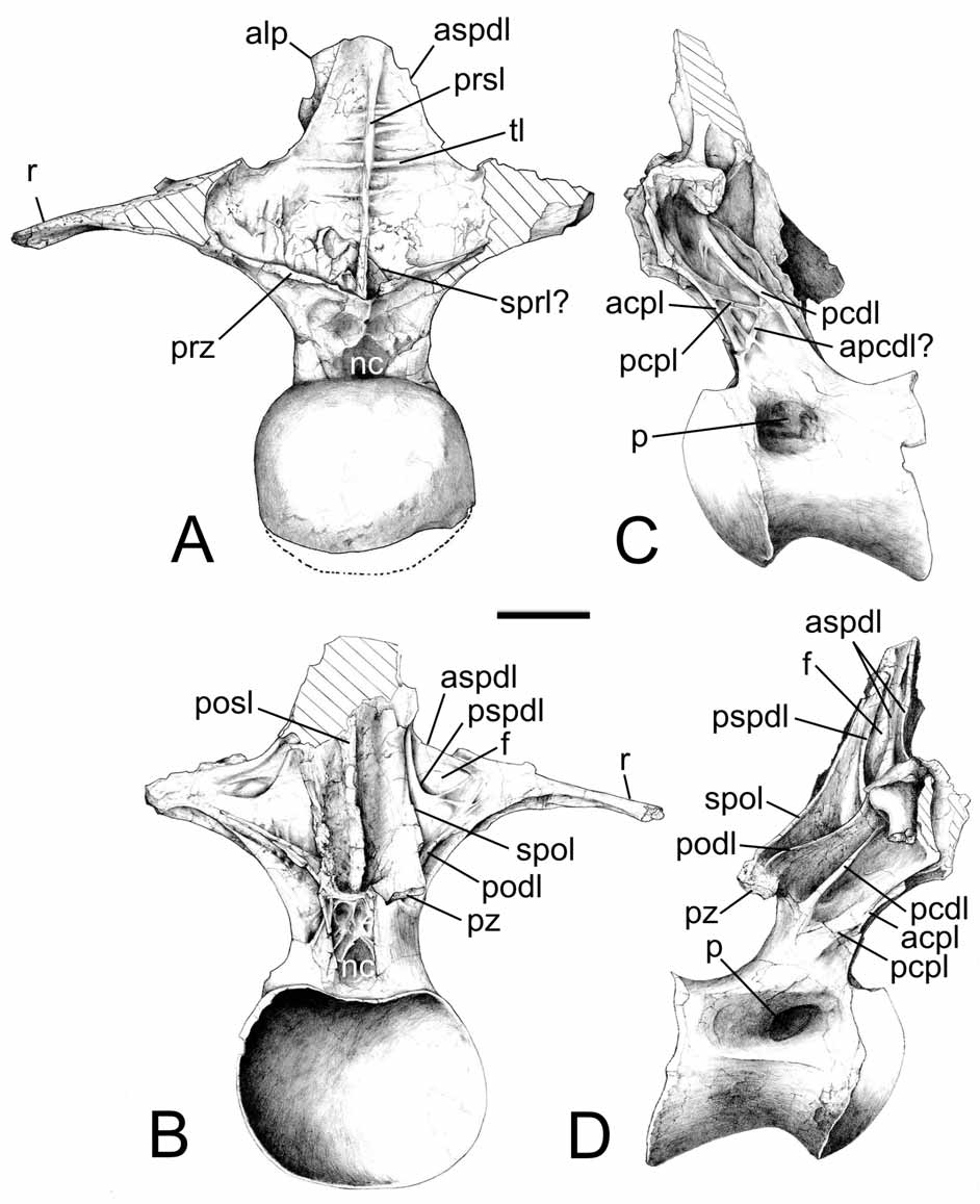

TABLE 1. Measurements of the holotype specimen, MCF-PVPH- 447 of Barrosasaurus casamiquelai gen. et sp nov. Measurements in mm.
| MCF-PVPH-447/3 | MCF-PVPH-447/1 | MCF-PVPH-447/2 | |
|---|---|---|---|
| Centrum length (including condyle) | 170 | 270 | 230 |
| Anterior centrum height | 160 | 180 | 185 |
| Posterior centrum height | -- | 200 | 200 |
| Posterior centrum width | -- | 270 | 280 |
| Height from iintraprezygapophyseal laminae to the spine apex | 80 | 380 | 320 |
| External width between prezygapophyses | 300 | 250 | 170 |
| Length of the neural arch base | 75 (preserved) | 120 | 110 |
| Anterior centrum width | 255 | 150 (estimated)| | 240 (estimated) |
| Spine maximum width (from aliform processes) | -- | 160 | 180 (estimated) |
| Distal neural spine height (from aliform process to top of neural spine) | -- | 180 (preserved) | 90 (preserved) |
| Total heigth | 290 (without neural spine) | 640 | 630 |
No known copyright restrictions apply. See Agosti, D., Egloff, W., 2009. Taxonomic information exchange and copyright: the Plazi approach. BMC Research Notes 2009, 2:53 for further explanation.
|
Kingdom |
|
|
Phylum |
|
|
Class |
|
|
Order |
|
|
Genus |