
Planopusa semenovi, Koretsky & Rahmat, 2021
|
publication ID |
https://doi.org/10.15407/zoo2021.02.143 |
|
DOI |
https://doi.org/10.5281/zenodo.6462354 |
|
persistent identifier |
https://treatment.plazi.org/id/039CDB41-9551-4C63-FF5F-FC0DFB16FCE0 |
|
treatment provided by |
Felipe |
|
scientific name |
Planopusa semenovi |
| status |
sp. nov. |
Planopusa semenovi , sp. n. ( figs 2–5 View Fig View Fig View Fig View Fig ; tables 1–3 View Table 1 View Table 2 View Table 3 )
Holotype. NMNHU-P 64-709 , rostral part of the skull with I3, C, P1, P2, and isolated P4 and M1; P3 is absent ( fig. 2 View Fig ). Collected by Yu. A. Semenov in 1995 and stored in the NMNHPM.
Type locality. Grytsiv , Shepetivskyi District Khmelnytskyi Region, Western Ukraine, karst deposits in limestone quarry on the right bank of Chomora River, 3 km west of village of Grytsiv; 49°58ˈ 05.2 N 27°10ˈ03 E (reef zone).
Formation and Age. Middle Sarmatian, middle-late Miocene; MN 9 , likely in the interval from 11.146 –11.056 Ma.
Diagnosis. Small seal with extremely short rostrum ( table 1 View Table 1 ), differing from all other fossil and extant phocines by: 1) flattened palatal process of maxilla; 2) P4 longer than M1; 3) alveoli form a straight line; 4) wider rostrum across canines compared to other small Phocinae (but narrower than in Monachopsis pontica ).
Description. The partial skull likely belongs to an adult, based on the fused sutures. However, teeth are not worn, characteristic for sub-adults. The maxilla has a very short convexity over P1 and convexity started over P2. The incisive bone is partially broken and palatal process of the maxilla is flattened from the level of the anterior alveolus of P2. The large grooves (fissura palatine) from anterior palatal foramina are directed towards the incisors (as in Enhydra lutris , fig. 3 View Fig ), in contrast to the condition stated by Wozencraft (1989) for other phocids, but similar to seals of the subfamily Devinophocinae ( Koretsky and Holec, 2002; Koretsky and Rahmat, 2015). The concave palatine is wider (18.0 mm) between canines, similar to that in Devinophoca emryi ( Koretsky and Rahmat, 2015) .
The hard palate is half the length of the smallsub-adult of Pusa caspica . Palatal grooves are shallow, but present without the posterior palatal foramen. Part of the palatal bone is missing. Posterior border of the anterior palatal foramen is situated at the level of P1 (in contrast to that in Pusa caspica where it is between C and P1). In coronal view, the space between the posterior border of the anterior palatal foramen and the anterior border for the choanae is 7.5 mm (11.0 mm in Pusa caspica ).
The lingual alveolar margins of canines and incisors are at the same level as those of the cheek teeth (as in D. emryi ), but alveolar margins of C and P1 are compressed towards the midline. From the anterior palatal foramina to the level of P2, the palatine is flat. A deep, antero-posteriorly aligned groove ( sulcus palatinus) is present posterior to P2, characterized as a derived condition among phocids according to Wyss and Flynn (1993) and in contrast to D. emryi .
Maxillary teeth: All teeth are very small with double roots (except C, P1). The incisors are arranged in a straight line. The roots of alveoli I3 are larger than I2, which in turn is larger than I1. The I3 crown is preserved with a small cingulum on the lingual side, round in cross-section. Cingula present only on lingual sides of the crown and do not extend the buccal (labial) side (in contrast to Devinophoca ) ( fig. 2 View Fig ).
Canines project ventrally, slightly worn (possibly sub-adult), but the anterior surface in both canines is worn or due to overbite with grinding of the lower canine ( figs 2 View Fig , 3 View Fig ). The diastema is absent between C and P1, which is located obliquely to the tooth row. The snout is shortened, but the canines are relatively large compared to the length of the snout and other teeth, supporting the overbite hypothesis where the lower canine wears the upper, similar to those in Gulo gulo (wolverine) (IZUAN 896, from the Archangelsk area).
Diastemata between postcanine teeth are absent. All alveoli are rounded and the posterior roots of P2, P4, and M1 are larger than anterior roots. Tooth crowns are not worn. The large paraconid (central cusp) is turned caudally, with one small anterior cusp and a posterior cusp that is larger and positioned higher. The third cusp is placed on the basal cingulum, located caudally to the posterior cusp, and it is much smaller than other cusps (especially smaller on M1).
The P1 has a single root, oval in cross-section, almost intact (similar to Praepusa ). Its crown is 5.0 mm long ( table 2 View Table 2 ), with a conical (triangular) central cusp and cuspidate lingual cingulum bearing diminutive two posterior cusps (in contrast to D. emryi ). Cinguli on P2, P4, and M1 are parallel to the lower enamel margin. On P2, P4, and M1, the gum line is parallel to the margin of the dentary. The complete crowns on postcanine teeth (except P1) are irregularly shaped in occlusal view (in contrast to D. emryi ). The P1 is flattened disto-lingually on its lingual side. The cingulum, bearing minute cuspules, encircles the crown on the lingual side of the P1.
The P2-M1 crowns are triangular in occlusal view and slightly worn. The P2-M1 teeth have two roots, both round in cross-section. The posterior root on P2 is wider and much larger than the anterior alveolus. The P2 is situated in parallel to the tooth row. The P2 crown has a very prominent lingual cingulum with a minute anterior cingular cusp and slightly larger than two posterior cusps. The cingulum on the labial side extends around the entire tooth (in contrast to D. emryi ) with carinae (sharp tooth edges) similar to those observed in Monachinae (Amson and Muizon, 2014) .
The buccal side of the P4 is straight. This tooth has the widest crown base. Posterior root is larger than anterior one, with one anterior cusp, two posterior cusps, and an additional cusp on the lingual side of the cingulum. The crown is triangular in occlusal view. The P4 is larger than M1.
The M1 is very small ( 5.5 mm long and 3.5 mm wide) with two fused roots. The crown is triangular in occlusal view, similar to other postcanines, but much smaller. The anterior cusp is absent, but there is a single posterior cusp and a gracile cingular posterior cusp. Cingulum on the labial side is absent; the tip of the tooth is turned caudally. Cingula are located transversely only on the lingual sides of the crown, not extending the labial (buccal) side (in contrast to those in Devinophoca ).
Comparisons with small-sized representatives of the family Phocidae
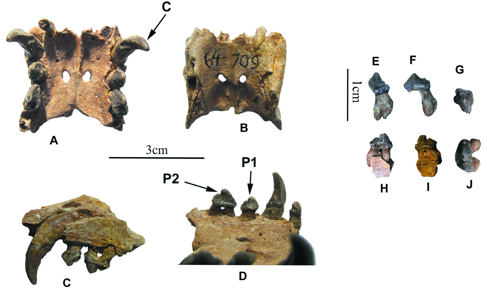
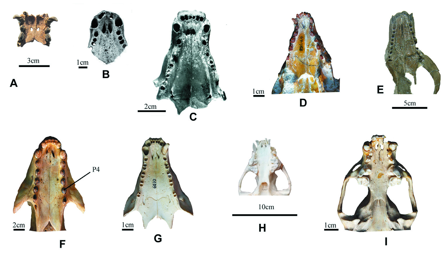
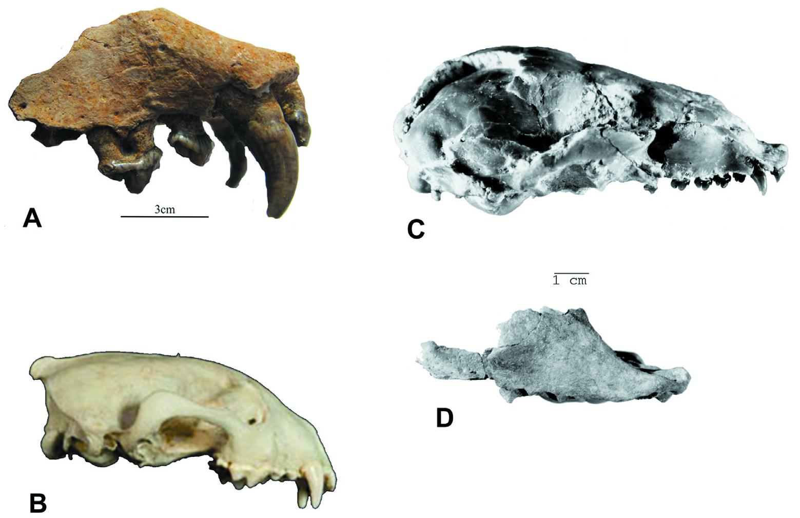
Table 1. Cranial measurements, mm
| Characters | Planopusa semenovi sp. n. | Histriophoca alekseevi | Monachopsis pontica | Prepusa vindobonensis (juv.) | Phocanella sp. , cf. Phocanella pumila | Leptophoca lenis | Devinophoca | |
|---|---|---|---|---|---|---|---|---|
| claytoni | emryi | |||||||
| 1. Total length | 116.0 | 220.0 | 119.9 | 119.5 | ||||
| 2. Condylobasal length | 127.0 | 208.0 | 119.3 | 118.0 | ||||
| 3. Length of palatine process | 55.5 | 65.0 | 83.0 | 71.0 | 81.0 | |||
| 4. Length of rostral part, measured from antero-upper corner of orbit | 52.0 | 47.0 | 63.7–69 | 49.0 | 60.0 | |||
| 5. Length of braincase, measured from posterior corner of orbit | 83.0 | 84.0 | 93.5 | 70.0 | ||||
| 6. Length of tympanic bulla | 23.0 | 29.0–35.0 | 38.5 | 33.6 | ||||
| 7. Length of toothrow, P1–M1 | 35.0 | 29.0 | 34.0 | 60.0 | 49.0–53.2 | 46.9 | ||
| 8. Length of toothrow, P2–P4 | 20.5 | 20.5 | 38.0–40.0 | 32.5–34.5 | 28.9 | |||
| 9. Maximum diameter of infraorbital foramen | 9.0–11.0 | 7.5–11.2 | 11.2–12.1 | |||||
| 10. Length of temporal fossa | 38.5 | 63.6–68.0 | 61.5 | 61.5 | ||||
| 11. Width of rostrum across canines | 30.5 | 24.5 | 19.5 | 40.0–40.5 | 33.0–36.0; 47.0 | 40.0 | 47.7 | |
| 12. Maximal infraorbital width | 16.0–22.0 | 25.5 | 22.0 | |||||
| 13. Minimal infraorbital width | 5.5 | 12.7–15.0 | 14.0 | 19.4 | ||||
| 14. Width of skull across of zygomatic process of squamosal | 66.0 | 112.0–116.0 | 124.0 | 127.7 | ||||
| 15. Width of braincase | 65.0 | 85.0–114.0 | 88.0 | 78.3 | ||||
| 16. Mastoid width | 68.5 | 92.0–140.0 | 113.0 | 102.3 | ||||
| 17. Width of palatine process between P1's | 15.5 | 13.5 | 17.0 | 9.0 | 20.0–21.0 | 15.5–19.0; 24.0 | 10.5 | 20.8 |
| 18. Maximum width of palatine process | 23.5 | 44.0 | 38.0 | 35.0 | 34.5–35.0 | 45.3–50.5 | 55.5 | 40.9 |
| 19. Maximum width of infraorbital foramen | 10.0 | 7.9 | 7.0 | 7.0–9.0 | 9.0–10.0 | 7.2 | ||
| 20. Width of tympanic bulla | 22.5 | 28.3–35.0 | 49.3 | 35.7 | ||||
| 21. Width of rostrum | 30.5 | 18.0 | 30.0 | 15.0 | 33.00 | 25.0–30.0; 40.0 | 37.0 | 45.8 |
| 22. Height of skull in region of tympanic bulla | 52.0 | 69.0–71.0 | 80.0 | 81.7 | ||||
| 23. Distance from center of stylomastoid foramen to center of postglenoid foramen | 14.5 | 19.5–20.0 | 15.1 | 19.2 | ||||
Table 2. Measurements of the upper dentition of Planopusa semenovi and other phocines, mm
| Teeth | Planopusa semenovi sp. n. | Monachopsis pontica | Phocanella sp. , cf. Phocanella pumila | Devinophoca | ||||||
|---|---|---|---|---|---|---|---|---|---|---|
| claytoni | emryi | |||||||||
| length | width | length | width | length | width | length | width | length | width | |
| I1 | 2.5 | 4.0–4.0 | 2.5–3.0 | 3.0 | 3.6 | |||||
| I2 | 3.5 | 5.0–5.5 | 4.0–4.0 | 3.0 | 3.7 | |||||
| I3 | 5.0 | 3.5 | 7.0–7.5 | 5.0–5.6 | 5.7 | |||||
| C | 6.5 | 5.0 | 13.2 | 10.5 | 11.8 | 11.5 | ||||
| P1 | 5.0 | 3.8 | 4.0 | 5.0 | 6.5–7.5 | 6.0–7.0 | 6.5 | 5.6 | 6.2 | 5.2 |
| P2 | 7.5 | 4.0 | 8.5 | 5.0 | 9.0–11.0 | 7.0–7.0 | 10 | 6.5 | 5.2 | 7.2 |
| P3 | 8.0 | 4.0 | 10.0 | 8.0 | 9.6 | 7.0 | ||||
| P4 | 7.0 | 4.0 | 4.0 | 4.0 | 10.6 | 8.2 | 11.0–11.1 | 5.7–7.1 | ||
| M1 | 5.5 | 3.5 | 3.0 | 3.0 | 9.3 | 7.5 | 8.0 | 4.7 | ||
No known copyright restrictions apply. See Agosti, D., Egloff, W., 2009. Taxonomic information exchange and copyright: the Plazi approach. BMC Research Notes 2009, 2:53 for further explanation.