
Agapetus, Curtis, 1834
|
publication ID |
https://doi.org/10.5281/zenodo.5353074 |
|
persistent identifier |
https://treatment.plazi.org/id/039C87FB-FFCD-AF16-FF47-FB30FA41F605 |
|
treatment provided by |
Felipe (2021-08-29 03:57:01, last updated by Plazi 2023-11-05 12:16:45) |
|
scientific name |
Agapetus |
| status |
|
Key to Agapetus View in CoL males of eastern and central North America.
(Males are unknown for A. aphallus .)
1. Segment X ( Fig. 13a View Figure 13-15 ), shaped like head of an ibis, beak pointing downward; inferior appendage shaped like a banana, extending posteriorly approximately half its length beyond the end of segment X .................................... Agapetus ibis Etnier, Baxter, and Parker n. sp., p. 21
— Segment X not like an ibis; inferior appendage not banana-shaped, shorter than length of X or extending only slightly posteriad of X ...................................................................................... 2
2(1). Inferior appendage with length at least 4.5 times maximum depth ( Fig. 5 View Figure 4-6 , 8 View Figure 7-9 , 15 View Figure 13-15 , 16 View Figure 16-18 , 22 View Figure 22-24 ) ........ 3
— Inferior appendage with length at most 4 times maximum depth ( Fig. 17, 18 View Figure 16-18 ) ......................... 7
3(2). Each inferior appendage (ventral view) with 2 denticles on inner face, no terminal denticle ( Fig. 5c View Figure 4-6 , 16c View Figure 16-18 ) ...................................................................................................................................... 4
— Each inferior appendage (ventral view) with 3 denticles on inner face, one of which is terminal ( Fig. 8c View Figure 7-9 , 15c View Figure 13-15 , 22c View Figure 22-24 ) ...................................................................................................................... 5
4(3). Denticle near middle of inferior appendage (ventral view) in middle of narrow triangular shelf ( Fig. 5c View Figure 4-6 ); preanal appendage ( Fig. 5b View Figure 4-6 ) typically curved in dorsal view (concave outer margin) ............................................... Agapetus baueri Etnier, Parker, and Baxter n. sp., p. 10 — Denticle near middle of inferior appendage at posterior end of broad, serrate, trapezoidal shelf ( Fig. 16c View Figure 16-18 ); preanal appendage nearly straight in dorsal view ( Fig. 16a View Figure 16-18 ) .................................... ............................................................................................. Agapetus jocassee Morse View in CoL , p. 25
5(3). Inferior appendages markedly sinuate on outer margin, denticles approximately evenly spaced along inner margin ( Fig. 8c View Figure 7-9 ) .... Agapetus flinti Parker, Etnier, and Baxter n. sp., p. 14
— Inferior appendages with outer margins nearly straight or with apex curved, denticles unequally spaced on inner margin ( Figs 15c View Figure 13-15 , 22c View Figure 22-24 ) .................................................................................... 6
6(5). Inferior appendages ( Fig. 22c View Figure 22-24 ) with proximal denticles close together, one slightly posterior and dorsal to the other; terminal denticle not darkly pigmented. Agapetus pinatus Ross View in CoL , p. 35
— Inferior appendages ( Fig. 15c View Figure 13-15 ) with proximal denticles rather widely spaced and all denticles darkly pigmented ........................................................................ Agapetus iridis Ross View in CoL , p. 24
7(2). Posterior portion of segment X truncate and vertical or nearly so, and with 3-15 large denticles or spines directed posteriad or dorsad ( Fig. 1 View Figure 1-3 , 14 View Figure 13-15 , 20 View Figure 19-21 , 23 View Figure 22-24 , 27 View Figure 25-27 ) ...................................................... 8
— Posterior margin of segment X may have small serrae, but never with more than 2 large denticles or spines ( Fig. 4 View Figure 4-6 , 24 View Figure 22-24 ), or if so X is definitely not truncate ...................................................... 12
8(7). Ten to fifteen denticles on posterior margin of X, similar in size, orientation, and spacing ( Fig. 14 View Figure 13-15 , 20 View Figure 19-21 , 23 View Figure 22-24 ) ....................................................................................................................................... 9
— Nine or fewer denticles on posterior margin of X, irregular in size, orientation, and spacing ( Fig. 1 View Figure 1-3 , 27 View Figure 25-27 ) ....................................................................................................................................... 11
9(8). Denticles on posterior margin of X do not extend above dorsal margin of X ( Fig. 14 View Figure 13-15 ); inferior appendage ( Fig. 14c View Figure 13-15 ) with two denticles connected by a darkened ridge .................................... ..................................................................................................... Agapetus illini Ross View in CoL , p. 23
— Denticles on posterior margin of X extend well above dorsum of X ( Fig. 20 View Figure 19-21 , 23 View Figure 22-24 ); inferior appendage ( Fig. 20c View Figure 19-21 , 23c View Figure 22-24 ) with two denticles, not connected by darkened ridge ..................................... 10
10(9). Dorsal setae of preanal appendage normal, not nearly as thick as denticles on posterior margin of X ( Fig. 20a View Figure 19-21 ); distal denticle of inferior appendage ( Fig. 20c View Figure 19-21 ) on ventral margin; denticulate posterior arms of X not flexible and with denticles in a double row ........................................... ............................................................................................ Agapetus minutus Sibley View in CoL , p. 31
— Dorsal setae of preanal appendage as thick as denticles on posterior arms of X ( Fig. 23a View Figure 22-24 ); distal denticle of inferior appendage ( Fig. 23c View Figure 22-24 ) submarginal; denticulate posterior arms of X flexible and with denticles in a single row ............................................................................................... ............................................... Agapetus ruiteri Parker, Etnier, and Baxter n. sp., p. 36
11(8). Posterior margin of X with 4-9 denticles; ventral margin of X curved ventrad on distal 1/4 ( Fig. 1a View Figure 1-3 ) ................................................................................. Agapetus alabamensis Harris View in CoL , p. 4
— Posterior margin of X with only 3 denticles; ventral margin of X horizontal or curved dorsad on its distal 1/4 ( Fig. 27a View Figure 25-27 ) ....... Agapetus tricornutus Etnier, Parker, and Baxter n. sp., p. 42
12(8). No denticles associated with segment X ( Fig. 3 View Figure 1-3 ); known only from Missouri ............................... ................................................................................................... Agapetus artesus Ross View in CoL , p. 7
— Denticles present on segment X ................................................................................................ 13
13(12). Inferior appendage with finger-like dorsal projection ( Fig. 9a View Figure 7-9 ) ... Agapetus gelbae Ross View in CoL , p. 16
— Inferior appendage not finger-like ( Fig. 4a View Figure 4-6 , 7a, 9a View Figure 7-9 , 12a View Figure 10-12 , 18a View Figure 16-18 , 21a View Figure 19-21 , 28a View Figure 28-29 ) ................................... 14
14(13). Greatest depth of inferior appendage at or beyond mid-length, not strongly tapered posteriad ( Fig. 4a View Figure 4-6 , 7a, 9a View Figure 7-9 , 12a View Figure 10-12 , 18a View Figure 16-18 , 28a View Figure 28-29 ) ........................................................................................................ 15
— Inferior appendage with greatest depth near base, strongly tapered ( Fig. 21 View Figure 19-21 ) ......................... 27 15(14). Inferior appendage with greatest depth near posterior end, not rhomboid ( Fig. 12 View Figure 10-12 , 24 View Figure 22-24 , 26 View Figure 25-27 ) .... 16
— Inferior appendage not as above; if slightly deeper near posterior end the appendage is rhomboid ( Fig. 6a View Figure 4-6 ) .................................................................................................................................. 20
16(15). Tip of inferior appendage concave to truncate ( Fig. 26a View Figure 25-27 ), or deeply incised ( Fig. 10a View Figure 10-12 ) ............. 17
— Tip of inferior appendage slightly produced near mid-depth ( Fig. 12 View Figure 10-12 , 24 View Figure 22-24 ) ................................ 18
17(16). X with prominent rounded lobe apicoventrally; end of inferior appendage concave, occasionally nearly truncate ( Fig. 26a View Figure 25-27 ) ......................................................... Agapetus tomus Ross View in CoL , p. 41
— X with narrow acute projection apicoventrally; end of inferior appendage deeply incised ( Fig 10a View Figure 10-12 ) .............................................. Agapetus harrisi Etnier, Parker, and Baxter n. sp., p. 17
18(16). Tip of inferior appendage concave both above and below median protrusion ( Fig. 12 View Figure 10-12 ); each inferior appendage with two terminal denticles ......... Agapetus hessi Leonard and Leonard View in CoL , p. 20
— Tip of interior appendage convex below median protuberance ( Fig. 24 View Figure 22-24 , 29 View Figure 28-29 ); each inferior appendage with a single terminal denticle............................................................................................... 19
19(18). Tip of each ventral arm of X with one (rarely two) denticles ( Fig. 29a View Figure 28-29 ); base of X with a laterally directed triangular denticle on each side ( Fig. 29c View Figure 28-29 ) .................................................................... .................................................................... Agapetus walkeri (Betten and Mosely) View in CoL , p. 46
— Tip of each ventral arm of X with a cluster of about 5 denticles ( Fig. 24a View Figure 22-24 ); denticles typically absent near base of X ( Fig. 24c View Figure 22-24 ) ........................ Agapetus spinosus Etnier and Way View in CoL , p. 38
20(15). Inferior appendage strongly rhomboid ( Fig. 6a View Figure 4-6 , 7a View Figure 7-9 , 11a View Figure 10-12 , 17a View Figure 16-18 ) .................................................. 21
— Inferior appendage with tip rounded ( Fig. 4a View Figure 4-6 , 28a View Figure 28-29 ), or truncate ( Fig. 18a View Figure 16-18 ) .............................. 24
21(20). Inferior appendage nearly a perfect rhombus (anterior, posterior, dorsal, and ventral margins subequal, Fig. 6a View Figure 4-6 , 7a View Figure 7-9 ) ............................................................................................................. 22
— Inferior appendage with dorsal and ventral margins at least 1.5 times longer than anterior and posterior margins ( Fig. 11 View Figure 10-12 , 17 View Figure 16-18 ) ............................................................................................... 23
22(21). Segment X with terminal denticle curved upward at posterioventral corner ( Fig 6a View Figure 4-6 ) .................. ............................................................................................... Agapetus crasmus Ross View in CoL , p. 12
— Segment X with blunt tooth at posterioventral corner, then posterior margin straight and sloping anteriad ( Fig. 7 View Figure 7-9 ) ........................................................ Agapetus diacanthus Edwards View in CoL , p. 13
23(21). Segment X (lateral) terminates in a darkened, long denticle, with a shorter, less darkened denticle above it ( Fig. 11a View Figure 10-12 ) .............. Agapetus hesperus Etnier, Baxter, and Parker n. sp., p. 18
— Segment X (lateral) terminates in a single denticle ( Fig. 17a View Figure 16-18 ) ...................................................... .......................................... Agapetus kirchneri Parker, Etnier, and Baxter n. sp., p. 27
24(20). Inferior appendage smoothly and rather symmetrically rounded at tip ( Fig. 4a View Figure 4-6 , 19a View Figure 19-21 , 28a View Figure 28-29 )..... 25
— Inferior appendage with tip obliquely truncate, sloping down and back from posteriodorsal corner ( Fig. 18 View Figure 16-18 ), known only from Arkansas .................................... Agapetus medicus Ross View in CoL , p. 29
25(24). Segment X ( Fig. 28a View Figure 28-29 ) with posterior margin truncate, nearly vertical ......................................... ..................................................................................................... Agapetus vireo Ross View in CoL , p. 44
— Segment X ( Fig. 4a View Figure 4-6 . 19a View Figure 19-21 ) sloping down and back ...................................................................... 26
26(25). Segment X ( Fig 19a View Figure 19-21 ) with posteriodorsal corner present; X terminating ventrally in a pointed projection ventral to ventral margin ........................................................................................... .................................... Agapetus meridionalis Etnier, Parker, and Baxter n. sp., p. 30
— Segment X ( Fig. 4a View Figure 4-6 ) lacking posteriodorsal corner; X terminating ventrally in a pointed projection level with ventral margin..................................................... Agapetus avitus Edwards View in CoL , p. 8
27(14). Tip of X with 2 sharp denticles, each with length subequal to depth of preanal appendage ( Fig. 25 View Figure 25-27 ) ............................................... Agapetus stylifer Etnier, Baxter and Parker n. sp., p. 39
— Tip of X with 2 short, blunt protuberances ( Fig. 21 View Figure 19-21 ) ...................................................................... .............................................. Agapetus pegram Etnier, Baxter and Parker n. sp., p. 33

Figure 13-15. Agapetus spp. male genitalia. 13) Agapetus ibis Etnier, Baxter, and Parker. 13a, lateral view; 13b, dorsal view; 13c, ventral view of IX and inferior appendages. 14) Agapetus illini Ross. 14a, lateral view; 14b, dorsal view; 14c, ventral view of IX and inferior appendages. 15 Agapetus iridis Ross. 15a, lateral view; 15b, dorsal view; 15c, ventral view of IX and inferior appendages.
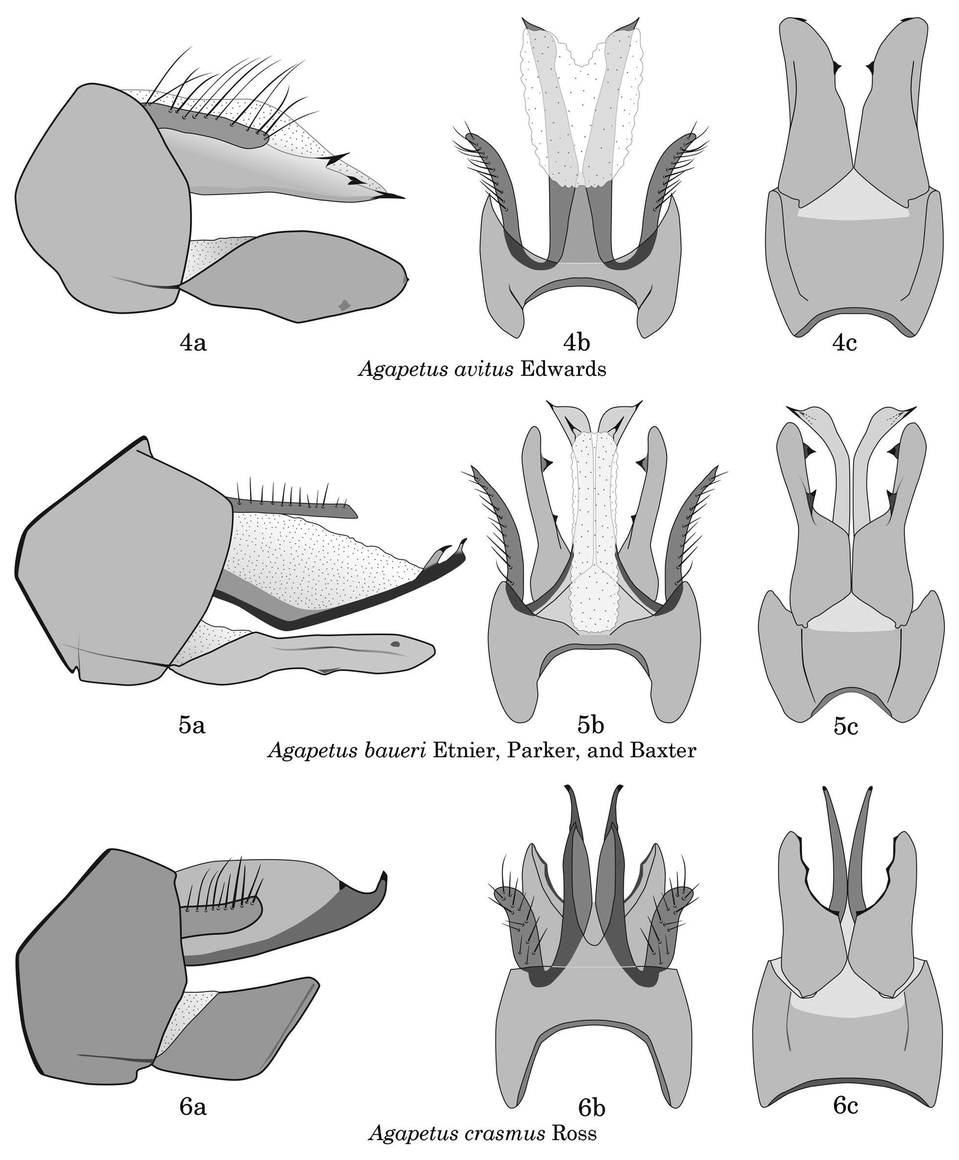
Figure 4-6. Agapetus spp. male genitalia. 4) Agapetus avitus Edwards. 4a, lateral view; 4b, dorsal view, inferior appendages not shown; 4c, ventral view of IX and inferior appendages. 5) Agapetus baueri Etnier, Parker, and Baxter. 5a, lateral view; 5b, dorsal view; 5c, ventral view, preanal appendages not shown. 6) Agapetus crasmus Ross. 6a, lateral view; 6b, dorsal view; 6c, ventral view, preanal appendages not shown.

Figure 7-9. Agapetus spp. male genitalia. 7) Agapetus diacanthus Edwards. 7a, lateral view; 7b, dorsal view; 7c, ventral view of IX and inferior appendages. 8) Agapetus flinti Parker, Etnier, and Baxter. 8a, lateral view; 8b, dorsal view; 8c, ventral view, segment X not shown. 9) Agapetus gelbae Ross. 9a, lateral view; 9b, dorsal view; 9c,

Figure 16-18. Agapetus spp. male genitalia. 16) Agapetus jocassee Morse. 16a, lateral view; 16b, dorsal view; 16c, ventral view of IX and inferior appendages. 17) Agapetus kirchneri Parker, Etnier, and Baxter. 17a, lateral view; 17b, dorsal view; 17c, ventral view of IX and inferior appendages. 18) Agapetus medicus Ross. 18a, lateral view; 18b, dorsal view; 18c, ventral view of IX and inferior appendages.
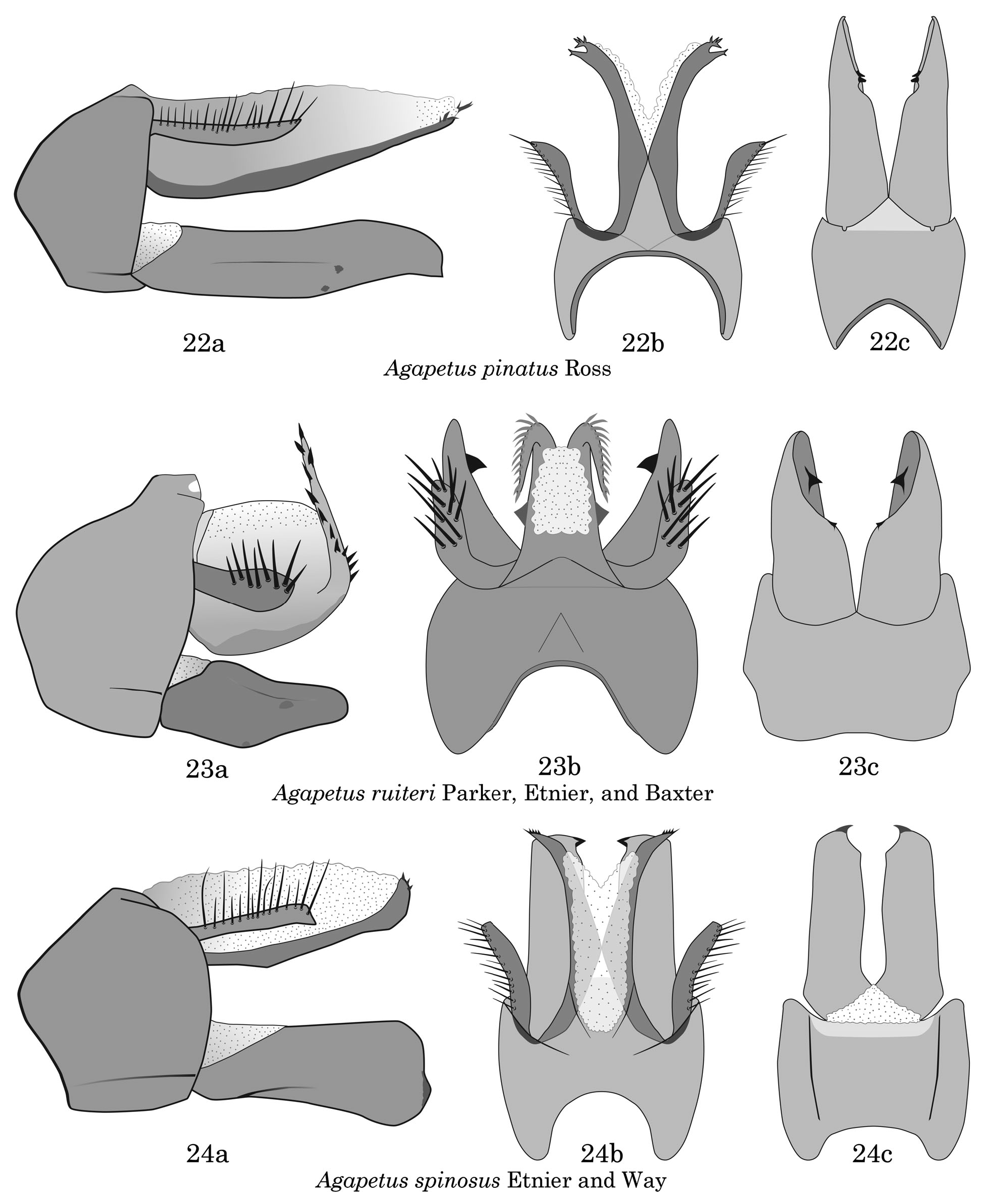
Figure 22-24. Agapetus spp. male genitalia. 22) Agapetus pinatus Ross. 22a, lateral view; 22b, dorsal view; inferior appendages not shown; 22c, ventral view of IX and inferior appendages. 23) Agapetus ruiteri Parker, Etnier, and Baxter. 23a, lateral view; 23b, dorsal view; 23c, ventral view of IX and inferior appendages. 24). Agapetus spinosusEtnier and Way. 24a, lateralview; 24b, dorsal view; 24c, ventral view of IX and inferior appendages.
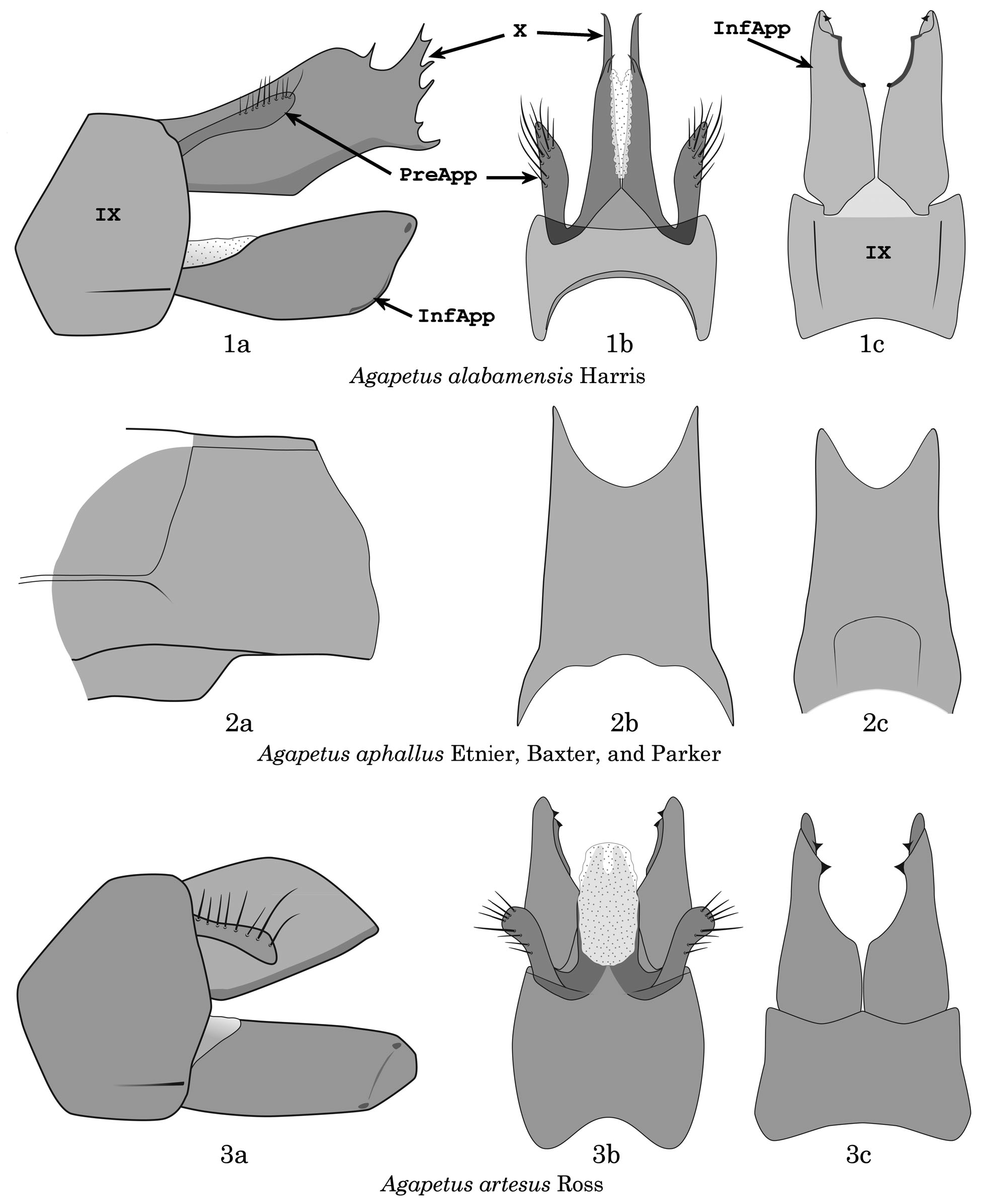
Figure 1-3. Agapetus spp. genitalia. 1) Agapetus alabamensis Harris, male genitalia. 1a, lateral view; 1b, dorsal view, inferior appendages and ventral portion of X not shown; 1c, ventral view of IX and inferior appendages. InfApp = inferior appendage; PrApp = preanal appendage; IX = segment IX; X = segment X. 2) Agapetus aphallus Etnier, Baxter, and Parker, female genitalia. 2a, lateral view; 2b, dorsal view; 2c, ventral view. 3) Agapetus artesus Ross, male genitalia. 3a, lateral view; 3b, dorsal view; 3c, ventral view of IX and inferior appendages.

Figure 19-21. Agapetus spp. male genitalia. 19) Agapetus meridionalis Etnier, Parker, and Baxter. 19a, lateral view; 19b, dorsal view, inferior appendages not shown; 19c, ventral view of IX and inferior appendages. 20) Agapetus minutus Sibley. 20a, lateral view; 20b, dorsal view; 20c, ventral view of IX and inferior appendages. 21) Agapetus pegram Etnier, Baxter and Parker. 21a, lateral view; 21b, dorsal view; inferior appendages not shown; 21c, ventral view of IX and inferior appendages.

Figure 25-27. Agapetus spp. male genitalia. 25) Agapetus stylifer Etnier, Baxter, and Parker. 25a, lateral view; 25b, dorsal view; 25c, ventral view, preanal appendages not shown. 26) Agapetus tomus Ross. 26a, lateral view; 26b, dorsal view; 26c, ventral view of IX and inferior appendages. 27) Agapetus tricornutus Etnier, Parker, and Baxter. 27a, lateral view; 27b, dorsal view, inferior appendages not shown; 27c, ventral view of IX and inferior appendages.

Figure 10-12. Agapetus spp. male genitalia. 10) Agapetus harrisi Etnier, Parker, and Baxter. 10a, lateral view; 10b, dorsal view; 10c, ventral view of IX and inferior appendages. 11) Agapetus hesperus Etnier, Baxter, and Parker. 11a, lateral view; 11b, dorsal view; 11c, ventral view, preanal appendages not shown. 12) Agapetus hessi Leonard and Leonard. 12a, lateral view; 12b, dorsal view; 12c, ventral view of IX and inferior appendages.
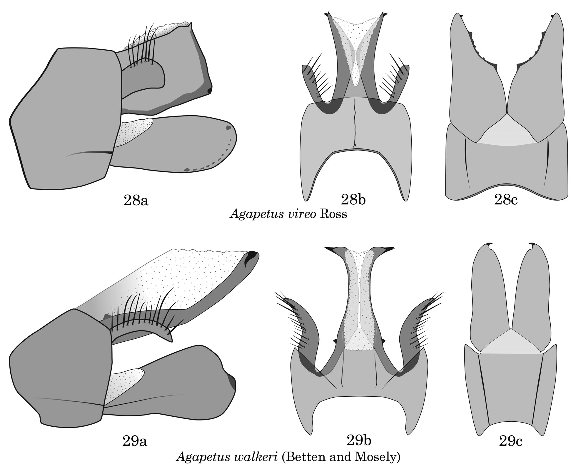
Figure 28-29. Agapetus spp. male genitalia. 28) Agapetus vireo Ross. 28a, lateral view; 28b, dorsal view, inferior appendages not shown; 28c, ventral view of IX and inferior appendages. 29) Agapetus walkeri (Betten and Mosely). 29a, lateral view; 29b, dorsal view; inferior appendages not shown; 29c, ventral view of IX and inferior appendages.
No known copyright restrictions apply. See Agosti, D., Egloff, W., 2009. Taxonomic information exchange and copyright: the Plazi approach. BMC Research Notes 2009, 2:53 for further explanation.
|
Kingdom |
|
|
Phylum |
|
|
Class |
|
|
Order |
|
|
Family |
|
|
Genus |