
Rhinagrion mima (Karsch)
|
publication ID |
https://doi.org/10.11646/zootaxa.3852.5.4 |
|
publication LSID |
lsid:zoobank.org:pub:F6CEDE9B-C6F5-40A2-BDDF-E02BC23422AF |
|
DOI |
https://doi.org/10.5281/zenodo.6136848 |
|
persistent identifier |
https://treatment.plazi.org/id/039B87CD-5E50-B91B-FF0E-3FE3FBCE19FE |
|
treatment provided by |
Plazi |
|
scientific name |
Rhinagrion mima (Karsch) |
| status |
|
Rhinagrion mima (Karsch) View in CoL
Figs. 1–5
Amphilestes mima Karsch 1891 : Entomol. Nachrichten. 17: 242–243. Rhinagrion mima: Calvert 1913 View in CoL . Proc. Acad. Nat. Sci. Phila. 65: 258.
Material. One ♀ F0 larva: THAILAND: Phetchabun Province, Nam Nao National Park, creek, 16° 44’ N, 101° 34’ E, 277 m, 3 May 2004, L-658, A. Vitheepradit leg. Deposited in Colección Entomológica del Instituto de Ecología, A.C., Xalapa ( IEXA).
Description. Larva small ( 12 mm total length including caudal lamellae), body slender, coloration mostly yellowish brown (Fig. 1), abdomen light brown, caudal lamellae dark gray.
Head: Wider than long, mostly yellowish-brown with small, pale spots on frons and vertex (Fig. 1a); labrum length 0.4 mm, its distal half covered with minute, sparse, dark spiniform setae, basal half glabrous, anterior margin flattened ventrally, with long, stiff, yellow setae; clypeus, frons and vertex glabrous, with three pale ocelli. Antennae 7-segmented (Fig. 1c), largely glabrous, scape short and thick, with basal half pale and apical half light brown, pedicel enlarged and thick, antennomeres 3–7 slender, scape and antennomeres 3–5 light brown with apices pale, 6–7 pale, size proportions: 0.4, 1.0, 1.0, 1.0, 0.7, 0.4, 0.3. Compound eyes large, strongly protruding laterally (Figs. 1a–c), with a row of 12–14 spines of different sizes on inferior margin, 6–7 of them strong, large and incurved (Fig. 1c). Cephalic lobes large (Figs. 1a–b), rounded, bulging, with a lateral, longitudinal, pale band and abundant spine-like tubercles each ending in a small, stiff seta, occipital margin widely concave. Mandibles (Fig. 2) with ventral margin of lateral surface with spine-like tubercles each ending in a small, stiff seta, with molar crest well developed, as a fleshy lobe on right mandible (Fig. 2a), sclerotized on left mandible (Fig. 2b), and with formula: L 12345 0 a (m1,2,3) b / R 1’1234 y a b, with a> b in both mandibles. Maxillae: Galeolacinia (Fig. 3) with seven moderately incurved teeth, three dorsal teeth long, approximately same length and robustness, three ventral teeth very short, apical tooth largest. Ventral pad of hypopharynx pentagonal (Fig. 4), whitish, with fleshy appearance, posterior margin concave, anterolateral corners with row of long, stiff, whitish setae. Labium: Prementum-postmentum articulation reaching posterior margin of procoxae (Fig. 1b). Prementum subpentagonal (Fig. 1b–c), 0.14 longer than its widest part, lateral margins almost straight and strongly divergent distally (Fig. 1b), with short spines on basal half and stout, long, incurved, sharp spines directed apically on distal half (Fig. 1c), each spine accompanied by a short, stiff seta; ligula large (Fig. 1c), prominent, three times as wide as long, with a PLATE I, FIGURES 1A–D. Rhinagrion mima , female F0 larva. a) Dorsal habitus; b) Ventral view; c) Details of head showing prementum and spines on inferior margin of compound eye, ventro-lateral view; d) Abdominal segments 7–10 and caudal lamellae, left lateral view.
deep, closed, median cleft, its apical margin slightly retroflexed and finely serrate; one long palpal seta (Fig. 5a), labial palp with row of small setae on basal half of dorsal margin, mesal margin very finely serrate, apical lobe ending in three teeth (Fig. 5b), ventral tooth triangular, the shortest, median tooth sharp and incurved, the longest, dorsal tooth bladelike half length of median tooth; ventral margin of ventral tooth, dorsal margin of medial tooth, and both margins of dorsal tooth very finely serrulate; movable hook long, sharp and strongly incurved, shorter than palp.
PLATE II, FIGURES 2–5. Detail of mouthparts of R. mima larva. 2) Mandibles: a, right mandible, dorso-basal view; b, right mandible, ventro-internal view, teeth 3–4 detached (broken); c, left mandible, internal view. 3) Right maxilla, ventral view; 4) Hypopharynx, ventral view. 5) Labial palp: a, dorsointernal view; b, external (frontal) view.
Thorax: Narrower than head (Fig. 1a); inferior border of propleura, lateral margins of pronotum and anterolateral surface of pronotal disc with row of short, stout, sharp spines, each spine with associated short, stiff seta; remainder of pronotum glabrous; anterior and posterior margins of pronotum convex, lateral margins triangular. Synthorax mostly glabrous, with minute, bristle-like setae more or less regularly spaced; ventral margins of meso- and metapleura with row of small spines; mesepisternum with large dorsal, oval, dark spot on each side of midline (Fig. 1a). Legs very long (Figs. 1a–b) with hind leg surpassing posterior margin of caudal lamellae; anterodorsal margin of procoxae with row of short, stout, blunt tubercles; femora light brown with three transverse, narrow, pale rings more or less regularly distributed (Fig. 1a), dorsal, lateral and ventral carinae serrate, each serration ending in a minute, bristle-like seta, the largest serrations on dorsal carina ending in scale-like, fan-shaped seta, remaining surface with sparse, minute, bristle-like setae; tibiae with color similar to that of femora, ventromesal carinae finely serrate, distal margins with row of strong spines, remaining surface with long, delicate setae; tarsi light brown, with basal, narrow, pale ring, with two ventral rows of short, stout setae; claws simple with pulvilliform empodium. Wing pads pale (Fig. 1a), glabrous, anterior and posterior wing pads reaching posterior margin of abdominal segments 4 and basal half of 5, respectively.
Abdomen: Cylindrical, slightly narrowing caudally (Figs. 1a–b), maximum width on S2, S10 narrowest. Tergites 1–4 pale yellow on basal half, grayish on middle third of distal half, tergites 5–9 light brown on basal half, grayish on distal half, tergite 10 yellowish brown, tergites 5–10 with a middorsal, longitudinal, pale stripe, which has in turn a thin, dark line along tergites 8–9 (Fig. 1a); tergites 1–10 covered with sparse, short and minute bristlelike, reddish setae mainly on lateral margins, lateral margins of S8–9 with row of stout spines (Fig. 1d); posterior margins of tergites 1–9 smooth, row of small spines on 10. Sternites yellow (Fig. 1b), with more or less abundant, small, bristle-like setae. Female gonapophyses very long (Figs. 1b, d), surpassing posterior margin of sternite 10 by half the length of valvae (Fig. 1d), dorsal and ventral margins of external valvae, in lateral view, strongly convergent on basal 0.65 and with row of small, reddish, bristle-like setae, distal 0.35 enlarged, smooth, digitiform, roundly pointed, internal valvae smooth, little longer than external valvae. Caudal lamellae broad (Fig. 1d), apically truncated, largely dark gray, with dendritic tracheation hardly visible, median lamella (epiproct) slightly shorter and paler than lateral lamellae (paraprocts); paraprocts slightly inflated in basal half (Figs. 1a–b), with three pale spots along apical margin (Fig. 1d) and some pale dots on dorsal and ventral margins, dorsal and ventral margins tending to be infolded and beset with numerous white, delicate, short setae; lateral, longitudinal carina on basal 0.75 the length of paraproct beset with abundant, stout, spiniform setae intermingled with stiff setae on basal 0.50 (Figs. 1b, d), remainder of carina with only stiff setae; epiproct much less pigmented than paraprocts, uniformly pale gray, with margins smooth, not inflated at basal half, tracheation quite visible as in R. viridatum ( cf. Lieftinck 1956, fig. 65).
Measurements: Total length (incl. app.) 18.1; abdomen 6.5; maximum width of head 4.2; hind femur 4.4. Caudal lamellae: Lateral (paraproct) 4.7, median (epiproct) 4.3.
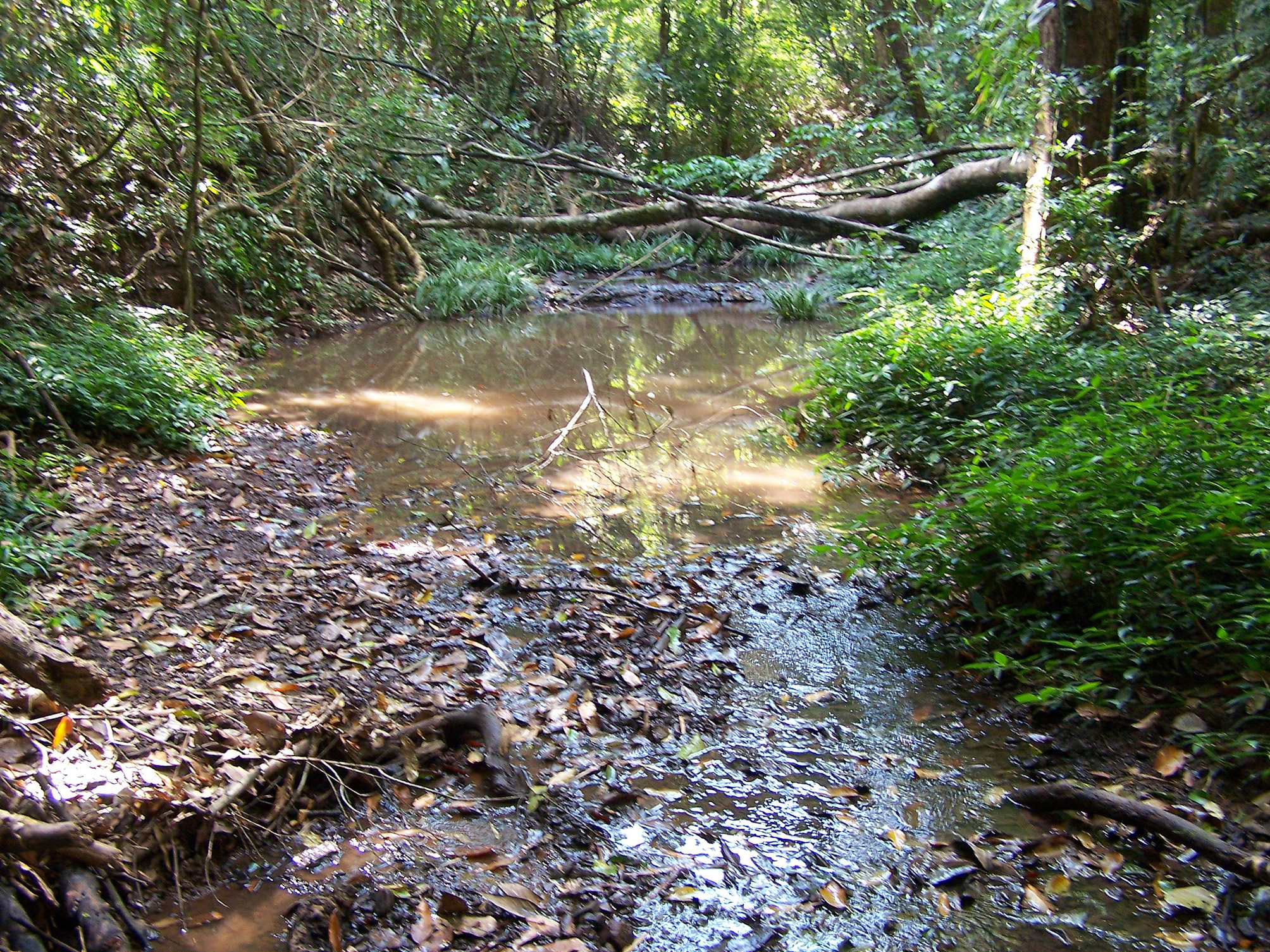
Habitat: The larva of R. mima was collected in a pool at the margin of a small creek ( Fig. 6 View FIGURE 6 ). Judging by the stage of development of the larva and the water level in the creek, the emergence period probably takes place at the end of the dry season. Lieftinck (1956) mentioned that larvae of Rhinagrion “live under varied conditions … one as genuine rheobiont ( R. tricolor [Krüger]), whereas R. borneense (Selys) does in shady forest brooks with a slow current”. Orr (2005) recorded larvae of R. mima “among accumulated leaf trash”.
No known copyright restrictions apply. See Agosti, D., Egloff, W., 2009. Taxonomic information exchange and copyright: the Plazi approach. BMC Research Notes 2009, 2:53 for further explanation.
|
Kingdom |
|
|
Phylum |
|
|
Class |
|
|
Order |
|
|
Family |
|
|
Genus |
Rhinagrion mima (Karsch)
| Novelo-Gutiérrez, Rodolfo, Sites, Robert W. & Vitheepradit, Akekawat 2014 |
Rhinagrion mima :
| Calvert 1913 |
Amphilestes mima
| Karsch 1891 |