
Gnathia grutterae, Ferreira, Maryke L., Smit, Nico J & Davies, Angela J, 2010
|
publication ID |
https://doi.org/ 10.5281/zenodo.199792 |
|
DOI |
https://doi.org/10.5281/zenodo.3507542 |
|
persistent identifier |
https://treatment.plazi.org/id/039B8794-6977-FFEC-5FE8-FF07771C6FB1 |
|
treatment provided by |
Plazi |
|
scientific name |
Gnathia grutterae |
| status |
sp. nov. |
Gnathia grutterae View in CoL sp. nov.
Material examined. Holotype. Male, 3.1 mm, off Lizard Island (14º40’54.68’’S, 145º26’53.72’’E), Australia, Museum of Tropical Queensland, Townsville, Australia (W31162). Paratypes. 9 males, 6 females and 9 third stage pranizae, off Lizard Island (14º40’54.68’’S, 145º26’53.72’’E), Australia, Museum of Tropical Queensland, Townsville, Australia (W31163).
Type host. Rhinecanthus aculeatus (Linnaeus, 1758) . Other hosts see Table 1.
Diagnosis. Males with large, bulbous eyes and slightly produced frontal border; superior fronto-lateral process conical, directed anteriorly and inferior mediofrontal processss shallow, conical notch dividing anterior part of mediofrontal process in two. Mandibles long, with dentate blade and armed carina; mandibles also with prominent internal lobe with small tubercles forming two rows from the lobe up to half the length of the mandible.
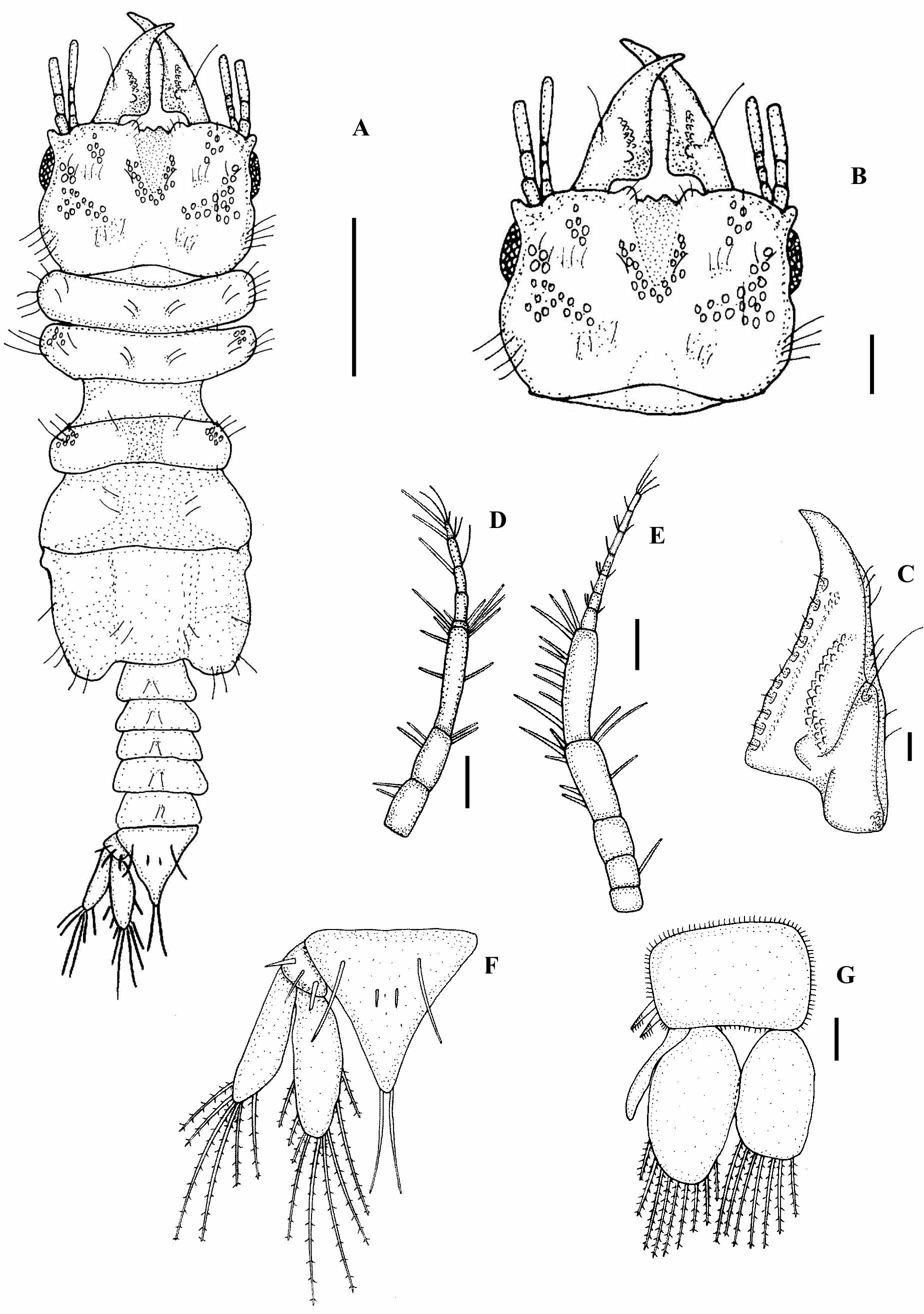
Male description ( Figs 1 View FIGURE 1 A–G, 2 A–C, 3, 6A–C, 7A).
Size: Total length of holotype: 3.1 mm. Total length of paratypes: 2.2–3.9 mm (3.1± 0.5 mm, n=9).
Cephalosome rectangular, 1.3 times as wide as long, wide and deep dorsal sulcus, almost same width as median inferior processes, third length of cephalosome, lateral margins straight ( Figs. 1 View FIGURE 1 A, B; 6A), posterior margin concave. Pappose setae, tubercles and short simple setae ventrally on lateral sides of buccal opening. Eye third the length of cephalosome. Cephalosome paraocular ornamentation with twelve to sixteen paraocular tubercles and five long pappose setae. Elliptical posterior median tubercle present ( Fig. 1 View FIGURE 1 B).
Frontal border slightly produced, superior fronto-lateral processes conical, directed anteriorly ( Figs 1 View FIGURE 1 A, B; 6B), 4 simple setae in a row on each process. Mediofrontal process inferior, shallow conical notch dividing anterior part of mediofrontal process in two. Lamina dentata not dorsally visible. External scissura shallow. Supraocular lobe not pronounced with single tubercle dorsally visible.
Pereon 2.5 times as long as wide, just as wide a cephalosome ( Fig. 1 View FIGURE 1 A) covered with numerous long setae, short simple hair-like setae and tubercles. Pereonites 2 and 3 of similar size, widest part of body, lateral margins pointing anteriorly. Long simple setae on dorsal sides of pereonites 2 and 3. Pereonite 4 with prominent anterior constriction separating it from pereonite 3. Tubercles as well as long simple setae on anterior lateral lobe of pereonite 4, median groove present. Pereonite 5 with areae laterales. Pereonite 6 posterior margin concave with lobi laterales. Pereonite 7 dorsally visible, small with rounded posterior margin, overlapping first pleonite. Long pappose setae on anterior, lateral and posterior margins of pereonites. Pectinated scales randomly distributed on pereonites 4 to 6.
Antenna 1 first article with two simple setae, second article with four simple setae distally and third article with two simple setae mid-dorsally and five simple setae dorsally. Flagellum articles 3 and 4 with one aesthetasc seta each, article 5 terminating in one aesthetasc and three simple setae ( Fig. 1 View FIGURE 1 D).
Antenna 2 article 2 with single simple seta, article 4 with three simple setae present mid-dorsally and five simple setae distally, article 5 with nine simple setae laterally. Flagellum article 7 terminating in four simple setae ( Fig. 1 View FIGURE 1 E).
Mandible two thirds length of cephalosome, 3 times as long as wide, narrow basal neck with three to five tubercles, curved inwards with eleven processes on dentate blade, cuticular extensions on processes ( Figs 1 View FIGURE 1 C; 6C). Apex cylindrical, distally raised in lateral view. Prominent incisor present. Single mandibular simple seta extending from middle of incisor process. Carina not armed. Sensory pits and small simple setae distributed from first to last process. Internal lobe on blade with small tubercles forming two rows from the lobe up to half the length of the mandible ( Fig. 1 View FIGURE 1 C).
Maxilliped proximal article largest with broad mediodistal endite not reaching article 3 ( Fig. 2 View FIGURE 2 A). Distal four articles bearing plumose setae on lateral margins in order 3-7-5-5, mesial border setose ( Fig. 2 View FIGURE 2 A). Distal article with three stout simple setae. Palp 1.7 times as long as wide. No coupling hooks.
Pylopod convex mesial border fringed with 29 plumose setae ( Fig. 2 View FIGURE 2 B), three simple setae present proximally. Six simple setae distributed over dorsal surface. Six simple setae present distally on posterior surface ( Fig. 2 View FIGURE 2 C). Second article oval shaped, 1.2 times as long as wide, margins densely setose, seven simple setae distally on posterior surface ( Fig. 2 View FIGURE 2 B, C), third article minute.
Pleon and pleotelson 1.8 times the total length of the pereon ( Fig. 1 View FIGURE 1 A). Five subequal pleonites dorsally visible, pappose setae on pleonites.
Pleotelson wider than long, lateral margins slightly concave, dorsal surface with two pairs of simple setae, pectinate scales on dorsal surface, distal apex terminating in pair of simple setae ( Fig. 1 View FIGURE 1 F).
Pereopod 2 ( Fig. 3 View FIGURE 3 ; P2) basis with five simple setae present posteriorly, four simple setae and three toothshaped tubercles anteriorly. Ischium with six simple setae posteriorly, four simple setae anteriorly. Merus with long simple setae and short single simple seta on bulbous protrusion, posterior margin with three simple setae and three tooth-shaped tubercles. Carpus with two simple setae and four tooth-shaped tubercles present posteriorly, and two simple setae anteriorly. Propodus with two robust serrate setae and two simple setae situated medially, robust seta on posterior side proximal to unguis, and two simple setae anteriorly. Dactylus terminating in sharp posterior-pointing unguis. Pereopods 3 to 6 similar in basic shape to pereopod 2 ( Fig. 3 View FIGURE 3 ; P3 to P6), but differing in setation as well as distribution of tubercles.
Pleopod endopod with nine and exopod with seven long pappose setae distally. Short simple setae on lateral and posterior margins of sympodite. Sympodite with retinaculae on medial margin. All pleopods similar, except pleopod 2 with appendix masculina on endopod, appendix masculina 1.6 length of rami ( Fig. 1 View FIGURE 1 G).
Uropod rami extending beyond apex of pleotelson, both with long pappose setae, ( Fig. 1 View FIGURE 1 F). Endopod with six plumose setae distally, three plumose setae laterally. Exopod with six plumose setae distally. Three simple setae on uropodal basis.
Penis prominent with two contiguous papillae, wider than long.
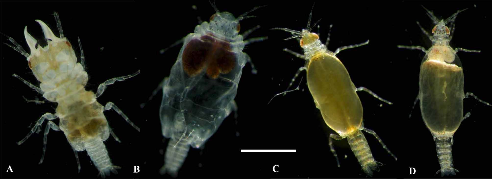
Externally, live male specimens of G. grutterae have distinct yellow-brown pigmentation on the cephalosome, stretching between eyes, and extending backwards to pereonite 6 ( Fig. 7 View FIGURE 7 A).
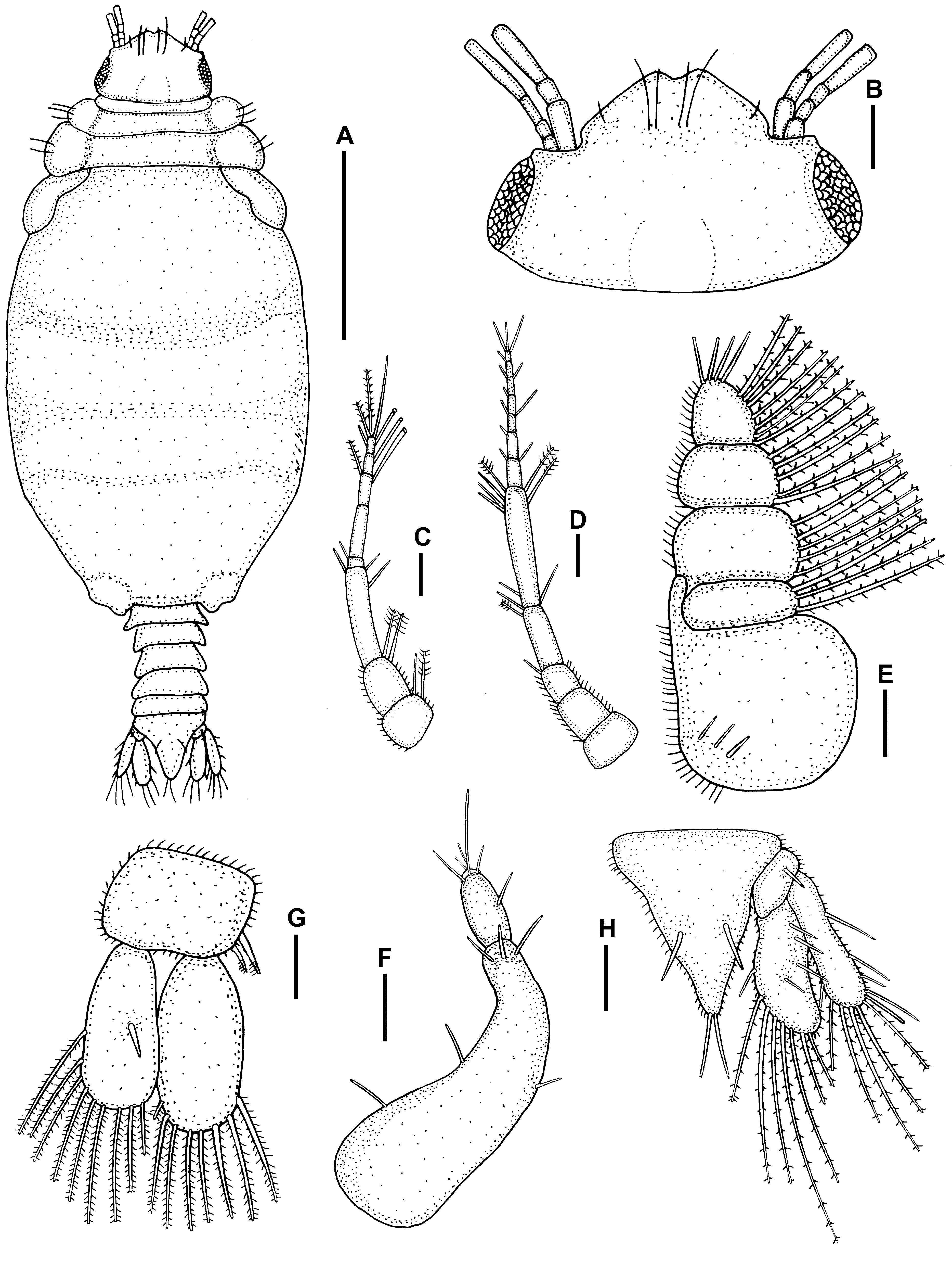
Adult female description ( Figs 4 View FIGURE 4 A–H, 7B).
Size: Total length of paratypes: 2.1–3.2 mm (2.7± 0.4 mm, n=6).
Cephalosome broad and rectangular, 1.6 times as wide as long, posterior margin straight ( Figs. 4 View FIGURE 4 A, B). Eyes 0.4 times the length of cephalosome. Elliptical posterior median tubercle present.
Frontal border broadly rounded, produced, slightly concave anteriorly, two pairs of long simple setae on mid-dorsal area ( Fig. 4 View FIGURE 4 A, B).
Pereon swollen, rounded, sutures between pereonites 5–7; 1.6 times as long as wide, wider than cephalosome ( Fig. 4 View FIGURE 4 A), Pereonite 7 dorsally visible, small with rounded posterior margin, overlapping first pleonite. Majority of setae on anterior and lateral margins of pereonites.
Antenna 1 article 1 with single simple and plumose seta on lateral margin, article 2 with two plumose setae, article 3 with three simple setae distally. Flagellum articles with aesthetasc setae similar to those of male, article 5 terminating in two plumose and two simple setae ( Fig. 4 View FIGURE 4 C). Antenna 2 article 3 with single simple seta dorsally, article 5 with three simple setae proximally and three plumose setae distally. Flagellum article 7 terminating in three simple setae ( Fig. 4 View FIGURE 4 D).
Maxilliped base and palp of four articles. Endite reaching article 2 of palp. Palp bearing plumose setae on lateral margins in order of 3-5-5-7 ( Fig. 4 View FIGURE 4 E). Coxa with attached oostegite just as broad as long.
Pylopod article 1 slender, curved anteriorly three simple setae distally. Article 2 with three simple setae distally ( Fig. 4 View FIGURE 4 F). Oval-shaped oostegite, covering mouthparts ventrally, not surpassing frontal border.
Pleon and pleotelson 2.6 times length of pereon total length ( Fig. 4 View FIGURE 4 A). Epimera present, short hair-like setae and pectinate scales randomly on pleonites.
Pleotelson longer than wide, lateral margins slightly convex, dorsal surface with pair of simple seta and pectinated scales, distal apex terminating in pair of simple setae ( Fig. 4 View FIGURE 4 H).
Pereopods similar in shape to male pereopods, but differing in setation and number of tubercles. Coxae of pereopods 4 to 6 with thin plate-like oostegites, enclosing brood pouch, oostegites overlapping.
Pleopod endopod and exopod both fringed distally with eight to nine pappose setae respectively ( Fig. 4 View FIGURE 4 G). Sympodite without retinacula, two plumose setae on lateral margin, Pleopods 2 to 5 similar to pleopod 1.
Uropod endopod with seven long pappose setae distally and four simple setae laterally. Exopod with six long pappose setae distally and five simple setae laterally. Uropodal basis with single simple seta ( Fig. 4 View FIGURE 4 H). Externally, live female specimens of G. grutterae have distinct black markings/spots on the cephalosome ( Fig. 7 View FIGURE 7 B).
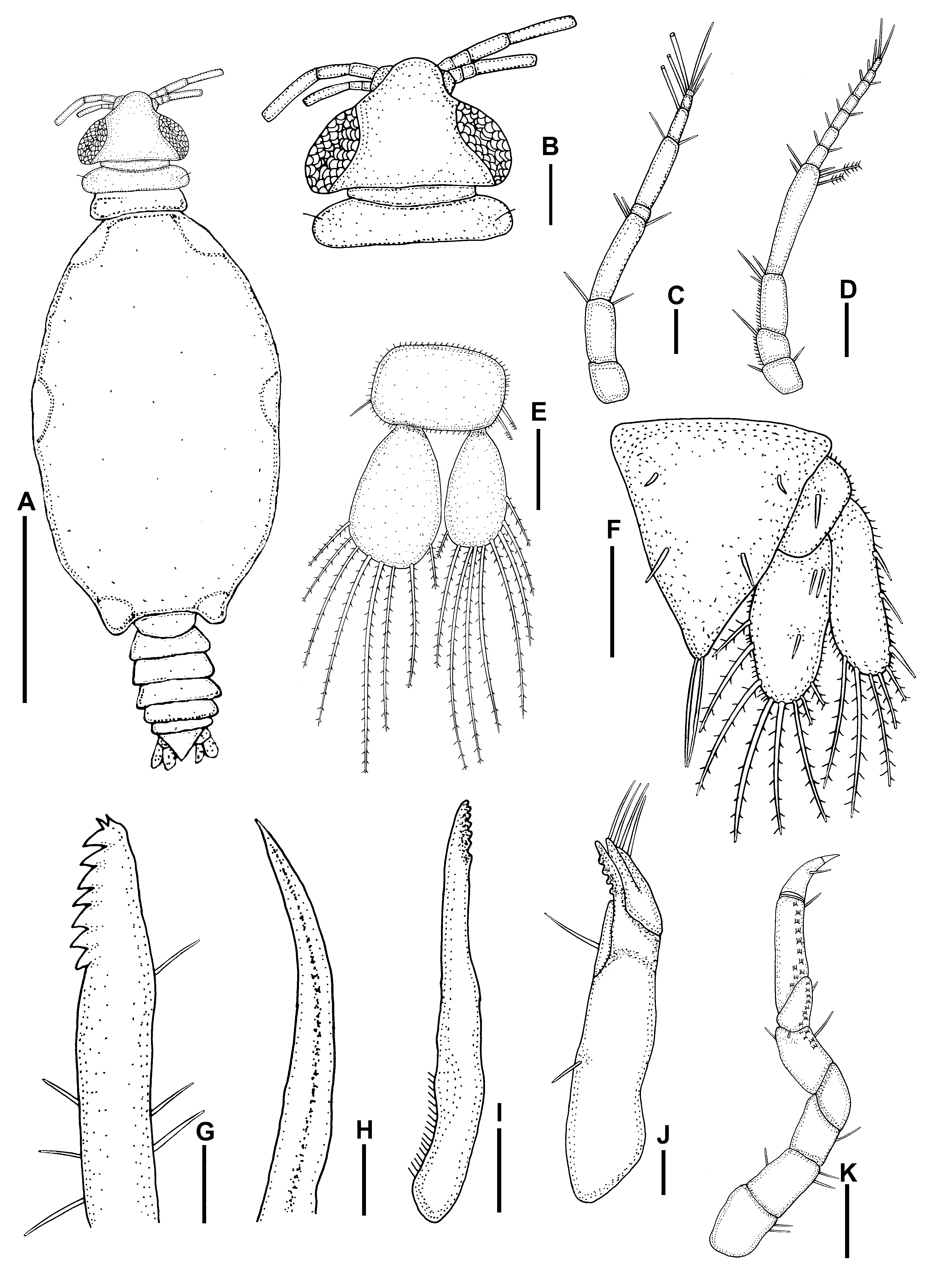
Third stage praniza description ( Figs 5 View FIGURE 5 A–K; 7C-D).
Size: Total length of paratypes: 2–3.2 mm (2.3± 0.3 mm, n=9).
Cephalosome posterior margin straight, wider than anterior margin, 1.8 times as wide as long lateral margins slightly concave ( Fig. 5 View FIGURE 5 A, B). Many sensory pits on dorsal surface of cephalosome. Eyes welldeveloped, oval-shaped, bulbous, on lateral margins of cephalosome, 1.4 times the length of the cephalosome. No sulcusses or tubercles on dorsal cephalosome. Medio-anterior margin of cephalosome straight with lateral concave excavations to accommodate first articles of antennae.
Labrum prominent, quarter the length of cephalosome, conical with apical process, truncated posterior margin, anterior margin concave ( Fig. 5 View FIGURE 5 B). Ventral part of labrum with central groove covering mandibles dorsally and laterally.
Pereon 1.9 times as long as wide, wider than cephalosome. Pereonite 2 with anterior constriction separating it medianly from pereonite 1. Pereonite 4 lateral sides tapering towards rounded posterior margin, posterior margin stretching over pereonite 5, lateral shields at leg attachment. Pereonite 5 consisting of elastic membrane fully expanded in praniza stage with blood meal, bulbous shields present on lateral sides at leg attachment. Pereonite 6 rectangular, posterior margin slightly concave, lateral shields at leg attachment. Pereonite 7 dorsally visible, small with rounded posterior margin, overlapping first pleonite ( Fig. 5 View FIGURE 5 A).
Antenna 1 article 2 with two simple setae distally and article 3 largest with three simple setae distally. Flagellum articles with aesthetasc setae similar to male, article 5 terminating in three simple setae ( Fig. 5 View FIGURE 5 C). Antenna 2 flagellum article 7 terminating in three to four simple setae, few setae on distal end of each article ( Fig. 5 View FIGURE 5 D).
Mandible stout, swollen at base, distal margin styliform with two small teeth on tip and seven large teeth on mesial margin, triangular and backwardly directed ( Fig. 5 View FIGURE 5 G). Six simple setae distributed randomly on surface.
Paragnaths elongated, terminate in sharp point, no teeth ( Fig. 5 View FIGURE 5 H).
Maxillule long, slender ( Fig. 5 View FIGURE 5 I), swollen base, stretching past distal margin of labrum. Six to seven small teeth on distal inner margin.
Maxilliped large, cylindrical, elongated base with short hair-like setae laterally, endite almost reaching palp with single long simple seta. ( Fig. 5 View FIGURE 5 J). Palp with three articles, first article acute with four to five small teeth, article three with four setae ( Fig. 5 View FIGURE 5 J).
Gnathopod smaller than pereopods, seven articles, few simple setae and pectinate scales distributed randomly on some articles ( Fig. 5 View FIGURE 5 K). Dactylus strongly hooked.
Pleon and pleotelson 1.3 times the length of pereon ( Fig. 5 View FIGURE 5 A). Single simple seta on posterior, lateral, side of each pleonite.
Pleopod endopod with eight plumose setae and exopod with nine plumose setae ( Fig. 5 View FIGURE 5 E). Sympodite with retinacula, single simple seta on lateral margin. Pleopods 2 to 5 similar to pleopod 1.
Pleotelson longer than wide, anterior half of lateral margins slightly concave, posterior half straight, setae similar to those of male ( Fig. 5 View FIGURE 5 F).
Uropod endopod with eight pappose setae, single simple seta at posterior end and one pair of simple setae on distal end. Exopod with five pappose setae ( Fig. 5 View FIGURE 5 F). Uropodal basis with single simple seta.
Pereopods similar in shape to male pereopods, but differing in setation and numbers of tubercles.
Externally, live praniza 3 specimens show differential pigmentation on dorsal surfaces of cephalosome of different sexes. Male pranizae with yellow pigment covering whole of cephalosome, yellow pigmentation on pleonites, and few black spots on pereonite 4 ( Fig. 7 View FIGURE 7 C). Female pranizae with brown spots on cephalosome, pereonites 3, 4, 6 and on pleonites ( Fig. 7 View FIGURE 7 D). Both sexes with yellow eyes.
Etymology. The species is named in honour of Alexandra Grutter, The University of Queensland, for her valuable contribution to gnathiid isopod and cleaner symbiosis research.
Remarks. Gnathia grutterae juveniles were collected at the same locality and on the same teleost hosts as those of Gnathia aureamaculosa (see Ferreira et al. 2009). However, the males of these two species, both moulted from the juveniles, differ in that G. aureamaculosa males have a slightly produced frontal border with a conical superior fronto-lateral process, mandibles with six processes on the dentate blade, a pseudoblade and a dentate internal lobe ( Ferreira et al. 2009). The two species also differ in the setation order of the distal most article of the maxillipedal palp, this being 3-7-5-6 for G. aureamaculosa (see Ferreira et al. 2009) and 3-7-5-5 for G. g r u t t e r a e. Gnathia grutterae males also differ from two other species, Gnathia grandilaris and Gnathia trimaculata described recently from Lizard Island elasmobranchs ( Coetzee et al. 2008; Coetzee et al. 2009). Gnathia grandilaris males (7.4 mm) and G. trimaculata (4.5 mm) are both larger than those of G. grutterae (3.1 mm). The frontal border and mandible morphology also differs in these species (see Coetzee et al. 2008; Coetzee et al. 2009).
Holdich & Harrison (1980) described five Gnathia spp. from Queensland littoral and shallow water habitats. Compared to these species, the male of G. grutterae is smaller (2.2–3.9 mm) than that of Gnathia biorbis Holdich & Harrison, 1980 (4.9 mm), but falls within the same size range as Gnathia calmani Holdich & Harrison, 1980 (2.6 mm), Gnathia falcipenis Holdich & Harrison, 1980 (3.54 mm), Gnathia cornuta Holdich & Harrison, 1980 (3.04 mm) and Gnathia meticola Holdich & Harrison, 1980 (2.7 mm). However, these species differ from G. grutterae in that males of G. calmani have inferior frontolateral processes that are acute, and half the length of the superior frontolateral process, G. falcipenis males have a quadrate cephalosome, mediofrontal processes that are conical and the same length as the superior frontolaterals, while males of G. c o r n u t a have cephalosomes that are quadrate, with a broad acute mediofrontal process. In G. meticola the superior fronto-lateral process is smooth, rounded and setose, and the inferior frontal border is straight and crenulated.
When compared to gnathiids species worldwide, similarities exist between G. grutterae and the South African species Gnathia nkulu Smit & Van As, 2000 ( Smit & Van As 2000) . The South African species’ frontal border is similar to that of G. grutterae , it is slightly produced, and frontolateral processes are conical with 3 long simple setae on these processes, the mediofrontal process is inferior and has a small concave median notch. However, these two species can be distinguished by mandible morphology, with G. grutterae males having eleven processes on the dentate blade and an internal lobe laterally on the blade, whereas G. nkulu males have seven to nine processes with pits on the dorsal and ventral sides of the mandible. Similarities also exist between G. grutterae males and males of the West Indian Ocean species from Aldabra, Gnathia glauca Kensley, Schotte & Poore, 2009 (see Kensley et al. 2009). Both species have distinct yellow/redbrown pigmentation present dorsally on their cephalosomes. Gnathia grutterae and G. glauca males also fall within the same size range ( G. glauca , 3.2 mm), both have a median notch, their superior frontolateral processes are conical and both have distinct paraocular ornamentation present (either as tubercles found in G. grutterae or granules in G. glauca ). The mandible of these two species however differ, whereas G. grutterae has a dentate blade, with an internal lobe laterally, G. glauca has a crenulated blade with a small structure present laterally (present on drawing, not mentioned in text). There are also differences in pleotelson setal numbers, the exopod of G. grutterae has 6 pappose setae, and the endopod 9, whereas the G. glauca exopod has 16 pappose setae and endopod 6.
Few Australian gnathiid females have been described in detail, thus G. g r u t t e r a e females can be compared only with those of G. aureamaculosa and G. trimaculata . Females of G. grutterae measure 2.7 mm, their frontal borders are broadly rounded, indented and produced, with two pairs of long simple setae on sides of mid-dorsal area. Gnathia aureamaculosa females are smaller (1.5 mm) and have a frontal border with 2 simple setae on the dorsal area near the bulbous eyes, two pairs of simple setae on the sides of mid-dorsal areas and a single pair of simple seta reaching median the tubercle ( Ferreira et al. 2009). Gnathia trimaculata females are bigger (4.1 mm) than G. grutterae females and have a slightly anteriorly concave frontal border with a pair of simple and pappose setae on sides of mid-dorsal area, as well as four long pappose setae on middorsal region ( Coetzee et al. 2008).
Gnathia grutterae View in CoL and G. aureamaculosa pranizae were collected from the same fish hosts (see Ferreira et al. 2009) and share two morphological characteristics. Firstly, both juveniles have nine teeth on their mandibles, two small teeth on the tip and seven, large, triangular, backwardly directed teeth on the mesial margin. Secondly, both juveniles have six to seven teeth on the distal inner margin on the maxillule ( Ferreira et al. 2009). However, the two species differ in number of setae found on the pereon, as well as in pereon shape and in the colour of live specimens ( Ferreira et al. 2009). Gnathia grutterae View in CoL pranizae also differ from those of G. trimaculata View in CoL and G. grandilaris View in CoL in the number of mandibular teeth, with G. trimaculata View in CoL having 8 and G. grandilaris View in CoL 9 teeth on the mesial margin. These species also differ in the pigmentation found on live specimens.


No known copyright restrictions apply. See Agosti, D., Egloff, W., 2009. Taxonomic information exchange and copyright: the Plazi approach. BMC Research Notes 2009, 2:53 for further explanation.
|
Kingdom |
|
|
Phylum |
|
|
Class |
|
|
Order |
|
|
Family |
|
|
Genus |