
Diaptomus, Westwood, 1936
|
publication ID |
https://doi.org/ 10.1080/00222933.2012.708450 |
|
persistent identifier |
https://treatment.plazi.org/id/039A87D0-FFDD-4453-FEB8-FDCAFF72FE61 |
|
treatment provided by |
Felipe |
|
scientific name |
Diaptomus |
| status |
sp. nov. |
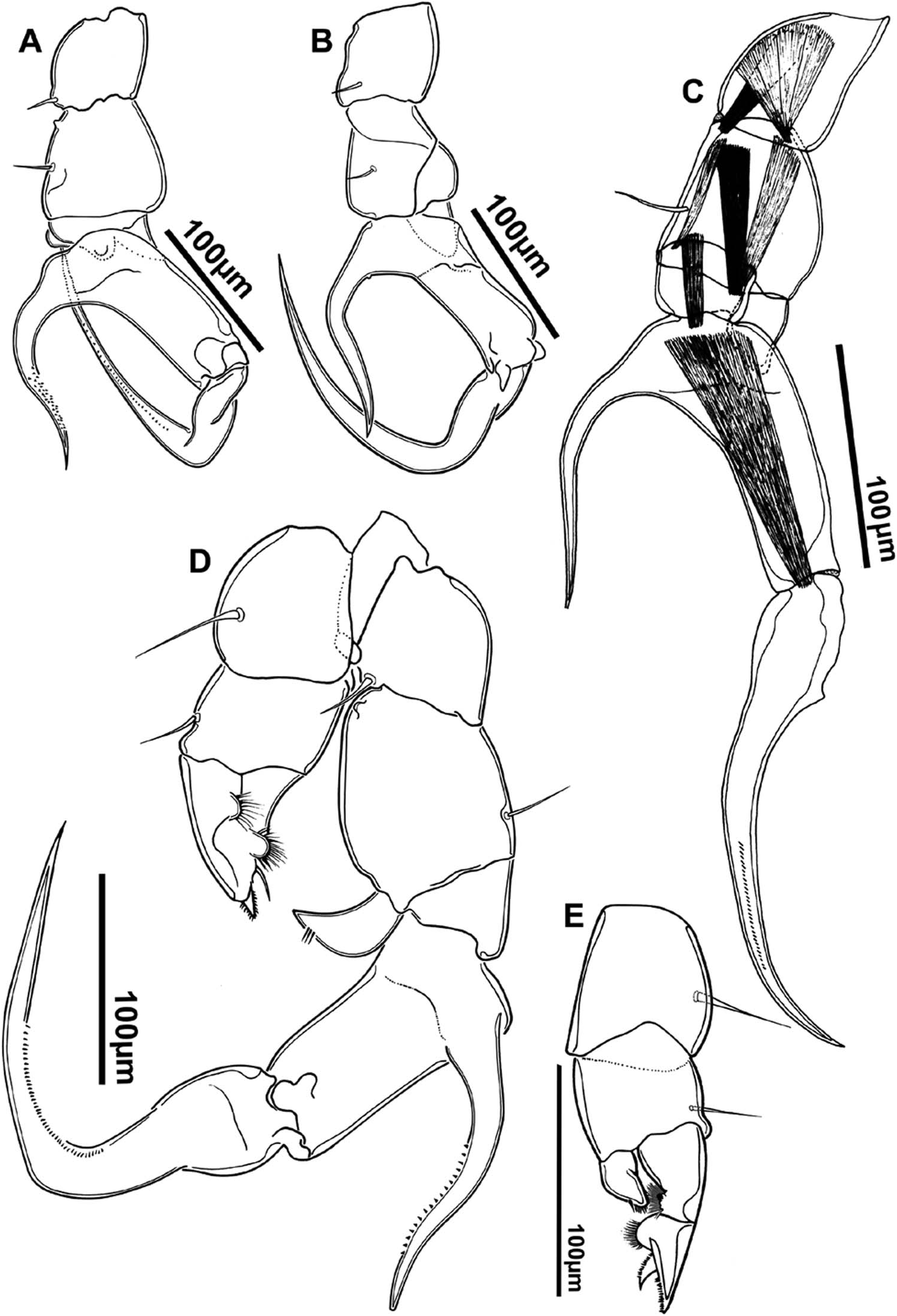
“ Diaptomus ” curvatus Perbiche-Neves, Boxshall and Paggi, sp. nov.
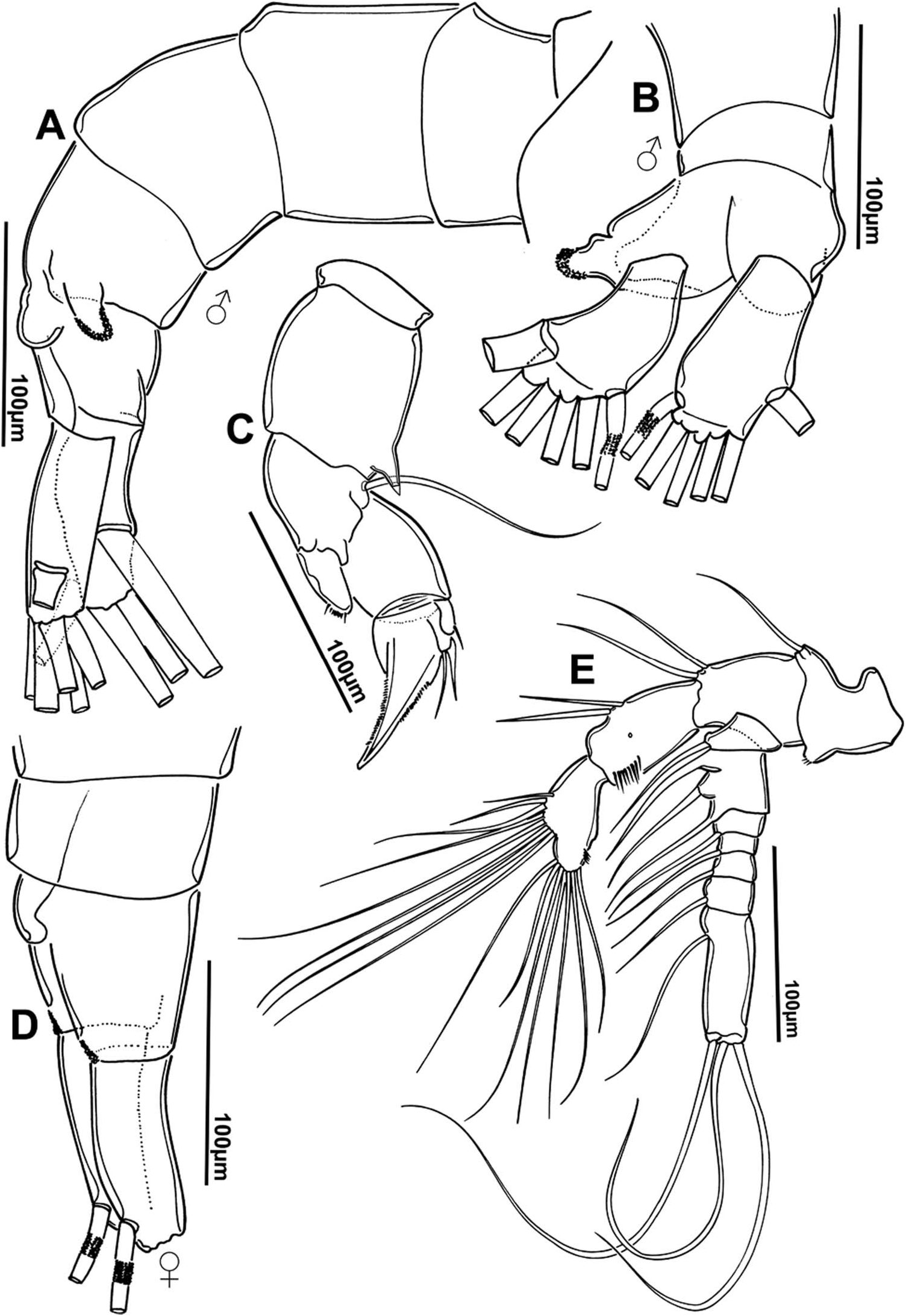
( Figures 1 View Figure 1 to 9)
Material examined
Holotype. One ♂, entire, alcohol + glycerine ( MZUSP 24476 View Materials ), Yacyretá Reservoir (27 ◦ 30 ′ 9.12 ′′ S; 56 ◦ 31 ′ 56.69 ′′ W), 15 km upstream from the dam, on border between Argentina / Paraguay. GoogleMaps
Paratypes. Two ♂ ( MZUSP 24478 View Materials ) , 1 ♀ ( MZUSP 24477 View Materials ) and 2 ♀ ( MZUSP 24479 View Materials ) entire, alcohol + glycerine; 5♂ and 5♀ ( NHM-UK 2011.1192 - 1201 ) ; 2♂ and 2♀ (1933 INPA). All collected from Yacyretá Reservoir, border between Argentina and Paraguay, 28 February 2010 and 5 June 2010 .
Etymology
The species name refers to the curvature in the strong lateral spine of the last exopod segment of the male right fifth leg.
Diagnosis
Male. Lacking ornamentation of spinules or denticles along sutures between prosome somites. Paired posterolateral wings on prosome subsymmetrical. Large subconical blunt dorsal process present on fourth urosomite, with chitinous protuberances on apex. Modified seta on segment 11 of right antennule of male longer than modified seta on segment 13. Segment 14 lacking process. Small processes on segments 15 and 16 of right antennule. Lateral hyaline lamella present on segment 20 of right antennule, with a small process distally. Small semicircular process present on proximal inner margin of basis of right leg 5. Endopod of right leg 5 well developed, triangular in outline. Lateral spine of fifth leg downwardly curved, strong and longer than the segment on which it is inserted.
Female. Lacking ornamentation of spinules or denticles on sutures between prosome somites. Posterolateral wings on prosome asymmetrical; left side of prosome better developed than right side; left prosome wing larger than right; apical sensilla on left side of genital double-somite carried on a semicircular process; no process on right side where lateral margin is smoothly convex.
Description – male
Prosome ( Figure 1A View Figure 1 ). Body length 923 µm, maximum width of 251 µm. Rostrum symmetrical with two filaments ( Figure 1C View Figure 1 ). Incomplete suture between pedigerous somites 4–5; lacking ornamentation of spinules or denticles on dorsal and lateral surfaces and along sutures. Posterolateral wings on prosome small and simple.
Urosome ( Figures 1A View Figure 1 ; 2A, B View Figure 2 ). Four-segmented, with large subconical blunt process dorsally on urosomite 4, with chitinous protuberances on margin of apex.
Caudal rami ( Figures 1A View Figure 1 ; 2A, B View Figure 2 ; 3A, B View Figure 3 ). Asymmetrical, right ramus longer than left; left ramus about two times longer than broad, right ramus 2.5 times longer than broad: inner margins of caudal rami with setules. Caudal rami bearing six setae with normal ornamentation.
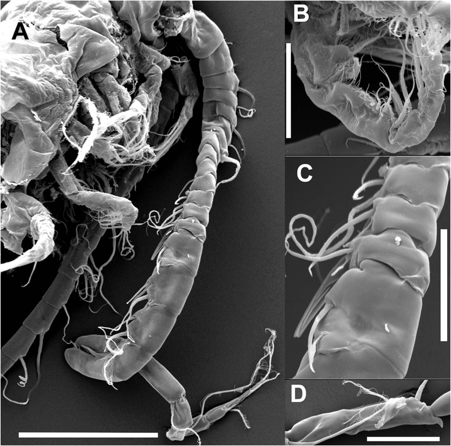
Right geniculate antennule ( Figures 4B View Figure 4 ; 5A, C, D View Figure 5 ). With 22 expressed segments, modified with enlarged segments in mid-section. Setal formula for setae (s), conical setae (cs), long setae (ls), modified setae (ms), vestigial setae (vs), aesthetascs (ae), and processes (p) on each segment as follows: (segment 1) 1s + 1ae, (2) 1cs + 1s + 1vs +1ae, (3) 1vs + 1ls + 1ae, (4) 1s, (5) 1s + 1vs + 1ae, (6) 1s, (7) 1s + 1ae, (8) 1s + 1cs, (9) 1s + 1ls + 1ae, (10) 1s + 1ms, (11) 1s + 1ms, (12) 1s + 1cs + 1ae, (13) 1s + 1ms + 1ae, (14) 1s + 1ls + 1ae, (15) 1ms + 1ls + 1p + 1ae, (16) 1s + 1ls + 1p + 1ae, (17) 1s + 1cs + 2ms, (18) 1ms + 1cs + 1s, (19) 2ms + 1s + 1ls + 1ae, (20) 2s + 2ls + 1p, (21) 1s + 1ls, (22) 3ls + 1s + 1ae. Spinous processes on segments 10 and 11 well developed, that on segment 11 longer than on segment 13, which has wide base. Process on segment 13 with minutely bifid apex ( Figure 5C View Figure 5 ). Segment 14 lacking process in contrast to segments 15 and 16, each with small process of similar length. Segment 20 with smooth hyaline membrane and small rounded tooth-like process distally, process sometimes acute ( Figure 5D View Figure 5 ).
Left antennule ( Figure 4A View Figure 4 ). With 25 expressed segments, number of setae (s), conical setae (cs), long setae (ls), vestigial setae (vs) and aesthetascs (ae) on each segment, as follows: (segment 1) 1s + 1ae, (2) 3s + 1ae +1vs, (3) 1ls + 1ae + 1vs, (4) 1s, (5) 1ls + 1vs + 1ae, (6) 1s, (7) 1ls + 1ae, (8) 1s + 1cs, (9) 1s + 1ls + 1ae, (10) 1s, (11) 1ls, (12) 1s + 1cs + 1ae, (13) 1s, (14) 1ls + 1ae, (15) 1ls, (16) 1ls + 1ae, (17) 1s, (18) 1ls, (19) 1s + 1ae, (20) 1s, (21) 1ls, (22) 1s + 1ls, (23) 1ls + 1s, (24) 2ls, (25) 4ls + 1ae.
Antenna ( Figure 2E View Figure 2 ). Biramous; coxa with one seta; basis with two setae. Endopod two-segmented: first segment with two setae at mid-level, and row of four or five spinules distally, with one pore next to row; compound second segment with 15 setae, eight on inner lobe and seven grouped around distal margin, plus spinule row (eight to ten spinules). Exopod seven-segmented, setation as follows: 1, 3, 1, 1, 1, 1, 4; segment 2 with ancestral segments partially or totally separated on one side only.
Mandible ( Figure 6C, D View Figure 6 ). Coxal gnathobase well sclerotized; cutting blade with subcaudal and triangular tooth and group of six multicusped teeth plus seta near toothed margin. Palp basis with four setae; endopod two-segmented with four and nine setae; exopod four-segmented with 1, 1, 1, 3 setae.
Maxillule ( Figure 6A View Figure 6 ). Coxal epipodite with nine setae. Coxal endite with four distal setae. Outer seta representing basal exite; proximal and distal basal endites each with four setae. Endopod two-segmented; with three setae on margin of proximal segment, and with five setae on distal. Exopod armed with six setae.
Maxilla ( Figure 6E View Figure 6 ). Proximal praecoxal endite with five setae and one spine (setules present on these setae but not illustrated here); distal praecoxal endite with three setae: proximal and distal coxal endites each with three setae; allobasis with three setae; free endopod with five setae in total.
Maxilliped ( Figures 5B View Figure 5 ; 6B View Figure 6 ). First syncoxal endite represented by one seta with row of spinules at base; second to fourth syncoxal endites with 2, 3, 3 setae from proximal to distal, distal angle of syncoxa extended into lobe with two rows of small spinules, pore present next to distal outer margin: basis with three setae and double row of setules proximally: endopod six-segmented, with 2, 3, 2, 2, 1 +1, 4 setae.
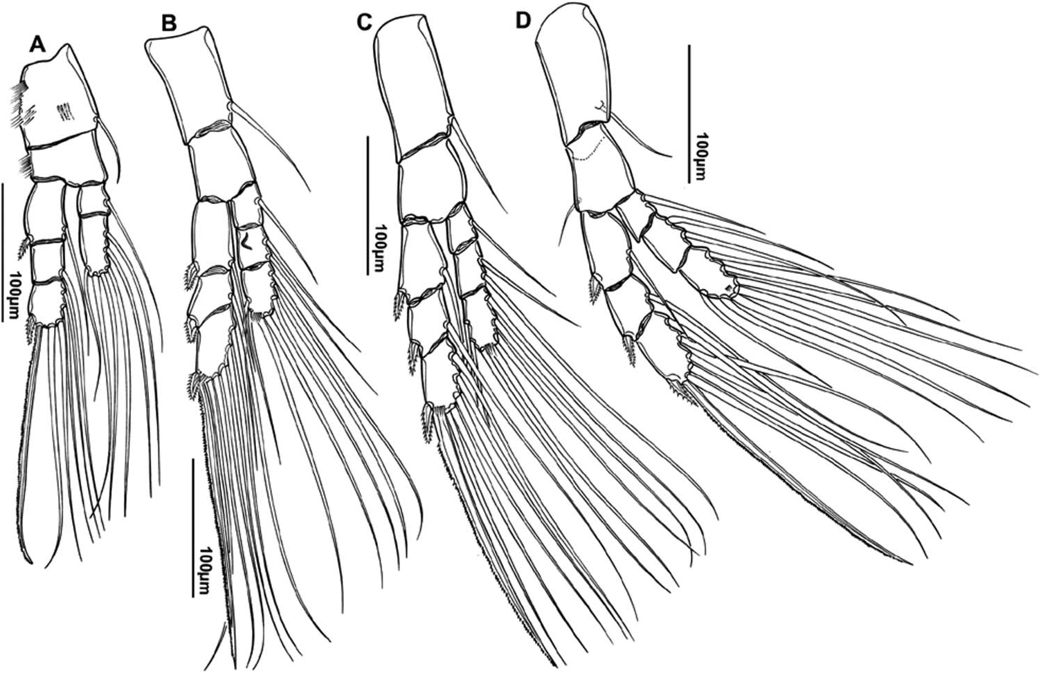
Swimming legs ( Figure 7 View Figure 7 ; Figure 9A View Figure 9 ). Exopods three-segmented; endopod threesegmented in legs 2–4 and two-segmented in leg 1; Schmeil’s organ present on second endopod segment of leg 2 ( Figure 7B View Figure 7 ). Row of setules present distally at base of terminal spine on endopod of each leg; setule rows present on basis of leg 1 ( Figure 9A View Figure 9 ). Spine and seta formula as in Table 1.
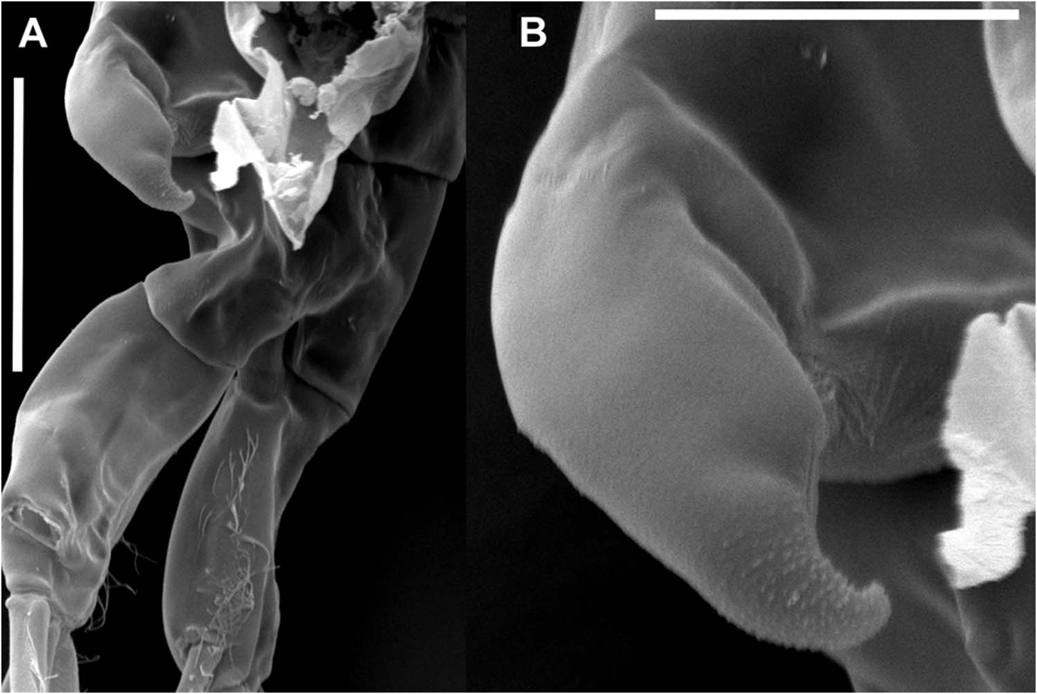
Right leg 5 ( Figures 8A–D View Figure 8 ; 9B–E View Figure 9 ). Coxa with inner distal process carrying seta at apex. Basis 1.3 times longer than wide; with small nodule-like process proximally on inner margin, and seta on posterior surface near distal margin. Endopod one-segmented subtriangular with curved sides with setules near apex, similar in length to width of last exopod segment ( Figure 9E View Figure 9 ). Exopod three-segmented; first segment wider than long; second segment twice as long as wide; lateral spine longer than segment, strongly curved, with tip reaching base of terminal claw. Terminal claw representing third exopod segment, 2.2 times longer than second segment. Row of small spinules present along inner concave margins of lateral spine and terminal claw.
Left leg 5 ( Figures 8D, E View Figure 8 ; 9B–D View Figure 9 ). Coxa about as long as wide, with seta located near outer margin. Basis 1.2 times wider than long, with small seta on outer margin. Endopod one-segmented, reaching midway along exopod, ornamented with row of small spinules apically. Exopod two-segmented; first segment with outer margin slightly curved; inner margin with rounded distal process bearing setules; semicircular process with setules present proximally on inner margin of second segment, armed with spinulate spine at middle of inner margin, and produced into distal spinous process ornamented with small spinules.
Description – female
Prosome ( Figure 1B View Figure 1 ). Body length 1120 µm, maximum width 261 µm. Body larger than male. Rostrum symmetrical, with paired rostral filaments ( Figure 1D View Figure 1 ). Cephalosome with complete dorsal suture; lacking ornamentation of spinules on dorsal and lateral surfaces. Posterolateral wings on prosome well developed, asymmetrical; right wing larger than left; both lateral wings with semicircular projection carrying spinous process on apex.
Urosome ( Figures 1B View Figure 1 ; 2D View Figure 2 ). Three-segmented; genital double-somite asymmetrical; right side smoothly convex with posteriorly directed sensilla located at widest point; left side with small semicircular process anteriorly bearing apical sensilla; left sensilla posteriorly directed. External genital area ventral: delimited anteriorly by broad symmetrical opercular pad, and laterally by posteriorly directed lateral processes. Paired gonoporal plates located adjacent to midline, between lateral processes. Urosomite 2 about twice as wide as long.
Caudal rami ( Figures 1B View Figure 1 ; 2D View Figure 2 ). About two times longer than broad; left ramus longer than right; setules present along outer and inner margins. Caudal rami bearing six setae with normal ornamentation.
Antennule. Symmetrical; extending beyond caudal rami but not as far as tips of caudal setae; setal formula similar to that of male left antennule.
Antenna, Mandible, Maxillule, Maxilla, Maxilliped, Swimming legs. Similar to male. Leg 5 ( Figure 2C View Figure 2 ). Symmetrical, coxa 1.2 times longer than wide, extended into spiniform process at outer distal corner, with conical sensilla at tip; basis triangular, with smoothly convex outer margin, bearing long seta reaching almost to tip of exopod. Exopod indistinctly three-segmented; first segment larger than second, unarmed; second segment with lateral spine and drawn out into large terminal claw ornamented with row of denticles laterally; offset third segment lobe-like and bearing two terminal setae, lateral seta smaller. Endopod one-segmented with discontinuity in cuticle; bearing two large setae on tip and ornamented with row of spinules between setae.


| INPA |
Instituto Nacional de Pesquisas da Amazonia |
No known copyright restrictions apply. See Agosti, D., Egloff, W., 2009. Taxonomic information exchange and copyright: the Plazi approach. BMC Research Notes 2009, 2:53 for further explanation.