
Helochares (Hydrobaticus) nipponicu, Hebauer, 1995
|
publication ID |
https://doi.org/ 10.5281/zenodo.4272324 |
|
DOI |
https://doi.org/10.5281/zenodo.4334962 |
|
persistent identifier |
https://treatment.plazi.org/id/039A87CB-FFC0-4936-FEBC-FBD4FD97EF12 |
|
treatment provided by |
Felipe |
|
scientific name |
Helochares (Hydrobaticus) nipponicu |
| status |
|
Helochares (Hydrobaticus) nipponicu s Hebauer, 1995
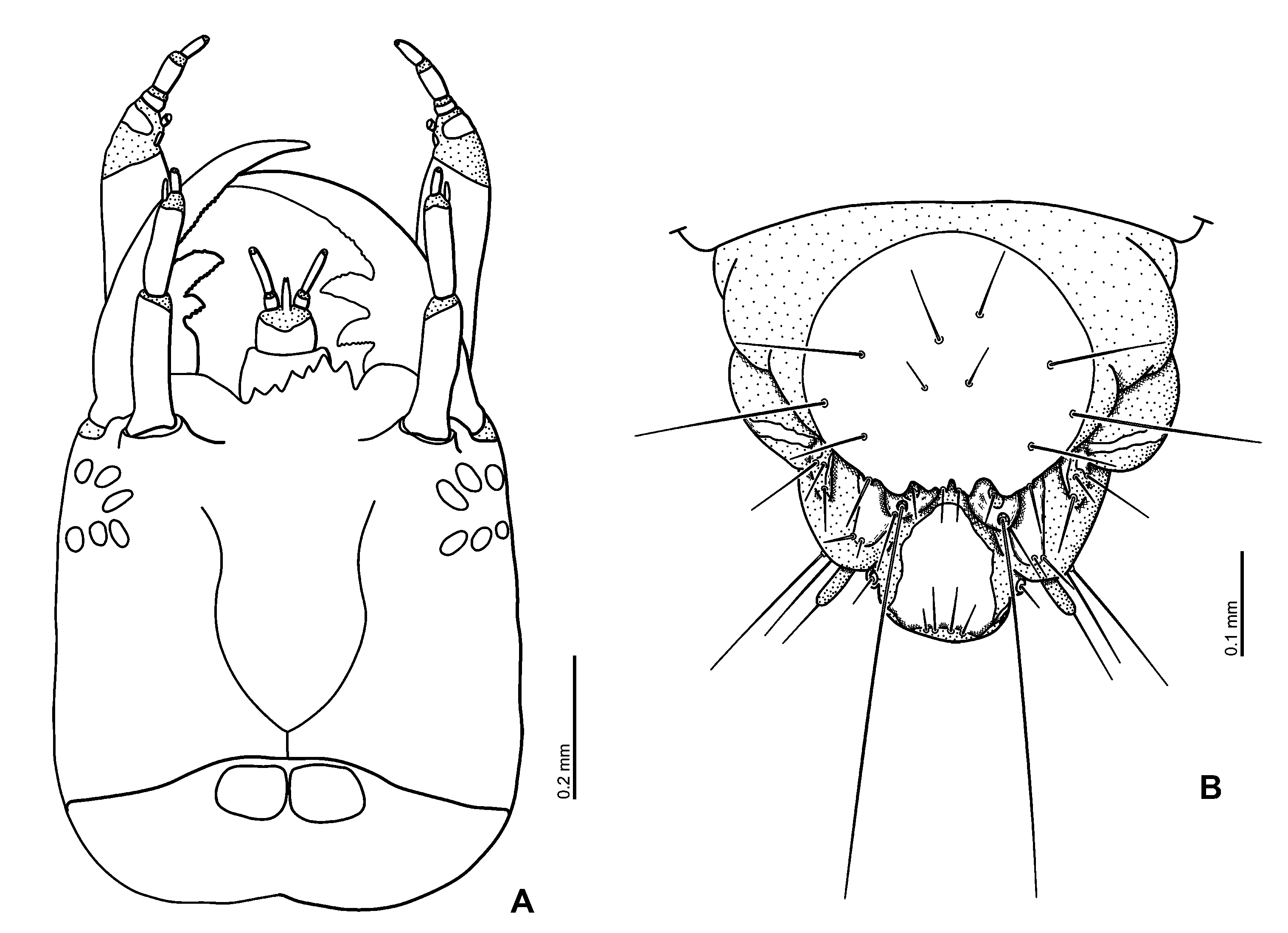
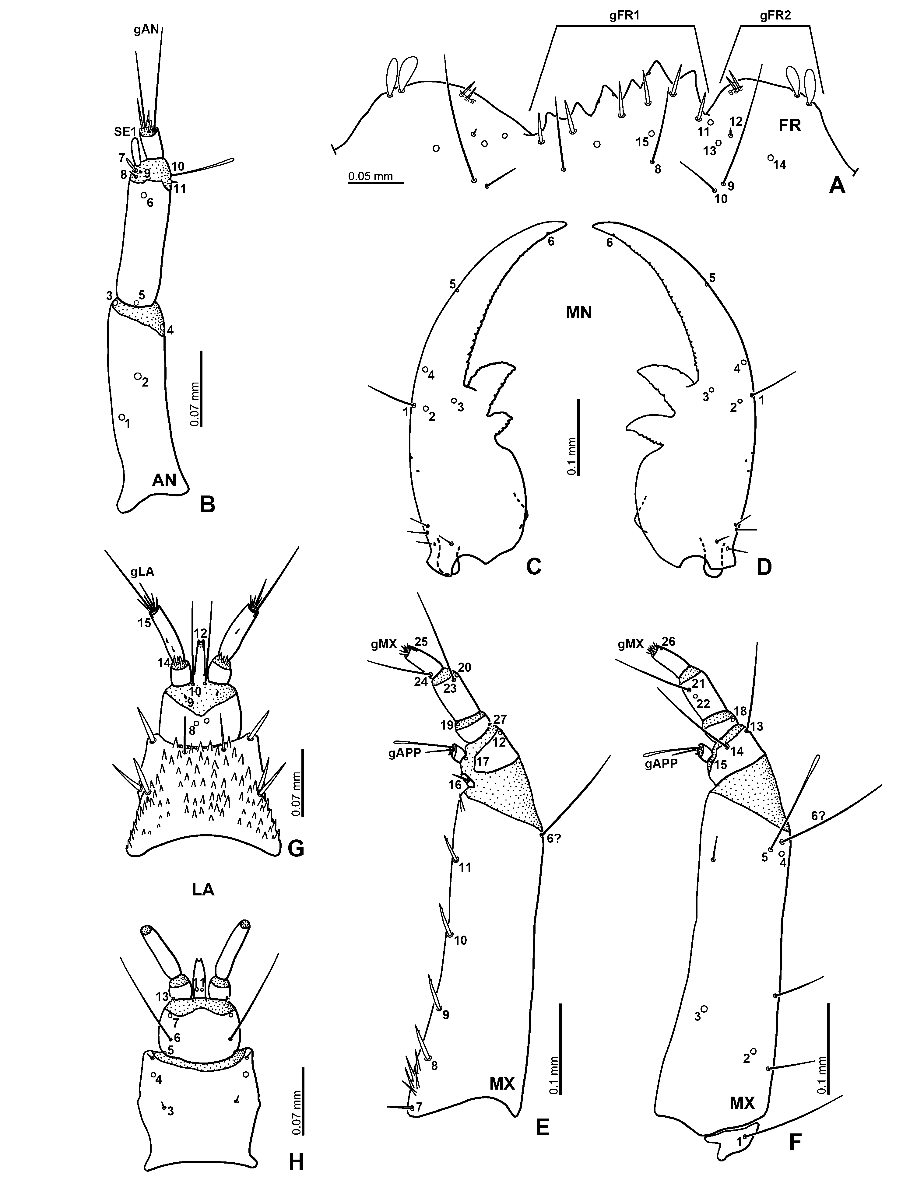
( Figs. 1F View Fig , 6B View Fig , 38–43 View Fig View Fig View Fig View Fig View Fig View Fig , 44C View Fig , 45D View Fig , 65C View Fig )
Material examined. JAPAN: HONSHÛ: Shimane-ken :5 L2, 6 L3, San-nôji, Daitô-chô, Un-nan-shi (pond), 17.vii.2008, MH ; 1 L3, Shakunouchi-kôen , Kisuki-chô, Un-nan-shi, 24.vi.2007, MH ; 1 L1, same locality, 17.vii.2008, MH ; 1 L1, 1 L2, 4 L3, same locality, 26.viii.2007, MH ; 1 L2, 1 L3, same locality, 23.ix.2007, MH ; 1 L2, same locality, 7.v.2008 (fixed), MH ; 76 L1, Wadakami , Oku-uga-chô, Izumo-shi (pond), 12.vii.2008 (egg cases carried by adults collected in the field), MH ; 13 L2, 6 L3, same locality, 12.vii.2008, MH ; 3 L1, 4 L2, 5 L3, same locality, 28.vii.2008, MH ; 2 L1, 4 L2, 1 L3, same locality, 19.viii.2008, MH .
General morphology. Third instar. Colour. Head and sclerotised parts light yellowish brown; membranous parts milky white ( Fig. 1F View Fig ).
Head. Nasale projecting slightly further than epistomal lobes ( Fig. 42A View Fig ). Epistomal lobes rounded, asymmetrical; right lobe projecting as far as left lobe.
Antenna short, rather slender ( Fig. 42B View Fig ). Scape longer than pedicel.
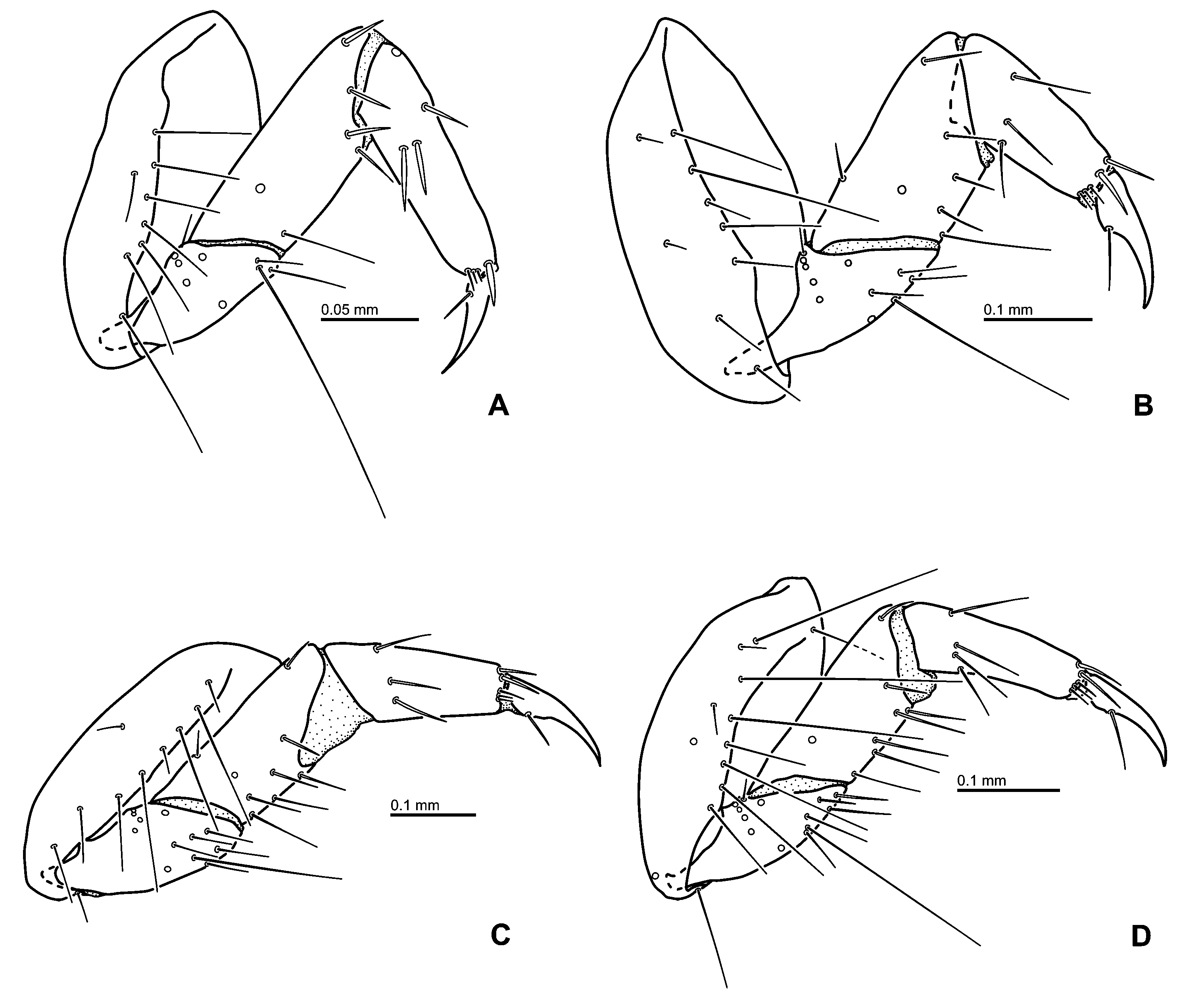
Mandibles: Inner teeth serrate ( Figs. 42 View Fig C–D); inner edge of mandibular apex serrate.
Maxilla ( Figs. 42 View Fig E–F): Maxillary palpomere 1 slightly shorter than palpomeres 3, palpomere 2 the shortest, palpomere 4 about as long as palpomere 1; palpomere 1 the widest.
Labium ( Figs. 42 View Fig G–H): Mentum bearing small, strong cuticular spines on dorsal surface. Labial palpi shorter than mentum, covered with narrow cuticular spines on dorsal surface of palpomere 2 and intersegmental membrane between palpomeres 1 and 2; palpomere 1 much shorter than palpomere 2.
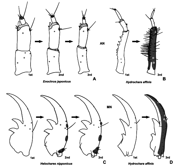
Abdomen. Segment 1 with four dorsal sclerites on each side; two on anteromedian part, anterior one small, posterior one larger than anterior one; remaining two sclerites very small, sometimes undetectable, situated behind former two sclerites, each bearing one rather long seta ( Fig. 6B View Fig ); segments 2 to 7 similar to segment 1 but with three dorsal sclerites on each side, one very small on anteromedian part, two hardly recognisable behind former sclerite.
Spiracular atrium ( Fig. 41B View Fig ): Segment 8 with large, oval dorsal plate; dorsal plate with four projections on posterior edge, each median projection weakly bifid, with two setae; each lateral part of dorsal plate with three long setae, median one longer than remaining ones; posterior part with two long and one short setae; procercus with one rather long stout seta and two rather long setae. Segment 9 trilobed; median lobe with two rather short and two long setae on posterior edge; each lateral lobe with two long setae on posterolateral surface.
Second instar. Similar to third instar, but more weakly sclerotised.
Head. Antenna: Scape about as long as pedicel ( Fig. 40B View Fig ).
Labium: Labial palpi about as long as mentum ( Figs. 40 View Fig G–H).
First instar. Similar to second instar, but more weakly sclerotised.
Head. Epistomal lobes weakly rounded, slightly asymmetrical ( Fig. 38C View Fig ).
Antenna proportionally short, rather stout ( Fig. 39A View Fig ). Scape distinctly shorter than pedicel.
Mandibles: Inner edge of basal tooth serrate ( Figs. 39 View Fig B–C).
Maxilla: Maxillary palpomere 1 slightly shorter than palpomeres 3 and 4 ( Figs. 39 View Fig D–E).
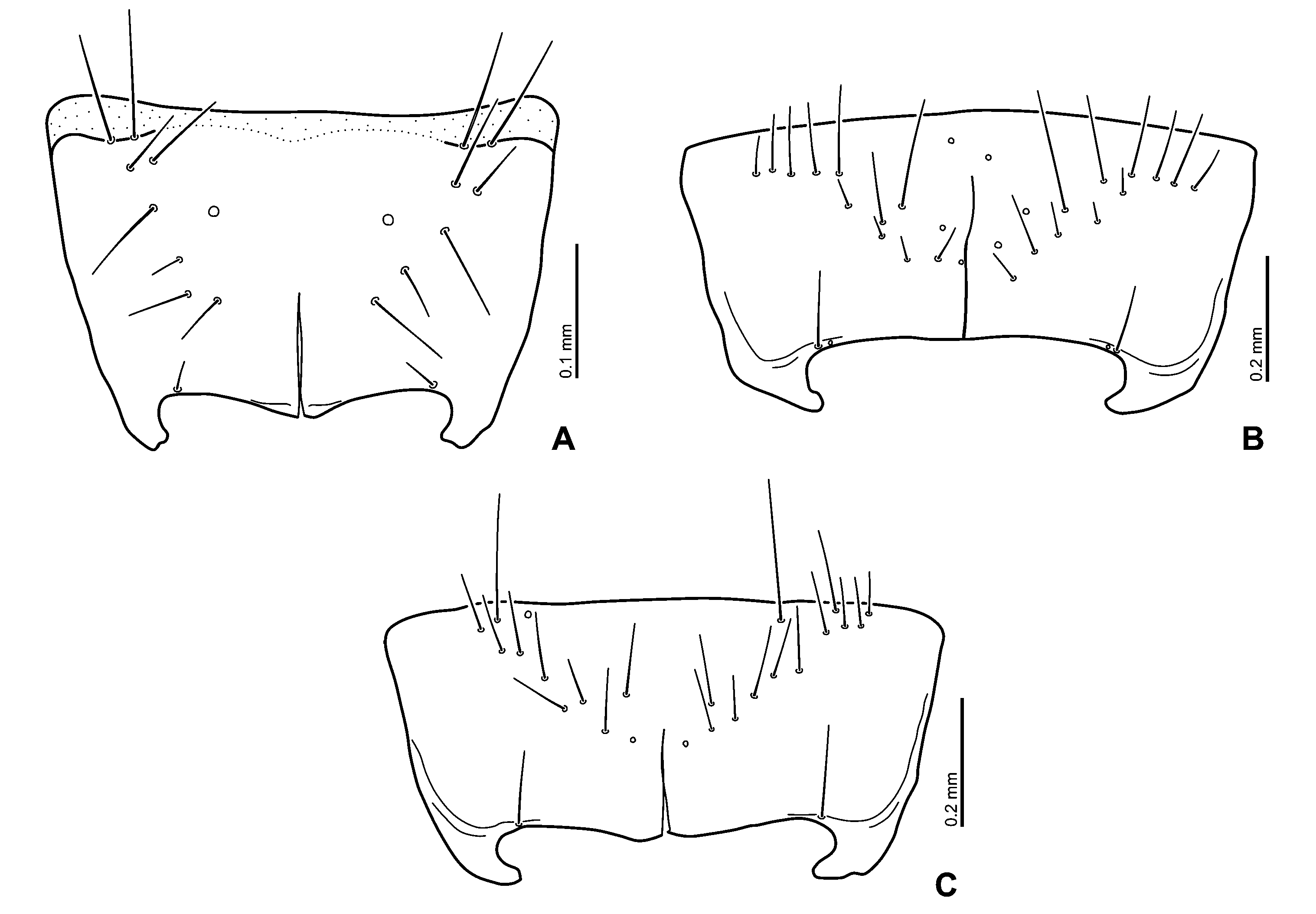
Thorax. Mesonotum with six long setae on each dorsal sclerite; metanotum with five long setae on each dorsal sclerite.
Abdomen. Segment 1 with three visible dorsal sclerites on each side; anterior one narrow, small; posterior two very small, weakly sclerotised, each with one long seta. Segments 2 to 7 similar to segment 1 but without anterior dorsal sclerites.
Spiracular atrium: Segment 8 with large, oval dorsal plate with four projections on posterior edge, each projection bifid at apex, looking like eight projections; procercus with one rather long stout seta and two rather long setae. Segment 9 trilobed; median lobe with two rather short and two moderately long setae on posterior edge of median lobe; each lateral lobe with two moderately long setae on posterior edge.
Primary chaetotaxy of head. Frontale altogether with 46 sensilla ( Figs. 38A, C View Fig ). FR 2 between FR 1 and FR 3. FR 5 short seta; FR 6 moderately long scale-like seta, close to FR 7; FR 7 moderately long, trichoid. Nasale with a group of six stout short setae and two very short setae (gFR1). Each epistomal lobe with two short setae on inner part and two short scale-like setae on mediolateral part (gFR2). FR 10 between FR 4 and FR 9; one pore-like sensillum ( FR 14) situated anteriorly to inner margin of antennal socket. Location of FR 11–13 asymmetrical. Right lobe with FR 11 situated anteriorly to FR 12 close to lateral seta on nasale, FR 12 between FR 11 and FR 13 but laterally of line connecting FR 11 and FR 13. Left lobe with FR 13 situated between FR 11 and FR 12, posteriorly to line connecting FR 11 and FR 12, FR 12 situated laterally of FR 11.
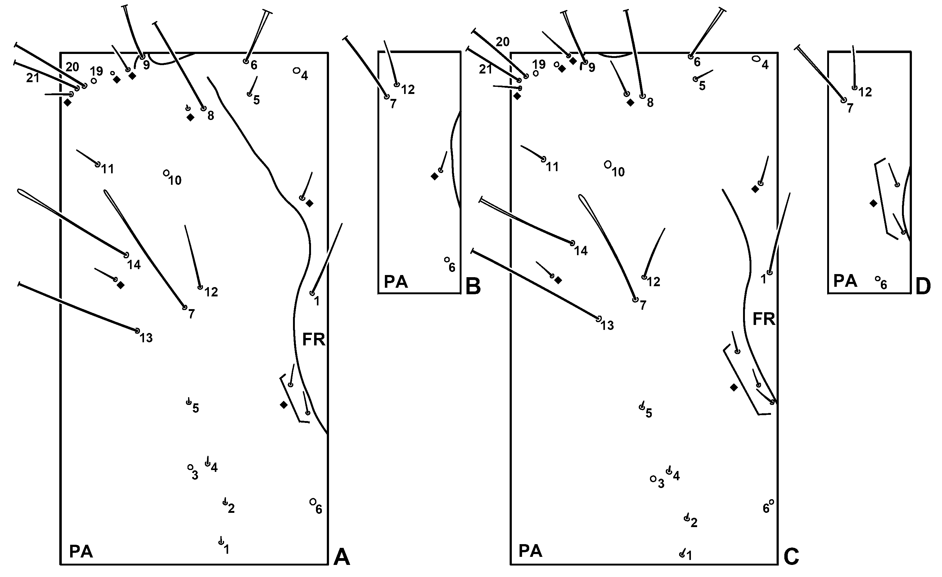
Parietale ( Figs. 38 View Fig A–B): PA 1–5 forming irregular row. PA 7 long scale-like seta; PA 12 rather long seta. PA 20 rather long seta. PA 17 close to PA 16, situated anteromesally to PA 16; PA 16 long, narrow scale-like seta. PA 26–28 in anterior 0.43 and lateral two-fifths of ventral surface of parietale. Two pore-like sensilla ( PA 29–30) located ventrally on about basal third of parietale.
Antenna ( Fig. 39A View Fig ): Antennomere 2 with one pore-like sensillum ( AN 6) situated dorsally close to distal margin of sclerite and five setae ( AN 7–11) on intersegmentary membrane between antennomeres 2 and 3. AN 7 rather short, AN 8 rather stout, shorter than AN 7, AN 9 minute; AN 7–9 on outer face of antenna next to sensorium SE 1; setae AN 10–11 on inner face of antenna; AN 10 long scale-like, AN 11 short, both setae close to each other.
Mandible ( Figs. 39 View Fig B–C): MN 2 located posteriorly to line connecting MN 3 and MN 4, between MN 1 and MN 3; MN 4 on lateral face. MN 5 small seta.
Maxilla ( Figs. 39 View Fig D–E): One pore-like sensillum ( MX 12) situated dorsally on outer face, close to distal margin of sclerite; distal part of ventral surface of sclerite with two very long setae ( MX 13–14); MX 13 on outer face, close to MX 12; MX 14 situated more mesally than MX 13. MX 23 on distal part of outer face of sclerite; MX 20 situated anteriorly to MX 23; MX 21 situated ventrally close to distal and lateral faces; MX 22 behind MX 21. MX 24 long seta.
Labium ( Figs. 39 View Fig F–G): LA 1 long; LA 2 short.
Secondary chaetotaxy of head. Second instar. Frontale ( Figs. 40A View Fig , 43A View Fig ): One rather short secondary seta between FR 1 and FR 5 close to frontal line. Scale-like setae of gFR2 wider than those of first instar.
Parietale ( Figs. 43 View Fig A–B): One to two secondary setae close to frontal line, mesally of line connecting PA 6 and PA 7. One short secondary seta close to PA 8, more laterally than PA 8. One pore-like secondary sensillum and one rather short secondary seta between PA 9 and PA 19, seta situated more mesally than pore-like sensillum. One rather short secondary seta close to PA 21. One rather short secondary seta located close to PA 14, posterolaterally to PA 14. One short secondary seta situated on lateral surface of parietale close to PA 16, more dorsally than PA 16. Lateroventral surface with three secondary setae; one rather long seta close to PA 17, situated anterolaterally to PA 16; one seta close to PA 18, situated anterolaterally to PA 16; remaining sensilla moderately long, between PA 16 and PA 30, situated mesally of line connecting PA 16 and PA 30.
Mandible ( Figs. 40 View Fig C–D): Three secondary sensilla situated medioposteriorly on outer face of mandible, one close to MN 1, remaining sensilla behind MN 1; basal part of mandible bearing four rather short secondary setae on outer part.
Maxilla ( Figs. 40 View Fig E–F): Outer face of stipes with three rather long to long secondary setae; one long seta on apical part; rather long seta on anterior third and two-thirds respectively; inner part of stipes with one rather long secondary seta on distal fourth of ventral surface of sclerite.
Labium ( Figs. 40 View Fig G–H): Dorsal surface of mentum with three rather short, stout secondary setae on each lateral part, one on apical part, two on median part; one pair of rather short moderately stout setae at midwidth of distal margin of mentum; one short secondary seta situated ventrally on each anterior corner.
Third instar. Similar to second instar larvae.
Frontale: Scale-like setae of gFR2 proportionally wider than those of second instar ( Fig. 42A View Fig ).
Parietale ( Figs. 43 View Fig C–D): Two to three rather short secondary setae close to frontal line, mesally of line connecting PA 6 and PA 7. Short secondary setae between PA 16 and PA 18, more close to PA 18 than those of second instar, sometimes situated mesally to PA 18.
Mandible ( Figs. 42 View Fig C–D): Three to four secondary sensilla in medioposterior part of outer face of mandible, one close to MN 1, remaining sensilla behind MN 1.
Maxilla ( Figs. 42 View Fig E–F): Outer face of stipes with three secondary setae, one long on distal part, two rather long in basal half.
Habitat. Standing water. Larvae were found in water ( HAYASHI 2009a).
Identification. First instar larvae of this species were reared from the egg case carried by an identified female. Second and third instar larvae were collected in the field and compared with the reared first instar larvae.


| MN |
Museu Nacional, Universidade Federal do Rio de Janeiro |
No known copyright restrictions apply. See Agosti, D., Egloff, W., 2009. Taxonomic information exchange and copyright: the Plazi approach. BMC Research Notes 2009, 2:53 for further explanation.
|
Kingdom |
|
|
Phylum |
|
|
Class |
|
|
Order |
|
|
Family |
|
|
Genus |