
Dendrochirus hemprichi
|
publication ID |
https://doi.org/ 10.11646/zootaxa.5446.1.1 |
|
publication LSID |
lsid:zoobank.org:pub:E0FDD64D-EA99-4AA9-A7E1-3EB074B3A0F0 |
|
DOI |
https://doi.org/10.5281/zenodo.11149058 |
|
persistent identifier |
https://treatment.plazi.org/id/039A87C4-FFCE-3D57-D3CC-A32DFF00FB42 |
|
treatment provided by |
Plazi |
|
scientific name |
Dendrochirus hemprichi |
| status |
|
Dendrochirus hemprichi View in CoL :
general morphology
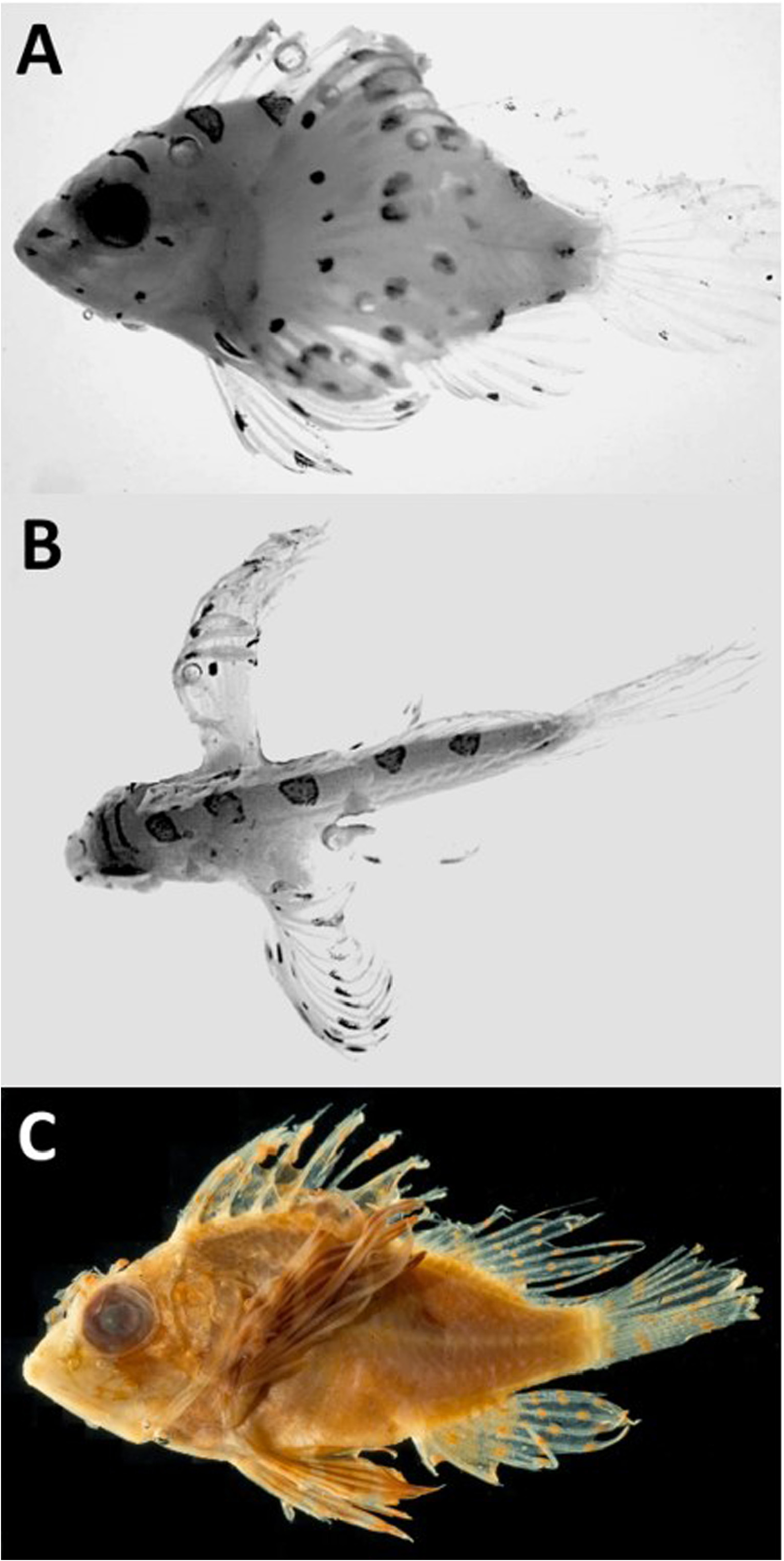
We examined images of two transforming larvae of D. hemprichi , one from Baldwin (2013: 540, fig. 40E) labeled D. brachypterus , and an 11.0 mm larva from the Red Sea ( Fig. 5a–b View FIGURE 5 ). These larvae were not available for examination and measurement; therefore, the following description of general morphology applies to the 21.0 mm juvenile only ( Fig. 5c View FIGURE 5 ). Head length 38% SL; OD about 1.5 times wider than snout. PPO-1 length 25% OD ( Table 2 View TABLE 2 ), and two times longer than PPO-2 through -5. Upper jaw terminates slightly beyond mid-orbit; maxilla swollen near its posterior margin. Preanal length 67% SL; BDc 43% SL; CP depth 36% BDc; CP length 75% of its depth; maximum width of pectoral-fin base 39% BDc ( Table 2 View TABLE 2 ). Seven BR.
Pigmentation
Transformers with pair of narrow stripes across dorsal surface of head: anterior stripe between posterior margin of orbits, posterior stripe between bases of parietal spines; a partial third stripe between lower margin of orbits. Lateral surface of snout with horizontal blotch near nares. Elsewhere, pigment near anterior margin and midway along lateral surface of lower jaw, below anteroventral margin of orbit, and near angle of lower jaw. Cheek with pigment behind posteroventral margin of orbit ( Fig. 5a View FIGURE 5 ), and diffuse blotch near anterior margin of gas bladder. Five roughly circular “saddles” along dorsal margin of body: anteriormost near origin of spinous dorsal fin, posteriormost near terminal margin of soft dorsal base ( Fig. 5 View FIGURE 5 a-b). Lateral midline of CP with short series of close-set melanophores. Ventrally, pigment near insertion of pelvic fins, near origin and termination of anal-fin base, and along posterior third of CP ( Fig. 5a View FIGURE 5 ). Presence or absence, and location of pigment along dorsolateral surface of hindgut uncertain, area concealed by pectoral fins in image ( Fig. 5a View FIGURE 5 ).
Pectoral- and pelvic-fin pigmentation pronounced in transformers. Pectoral fin with series of roughly circular to oblong blotches in uneven rows near mid-fin and toward its outer margin; single blotch near base of rays 12–13. Pelvic fin with blotch on membrane between spine and outermost ray about halfway between inner and outer margins, another blotch near outer margin of fourth ray, and near origin of fin base. Dorsal fin with pigment on webbing or along shaft of second and third spines, and near outer margin of anterior few rays; anal fin with pigment near outer margin of first and second rays; primary rays of caudal fin with pigment on membrane or along shaft of numerous rays near mid-fin and outer margin ( Fig. 5a View FIGURE 5 ).
Pigment faded, but juvenile with three bands on lateral surface of head: anterior band near anteroventral margin of orbit down across upper lip; diagonal medial band from supraocular cirrus across eye to posterior margin of interopercle; posterior band from margin of orbit across PPO. Snout with narrow bar or band from rim of lower nostril to dorsal margin of lower lip. Operculum and sides of upper and lower jaws mottled. Abdomen of juvenile with series of faint saddles along dorsal and ventral margins as described for transformers; mid-CP with partial band across dorsal margin. Dorsal and anal fins with series of circular to oblong blotches on shaft and webbing of most elements. Caudal fin with series of pigments in three irregular bands, indication of fourth band near outer margin of existing rays. Pelvic fins with three broad bands of pigment ( Fig. 5c View FIGURE 5 ). Folding of rays and loss of pigment due to long-term preservation makes pectoral-fin pigmentation pattern uncertain.
Fin development
Transformers with full complement of primary elements in all fins. Spinous and soft dorsal-fin bases continuous, spinous base about three times longer than rayed portion. Tips of most dorsal- and all anal-fin rays extend to or beyond posterior margin of CP. Third element of anal fin transformed into third spine in juvenile ( Table 3 View TABLE 3 ).
Juvenile with 7+7 primary caudal rays (outermost on each side unsegmented), 3+3 secondary elements (20 total). Gap between first and second dorsal spines narrower than between subsequent spines; second spine about 25% longer than first; spines two through seven similar in length; 12 th shortest; 13 th about three times longer than 12 th. First dorsal pterygiophore inserted above posterior margin of APO. Second spine of anal fin two times longer than first ( Table 2 View TABLE 2 ); third spine slightly longer than second. Pterygiophore of first anal spine inserted below 12 th dorsal spine; first “true” ray of anal fin inserted below first to second pterygiophore of soft dorsal-fin base; terminal pterygiophore of anal-fin base inserted below sixth to seventh pterygiophore of soft dorsal-fin base. Tips of dorsal and anal rays initially bifurcate, not those in pectoral and pelvic fins. Juvenile with pectoral fins relatively shorter than in transformers, tips of longest rays extend to, but not beyond, terminal margin of dorsal-fin base. Tips of longest pelvic rays extend to third anal spine; tip of pelvic spine short of anus when pressed against body. Membrane attaches innermost pelvic ray to abdominal wall for about 40% of its length. Terminal ray of dorsal fin attached by membrane to body wall for about 15% of its length; terminal ray of anal fin free from body.
Cephalic and opercular spination, and cephalosensory canals
Images of transformers reveal supraocular, postocular, parietal, and nuchal ridges, each with spine. Pterotic and posttemporal ridges also present, and small spine on IO 1-L1 and IO 2-L1. Unable to determine presence or absence of cephalosensory canals or details of PPO spination from images, but 9.2 mm larva in Matsunuma et al. (2017) has five spines.
Description below applies to juvenile only. Spines along APO subdermal or resorbed. Five PPO’s, upper three exposed, lower two subdermal. PPO-1 and -2 along upper margin; PPO-3 slightly below shelf angle; PPO-4 and -5 along lower margin. PPO-1 nearly aligned with lower rim of orbit, oriented slightly upward, its length about 25% OD. PPO-1 nearly two times longer than PPO-2 and -3, small supplemental spine at base of PPO-1. PPO-2 oriented slightly outward from axis of head, similar in length to PPO-3, but more acute; PPO-3 directed posteriorly. Opercle, interopercle, and subopercle lack spines. Cephalosensory canals and pores obscured by epithelium in juvenile, but series of three-tubed scales along trunk lateral line immediately behind head. Numerous scales and scale pockets on shoulder, abdomen, and CP suggest scale development complete or nearly so by 21.0 mm.
Juvenile with most cranial ridges and spines low, subdermal. Parietal ridge terminates in short, dorsoposteriorly directed parietal and nuchal spines, elevation ≤15° above longitudinal axis of head; nuchal spine attached to outer margin of parietal base, nuchals behind, slightly smaller than parietals. Pterotic and lower posttemporal ridges laterally swollen, both terminate in small spine. Lower posttemporal ridge elevated posteriorly, higher, but shorter, than pterotic ridge. Supraocular ridge smooth, postocular ridge rises to low, obtuse lateral peak near mid-orbit, descends along posterior rim. Entire IO ridge elevated, lateral margin smooth, slight indentation between IO 1 and IO 2. Spine on IO 1-L1 overlaps dorsal margin of upper lip. Coronal and tympanic ridges elevated, tympanic ridge two times higher and longer than coronal; lacks medial interorbital ridge. Vertical pair of short, parallel, weakly dentate sphenotic ridges behind orbit. Elevated supracleithral ridge terminates in short spine.
Dermal flaps, and supraocular and nasal cirri
Transformers with short supraocular and nasal cirri; nasal cirrus along dorsal rim of anterior nostril. Juvenile with supraocular cirrus about 85% OD, and encircled by three bands; tip spade-shaped. Nasal cirrus slender, encircled by narrow band about mid-length, tip extends beyond mid-orbit if pressed against forehead. Juvenile with pair of filamentous barbels just above upper lip, barbels shorter than nasal cirrus. Small pigmented flap on anterodorsal surface of eye, and minute flap on anteroventral margin of IO 1–L1. Leafy flap behind IO 2-L1 with many bands, margin double-toothed; another flap along posteroventral margin of PPO adjacent to lowermost ray of pectoral fin. Location, presence or absence, size, and shape of dermal flaps and snout barbels difficult to determine without stain.
| IO |
Instituto de Oceanografia da Universidade de Lisboa |
No known copyright restrictions apply. See Agosti, D., Egloff, W., 2009. Taxonomic information exchange and copyright: the Plazi approach. BMC Research Notes 2009, 2:53 for further explanation.