
Tyrannosaurus rex, Osborn, 1905
|
publication ID |
https://doi.org/ 10.5281/zenodo.3676505 |
|
DOI |
https://doi.org/10.5281/zenodo.3717027 |
|
persistent identifier |
https://treatment.plazi.org/id/039987D5-DD7C-8D38-0FFA-F9A8F0899C30 |
|
treatment provided by |
Jeremy |
|
scientific name |
Tyrannosaurus rex |
| status |
|
Skulls
Six partial and complete skulls are now known for Tyrannosaurus rex ,
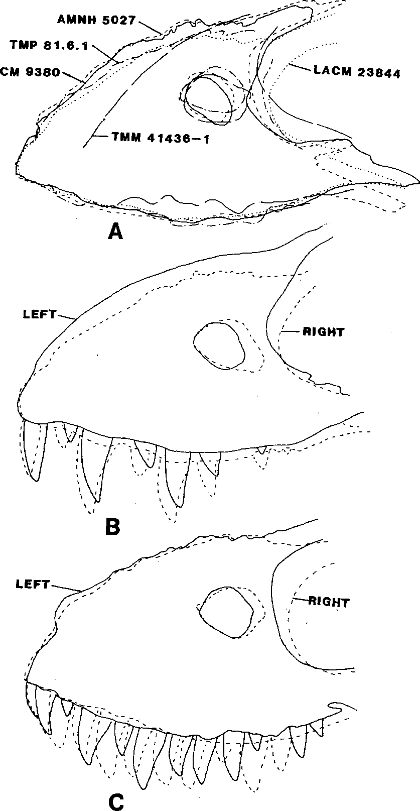
including the holotype CM 9380 (formerly AMNH 973) View Materials , AMNH 5027 View Materials , LACM 23844 View Materials , MOR 009 , SDSM 12047 , and TMP 81.6.1 . Two additional specimens, AMNH 5029 View Materials and AMNH 5117 View Materials , are braincases ( Osborn 1912). Five of the skulls are shown in Figure 10.1 View Figure 10.1 . As may be seen, there is a considerable amount of variation in the size and shape of all the cranial openings (e.g., orbit and lateral temporal fenestra), as well as all the individual elements, including the lacrimal, postorbital, quadratojugal, jugal, and surangular. In fact, no two specimens are identical.
Seven maxillae were available for comparison. Five of these were overlain in Figure 10.2A View Figure 10.2 using the anterior-most margins of the maxilla and maxillary fenestra as standard lengths. Most of the maxillae resemble one another, except for TMM 41436-1 , the specimen reported from the Tornillo Formation by Lawson (1976).
Variation can be seen in the depth of the maxilla, size and shape of the maxillary and antorbital fenestrae, position of the lacrimal processes, and the position and shape of the jugal process ( Fig. 10.2A View Figure 10.2 ). Differences in the depth of the maxilla affect the position of the lacrimal process, which in tum influences the height and shape of the antorbital fenestra. There is also a considerable amount of variation in position of the jugal process that forms the lower rim of the antorbital fenestra, but this does not seem to correlate with the size of the antorbital process. The shape of the maxillary fenestra ranges from almost oval ( CM 9380 View Materials ) to subtriangular ( LACM 23844 View Materials and TMP 81.6.1 ) to almost square ( AMNH 5012 View Materials ). There is little or no difference in the position of the largest teeth and therefore this feature is not affected by the depth of the maxilla or the shape of the antorbital fenestra. Even the left and right maxilla of the same skull show variation ( Figs. 10.2B,C View Figure 10.2 ).
The maxilla from the Tornillo Formation lacks the anterior-most margin and was scaled to the other maxilla using height and the anterior margin of the maxillary fenestra ( Fig. 10.2A View Figure 10.2 ). The nasal, or dorsal, margin of the maxilla arcs sharply down suggesting a face considerably shorter than in T. rex . Other differences include a slightly larger maxillary fenestra in proportion to the size of the maxilla and a much deeper jugal process. The antorbital fenestra appears to be smaller, but the lacrimal process is too incomplete to be certain. These differences are great enough to suggest that TMM 41436-1 falls outside the range of individual variation for Tyrannosaurus rex . It may belong to a new genus, but no name should be proposed until additional material is available. Because it is unusual, it is excluded from discussions on variation.
Variation in the dentary is demonstrated by six specimens ( Fig. 10.3 View Figure 10.3 ). The distance between the anterior tip and the highest part of the dentary were used for the standard length. The greatest difference is the depth of the posteroventral margin of the dentary. It is relatively shallowest in SDSM 12047 and deepest in AMNH 5027 View Materials ( Fig. 10.3 View Figure 10.3 ). However, AMNH 5027 View Materials also has a tooth row that curves more ventrally than the others and this may have had an influence on the posteroventral margin. The posterior margin of the dentary is thin and easily damaged, although three of the dentaries have complete margins ( CM 9380 View Materials , AMNH 5027 View Materials , and TMP 81.6.1 ). The shape of the anterior portion of the external mandibular fossa (expressed as a notch at the lower edge of the posterior margin, see Fig. 10.1 View Figure 10.1 ) is variable. The teeth seem to show more variation in positional size, but this may be due to how the dentaries were overlain.
Cervicals
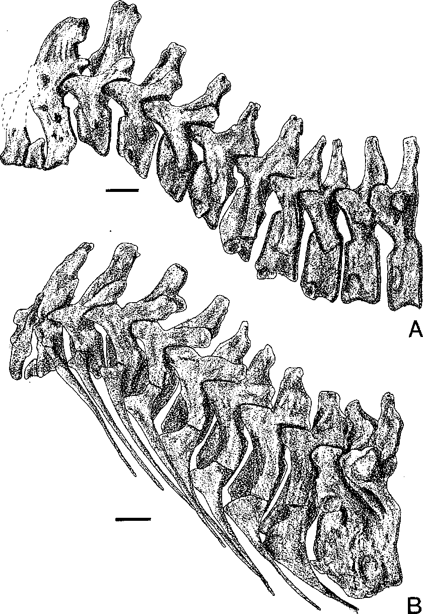
Complete cervical series are known for only two specimens, AMNH 5027 View Materials and BM(NH) R7994 View Materials . There is a considerable amount of variation in the shape of the neural spines. Similar variation appears to be present in the tyrannosaur Albertosaurus libratus , so the use of neural spine shape in the diagnosis of the tyrannosaur Daspletosaurus torosus ( Russell 1970) is suspect.
The neck is more robust in BM(NH) R7994 View Materials than in AMNH 5027 View Materials ( Fig. 10.4 View Figure 10.4 ). This is especially evident in the atlas and its intercentrum, and in the neural spines of cervicals two and three.
Asymmetrical co-ossification of the last cervical and first dorsal in AMNH 5027 View Materials has not been observed in any other specimen, and is probably pathological.
Ischiae
Three ischia were available for comparison. Length was standardized and the articular surface of the iliac peduncle was used to determine orientation in Fig. 10.5 View Figure 10.5 . Points of variation include the relative size of the iliac peduncle, the relative size and position of the pubic peduncle, the position and size of the obturator process, and the development of the insertion scar for the M. flexor tibialis internus part 3. This scar is best developed in CM 9380 View Materials where it forms a prominent ridge.
Of the two types of ischia, one ( Fig. 10.5A View Figure 10.5 ) is oriented more ventrally from the horizontal articular surface of the iliac peduncle. I suspect that CM 9380 View Materials and TMP 81.6.1 are females because the greater angle between the sacral vertebrae and distal end of the ischium would permit the passage of eggs (or live young) more readily than that of AMNH 5027 View Materials . The more divergent ischia are associated with robust skeletons, whereas the less divergent ischium is from a gracile skeleton.


No known copyright restrictions apply. See Agosti, D., Egloff, W., 2009. Taxonomic information exchange and copyright: the Plazi approach. BMC Research Notes 2009, 2:53 for further explanation.