
Zorotypus asymmetricus Kocarek
|
publication ID |
https://doi.org/ 10.11646/zootaxa.4286.2.11 |
|
publication LSID |
lsid:zoobank.org:pub:DC8D1D74-FB6B-4E8E-96F6-C4B2CDCB7388 |
|
DOI |
https://doi.org/10.5281/zenodo.6046316 |
|
persistent identifier |
https://treatment.plazi.org/id/E748C008-F5CD-4A1B-88DD-16A764A02F87 |
|
taxon LSID |
lsid:zoobank.org:act:E748C008-F5CD-4A1B-88DD-16A764A02F87 |
|
treatment provided by |
Plazi |
|
scientific name |
Zorotypus asymmetricus Kocarek |
| status |
sp. nov. |
Zorotypus asymmetricus Kocarek View in CoL , sp. nov.
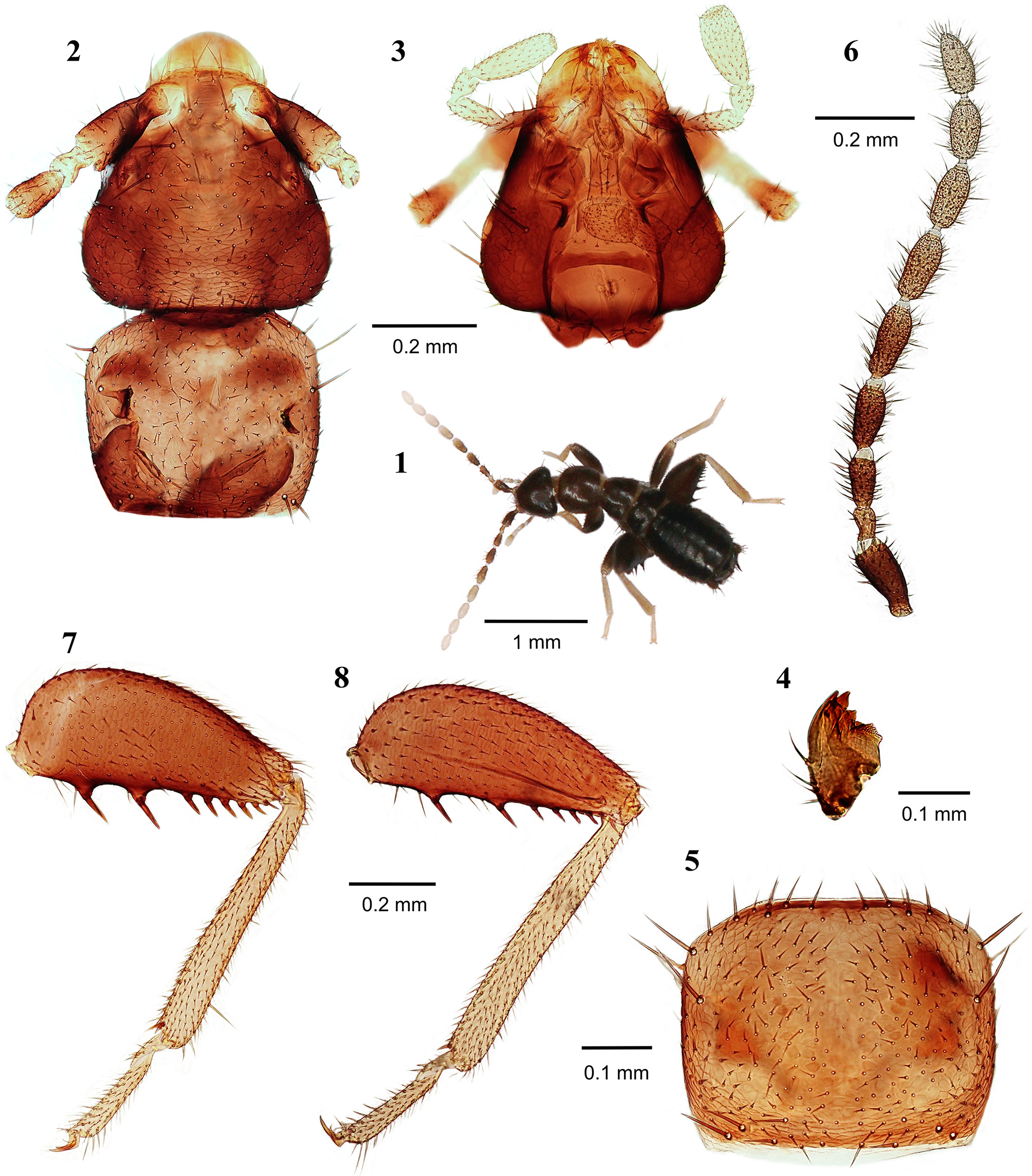
( Figs. 1‒17 View FIGURES 1 – 8 View FIGURES 9 – 16 View FIGURES 17, 18 )
Type material. Holotype apterous male, labelled ' Brunei Darussalam, 9.i.2014, Ulu Temburong NP, Sungai Esu stream, 150 m a.s.l., GPS: 04°32'14.1"N, 115° 9'47.1"E, P. Kočárek leg.' ( NMPC), under bark of rotting log GoogleMaps . Paratypes: same data as for holotype, 1 apterous female ( NMPC), 1 apterous male, 1 apterous female ( UBDC), 1 apterous male, 1 apterous female ( BMNH), 3 apterous males, 4 apterous females ( PKCO) GoogleMaps .
Diagnosis. The new species is similar to Z. sinensis Hwang, 1974 , Z. medoensis Hwang 1976 , Z. impolitus Mashimo, Engel, Dallai, Beutel & Machida 2013 and Z. weiwei ( Hwang 1974, 1976; Mashimo 2013; Wang et al. 2016), but it can be easily distinguished from them by the asymmetrical cerci, with the right cercus noticeably enlarged and sickle-shaped ( Figs. 9–11 View FIGURES 9 – 16 ), the species-specific shape of the male genitalia ( Fig. 13 View FIGURES 9 – 16 ), and the presence of 7‒8 stout, long spines on the ventral surface of metafemur. The body is typically matte dark brown with the exception of pale yellowish gray tibiae and tarsi on all legs and antennomeres VI‒IX.
Description of apterous male ( Figs. 1‒4, 6, 7 View FIGURES 1 – 8 , 9‒13 View FIGURES 9 – 16 , 17 View FIGURES 17, 18 ). Total body length 2.39 mm, head width 0.43 mm, antenna length 1.45 mm, pronotal width 0.41 mm, pronotal length 0.36 mm, metafemur length 0.644 mm. Body color matte dark brown except membranous regions; antennal flagellomeres VI‒IX and tibiae and tarsi of all legs pale yellowish gray ( Figs. 1 View FIGURES 1 – 8 , 17 View FIGURES 17, 18 ). Head subtriangular, slightly wider than pronotum ( Figs. 2, 3 View FIGURES 1 – 8 ); cephalic setae ( Fig. 2 View FIGURES 1 – 8 ) short and sparse, not grouped; compound eyes and ocelli absent; antennae 9-segmented, distal four antennomeres paler ( Figs. 1, 6 View FIGURES 1 – 8 ), antennomere I slightly curved outward, antennomere II slightly curved, short, about one-half length of antennomere III, antennomeres III–IX longer than wide, length subequal to that of antennomere I ( Fig. 6 View FIGURES 1 – 8 ). Mandibles asymmetrical, each mandible with four apical teeth and well-developed molar region ( Fig. 4 View FIGURES 1 – 8 ); maxillary palpus five-segmented, labial palpus three-segmented. Pronotum subrectangular, only slightly wider than long, slightly narrowed posteriorly and setose ( Fig. 2 View FIGURES 1 – 8 ); mesonotum trapezoidal, slightly shorter than pronotum; metanotum trapezoidal, distinctly wider than long, shorter than mesonotum. Legs with short setae; tibiae and tarsi of all legs pale yellowish gray ( Fig. 1 View FIGURES 1 – 8 ); posterior surface of profemur covered with short setae, anterior and dorsal surfaces covered with longer setae; protibia with short setae, spines arranged as comb in distal two-thirds along ventral surface, with two apical spurs; mesofemur slightly narrower than profemur, anterior surface broadly setose, posterior and dorsal surfaces covered with setae only distally; mesotibia covered with short setae and two apical spurs; metafemur broader than profemur, swollen proximally ( Figs. 7, 8 View FIGURES 1 – 8 ), anterior surface broadly setose, posterior surface sparsely setose, middle part without setae, distal half of lower edge lined with one row of longer setae ( Figs. 7 View FIGURES 1 – 8 , 9 View FIGURES 9 – 16 ), ventral surface with 7‒8 stout spines, proximal first and second spines (spine I and II) longer than others ( Fig. 7 View FIGURES 1 – 8 ), distance between spine I and II and between spines II and II equal to length of spines 1 and 2; spines III‒VIII shorter and closer to each other ( Fig. 7 View FIGURES 1 – 8 ); metatibia with short setae, subapically with one strong spine ventrally and one finer spine dorsally; basal tarsomere with stronger two spurs ventrally ( Figs. 7 View FIGURES 1 – 8 , 9 View FIGURES 9 – 16 ).
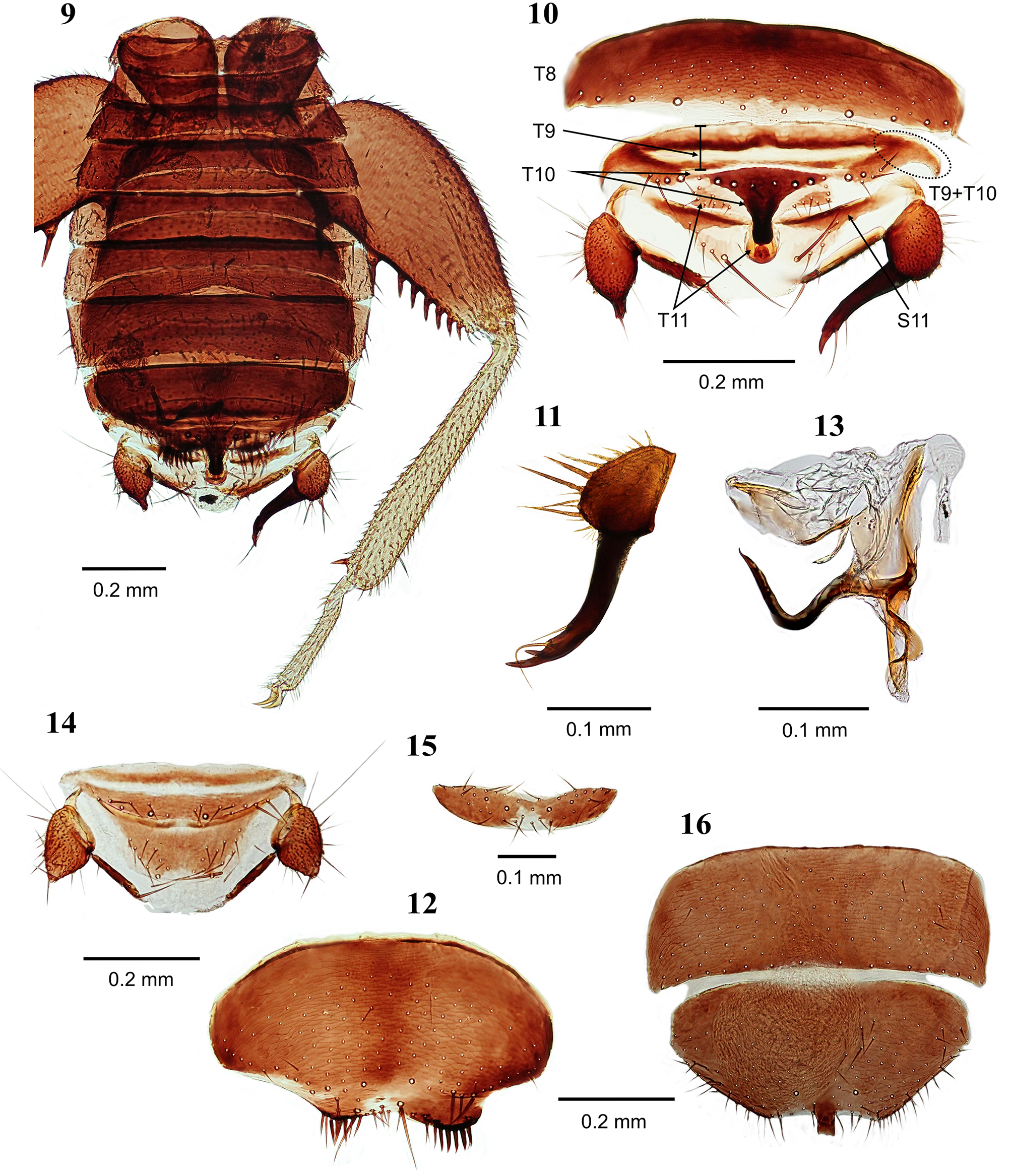
Abdominal tergite 1 (T1) with single transverse row of setae flanking distal edge, and one pair of short setae in antero-posterior margin; T2 with two parallel rows of setae in distal third of the tergite with regular distances between individual setae; T3 with three irregular rows of setae in distal half; T4‒5 with 4‒5 irregular rows of setae filling distal half of the tergite, setae in the most distal row longer; T6‒7 with 6‒7 irregular rows of setae occupying two-thirds of the tergite, setae in the most distal row longer; T8 with 4 irregular rows of setae in posterior half of tergite and with ten longer setae on the posterior edge ( Fig. 10 View FIGURES 9 – 16 ); T9 short, strongly sclerotized anteriorly and scarcely sclerotized posteriorly, well delimited from T 10 in the middle, but fused with T 10 in lateral fifths ( Fig. 10 View FIGURES 9 – 16 ); T10 separated into anterior and posterior parts, anterior part scarcely sclerotized, with setae laterally, posterior half strongly sclerotized with medial spatula-like upcurved projection, several short setae on the base of projection and two short setae on the obtuse top ( Fig. 10 View FIGURES 9 – 16 ); T11 with long and strongly upcurved median projection with a tiny apical hook ( Fig. 10 View FIGURES 9 – 16 ) and two smaller, lateral sclerites each bearing two longer setae; projection of T11 narrower in the middle and longer than that of T10 ( Fig. 10 View FIGURES 9 – 16 ); epiproct and paraproct unsclerotized.
Cerci ( Figs. 9‒11 View FIGURES 9 – 16 ) unsegmented, left cercus conical with pointed apex, right cercus enlarged with sickleshaped, pointed appendage terminating in one larger apical tooth and one smaller subapical tooth on outer margin; right cercus with two long distal setae, the the more apical seta longer than and extending past distal tooth ( Fig. 11 View FIGURES 9 – 16 ). Outer basal margin of both cerci with short setae and several long and fine setae ( Figs. 9‒10 View FIGURES 9 – 16 ); surface covered with numerous minute spicules except base and apex; pointed appendage of right cercus without spicules.
Abdominal sternite 1 (S1) scarcely sclerotized; S2 weakly sclerotized with a few short setae on each side and four setae in the middle posterior margin; S3 with two irregular parallel rows of short setae in distal third of the sternite; S4 with three irregular rows of short setae in distal half; S5 with four irregular rows of short setae occupying posterior two-thirds of the sternite; S6 with five irregular rows of short setae occupying all but anterior fourth; S7 with 4‒5 irregular rows of short setae on posterior two-thirds; S8 and S9 fused ( Fig. 12 View FIGURES 9 – 16 ), with shallow, partly visible furrow, S8 with evenly scattered fine setae, posterior margin with moderate-length to long setae, S9 asymmetrical with two posterior lobes, left lobe more prominent, each lobe with one row of strong setae; S10 invaginated beneath S8+S9, not visible externally; sternite S11 with two lateral sclerites, each with one row of small setae.
Genitalia asymmetrical, with hooked and strongly sclerotized aedeagus, without elongate coiled flagellum; spatula-like basal plate present beneath aedeagus ( Fig. 13 View FIGURES 9 – 16 ).
Apterous female. Generally as in male except as follows: pronotum transverse, apparently wider than long and only slightly narrowed posteriorly ( Fig. 5 View FIGURES 1 – 8 ); metafemur slender, ventral surface with 7 stout spines, thinner than in males, proximal first and second spines longer than others, but the distance between them twice the length of spines 1 and 2; distance between second and third bristles equal to length of spine 2 ( Fig. 8 View FIGURES 1 – 8 ); Abdominal T10 uniformly sclerotized with 6‒8 short setae on each side and a pair of longer setae ( Fig. 14 View FIGURES 9 – 16 ); T11 trapezoidal, weakly sclerotized at distal end, with small setae laterally and one pair of longer setae ( Fig. 14 View FIGURES 9 – 16 ); both cerci short, conical with slightly pointed distal apex, surface covered with numerous minute spicules except on base and apex, and shorter and longer setae ( Fig. 14 View FIGURES 9 – 16 ); S8 strongly trapezoidal, wider than long, with short setae evenly scattered and longer setae flanking the distal and lateral edges; distal end slightly concave with peg-like projection in the middle ( Fig. 16 View FIGURES 9 – 16 ); S9 short and trapezoidal with several small setae along posterior margin ( Fig. 15 View FIGURES 9 – 16 ).
Alate males and females. Unknown.
Etymology. The name refers to the asymmetrical cerci and asymmetrical abdominal sternite S 9 in males.
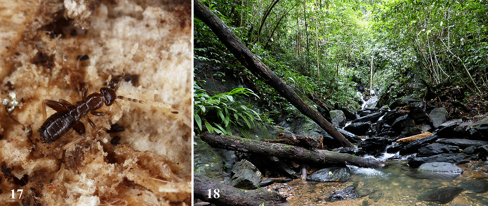
Distribution and occupied habitat. Zorotypus asymmetricus sp. nov. was collected under the bark of rotting logs in shade in the valley of Sungai Esu stream ( Fig. 18 View FIGURES 17, 18 ). The species is currently known only from Ulu Temburong National Park in Brunei Darussalam, but we expect its occurrence in similar habitats throughout Borneo.
| NMPC |
National Museum Prague |
No known copyright restrictions apply. See Agosti, D., Egloff, W., 2009. Taxonomic information exchange and copyright: the Plazi approach. BMC Research Notes 2009, 2:53 for further explanation.
|
Kingdom |
|
|
Phylum |
|
|
Class |
|
|
Order |
|
|
Family |
|
|
Genus |