
Sarcophaga (Heteronychia) infixa Böttcher, 1913
|
publication ID |
https://doi.org/ 10.5281/zenodo.185607 |
|
DOI |
https://doi.org/10.5281/zenodo.6217768 |
|
persistent identifier |
https://treatment.plazi.org/id/0397C163-D94B-435B-4284-FB334918A0DE |
|
treatment provided by |
Plazi |
|
scientific name |
Sarcophaga (Heteronychia) infixa Böttcher, 1913 |
| status |
|
Sarcophaga (Heteronychia) infixa Böttcher, 1913 View in CoL sp. rev.
Sarcophaga infixa Böttcher, 1913a: 124 View in CoL . Type locality: Hungary, “Gegend von Budapest”.
Type material. Holotype ɗ: Gyón [ Hungary] / 16.VI.02 // Sarcophaga / infixa / i. lit. Villen. // G. Böttcher ( SMF) // HOLOTYPE / det. D. Whitmore, R. Richet, T. Pape & R. Blackith 2008 [holotype in moderate condition with torn wings, somewhat abraded chaetotaxy and front right leg glued to a piece of card pinned with the specimen; the terminalia were removed from the abdomen, dissected and placed in glycerine in a small plastic tube pinned with the rest of the specimen].
Other material. Austria. 1 ɗ: Austria inf., Obenweiden, 29.6.93, Mik, S. infixa Villen. ɗ det. G. Böttcher ( NMW). Hungary. 1 ɗ: [ Hungary], Pócsmegyer, Fegyveres-sz., 1958. V.20, leg. Kakassné ( HNHM); 1 ɗ: [ Hungary], Ágasegyháza homokbuckás, 1959. VII.9, leg. Mihályi ( HNHM); 1 ɗ: [ Hungary], Mecsek-hg., Tubes, 1960. VI.15, leg. Soós ( HNHM); 1 ɗ: [ Hungary], Tompa, Zsiros-kuti erdő, 1962. V.8, leg. Zsirkó ( HNHM); 1 ɗ: [ Hungary], Csévahaszrt borókás, 1971. VIII.11, leg. Mihályi ( HNHM).
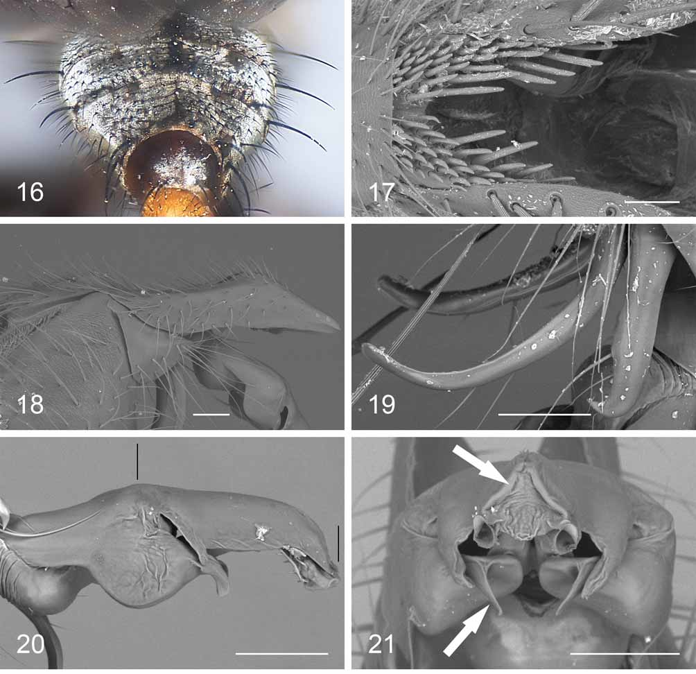
Diagnosis (male). Parafacial narrow; scutellum with a pair of apical setae; mid tibia with one anteroventral seta; hind trochanter with a brush of short spine-like setae; hind femur with a strong subapical seta but no additional anteroventral setae; hind tibia with a row of fine setulae on anteroventral surface; wing vein R1 setose dorsally; abdominal tergite 3 without median marginal setae; protandrial segment with a row of marginal setae and a large spot of grey microtrichosity; epandrium red; cercus with a dorsal inflexion when viewed in profile and with a slight dorsal excavation medially; pregonite with a rounded tip, not widening apically; phallus: apical process of harpes short, with main width oriented in the same plane as median longitudinal plane of distiphallus; juxta long, 1.11–1.47 times the length of basal part of distiphallus; juxta with short, thin, parallel-sided appendages at base, variable in length and thickness.
Redescription. Male (measurements refer to the holotype, with the variation range of the species given in square brackets).
Length: 4.8mm [4.8–7.5].
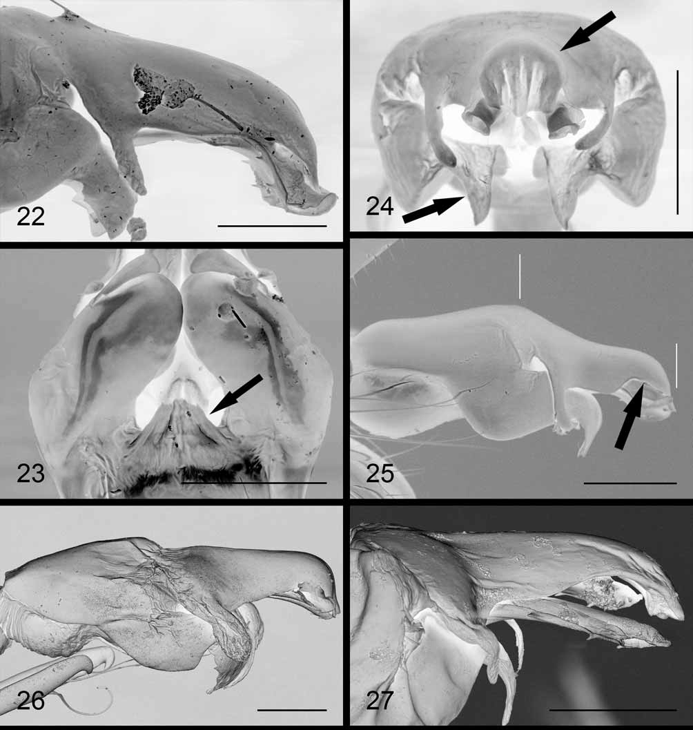
Colour. Head black, with dense silver-grey microtrichosity on parafacials and fronto-orbital plate, changing with the incidence of light. Frontal vitta black. Gena, face and occiput grey-microtrichose. Antenna: pedicel black, brown at tip; postpedicel black. Prementum dark brown, palpus brown. Ground colour of thorax black, grey-microtrichose with three longitudinal dark vittae; legs black; tegula black, basicosta light yellow. Abdomen black, densely grey-microtrichose; when viewed posteriorly, abdomen almost entirely light grey with central dark stripes (wider on tergites 3 and 4) and small lateral dark spots (often very reduced) on tergites 3–5 ( Fig. 16 View FIGURES 16 – 21 ). Protandrial segment black with a large spot of grey microtrichosity distally, occupying 2/3 of the width of the segment; epandrium red; cercus black; surstylus brown; phallus, pre- and postgonites brown. Head. Arista thickened on basal 1/4 [1/4–1/3]. Postpedicel 2.00 [1.82–2.00] times as long as pedicel. Frons at its narrowest point 0.47 [0.47–0.55] times as wide as an eye in dorsal view. Frontal vitta 0.56 [0.48–0.56] times as wide as frons at its narrowest point, widening towards antennal insertion. Lateral vertical seta strong, about 1.5 times as long as longest postocular setae. Five to 6 frontal setae, not descending below level of middle of pedicel. Fronto-orbital plate with a few scattered, fine setulae. Parafacial with a row of fine hair-like setulae near eye margin. Parafacial at its narrowest point 0.13 [0.13–0.21] times as wide as eye width in strict lateral view. Lower facial margin slightly visible in lateral view below vibrissa. Facial ridge above vibrissa with a few decumbent setulae. Gena in profile 0.25 [0.25–0.31] times the vertical height of eye (measured in the same vertical plane as height of head); gena entirely covered with black setulae; postgenal setulae white. Two rows of black occipital setulae behind postocular row, remaining occipital setulae white. Prementum about three times as long as wide. Thorax. Postpronotum with 3 stronger setae forming a triangle. Scutum with 2–3 + 1 (prescutellar) acrostichal, 4 + 3 dorsocentral, 2 intraalar, 1 posthumeral (sometimes an additional weak outer seta present), 1 presutural, 4 notopleural and 3 supraalar setae; postalar callus with 2 setae. Katepisternum with 3 setae. Katepimeron with fine setulae on posterior half. Scutellum with 3 pairs of marginal setae (basal, subapical, apical) and one pair of discal setae. Legs. Fore tibia with 2–3 anterodorsal and 1 posterior setae. Mid femur with 2–5 anterior setae near middle, 2–4 anteroventral setae, 2–3 subapical posterodorsal setae, only fine, long setulae on posteroventral surface, some with wavy tip; mid femur without a subapical posteroventral comb. Mid tibia with 2–3 (sometimes a few additional short) anterodorsal, 2–3 posterodorsal, 1 posterior and 1 anteroventral setae. Hind trochanter with a posteroventral brush of widely spaced, short, spine-like setae. Hind femur with a strong subapical seta but no additional anteroventral setae (although always with a few stronger setulae), and with several stronger posteroventral setulae in basal third. Hind tibia with a row of anterodorsal setae of irregular length, 2 posterodorsal and 2–3 anteroventral setae; hind tibia usually with a row of fine setulae on posterovental surface, usually in a more or less straight line (such setulae shorter in smaller specimens, e.g. in the holotype). Wing. Costal spine well developed, about as long as crossvein R-M or slightly longer. Vein R1 with several setae along dorsal surface. Setae on dorsal surface of vein R4+5 extending over halfway to crossvein R-M. Second costal section [0.87–0.95] times fourth costal section [not measurable in holotype, which has a damaged costa]. Small spines on costa reaching about 2/3–4/5 of the way across fourth costal section. Wing cell r4+5 open at wing margin (closed in holotype but without a petiole). Abdomen. Syntergite 1+2 and tergite 3 without median marginal setae. Tergite 4 with a pair of strong median marginal setae and 2–3 lateral marginal setae. Tergite 5 with a complete row of marginal setae. Terminalia. Sternite 5 ( Fig. 17 View FIGURES 16 – 21 ) strongly indented, v-shaped, with tightly spaced stout, short setae at base of and along each of its processes, the innermost ones visibly longer. Protandrial segment with a row of setulae along posterior margin. Epandrium with a gently curved dorsal margin, about as long as high. Cercus ( Fig. 18 View FIGURES 16 – 21 ) with a dorsal inflexion and downcurved apex when viewed in profile, and with a slight dorsal excavation medially. Surstylus ( Fig. 18 View FIGURES 16 – 21 ) sub-triangular. Pregonite ( Fig. 19 View FIGURES 16 – 21 ) thin and curved with a rounded, not enlarged tip and with fine setulae along dorsal surface. Postgonite ( Fig. 19 View FIGURES 16 – 21 ) with a hooked tip. Distiphallus ( Fig. 20 View FIGURES 16 – 21 ): apical process of harpes short, more or less ventrally directed, with main width oriented in the same plane as median longitudinal plane of distiphallus ( Fig. 21 View FIGURES 16 – 21 ); juxta long, approximately 1.47 [1.11–1.47] times the length of basal part of distiphallus (see Fig. 20 View FIGURES 16 – 21 ) with short, thin (parallel-sided) appendages arising from base, somewhat variable in length and thickness (see Figs 20 View FIGURES 16 – 21 , 22 View FIGURES 22 – 27. 22 – 23 ); juxta not separated from rest of distiphallus by a distinct suture; juxta, in apical view ( Fig. 21 View FIGURES 16 – 21 ), with two small, upturned processes at sides (see Figs 20–22 View FIGURES 16 – 21 View FIGURES 22 – 27. 22 – 23 ) and a deep subtriangular median depression delimited by two conspicuous folds converging dorsally ( Fig. 21 View FIGURES 16 – 21 ); lateral styli funnel-shaped, slightly widening apically ( Fig. 22 View FIGURES 22 – 27. 22 – 23 ); vesica small, laminar ( Fig. 23 View FIGURES 22 – 27. 22 – 23 ).
Female unknown.
Biology. Unknown.
Distribution. Austria, Czech Republic, Germany and Hungary (cf. Pape 1996; Povolný & Verves 1997). Mentioned from Germany by Verves (1986) and later also by Pape (1996) and Rudzinski (1999); Povolný and Verves (1997) were more precise in listing the species from “Southern Bavaria”, but they did not give a reference for this record and its origin could not be retraced by us. Sarcophaga infixa seems to be rare throughout its range, and very few specimens exist in museum collections.
Remarks. Sarcophaga infixa was described on a single male from Hungary (see above). Based solely on Böttcher’s (1913a: 124) very schematic original illustration, Rohdendorf (1937) placed S. infixa in Pierretia subgenus Pandelleola Rohdendorf, 1937 (= what we consider the “ filia -group”). Mihályi (1979) studied new material from Hungary (probably the specimens from HNHM listed above) and provided a more detailed illustration of the phallus showing the short appendages at the base of the juxta (not visible in Böttcher’s original illustration). Verves (1986) still placed S. infixa in subgenus Pandelleola , but placed the subgenus under genus Heteronychia . He later transferred S. infixa to Heteronychia sensu stricto after examining the male terminalia ( Verves 1989). Povolný and Verves (1997) reproduced Mihályi’s drawing and gave a brief description of S. infixa without discussing affinities with other species. Pape (1996) moved Heteronychia to Sarcophaga , recognizing it as a subgenus with Pandelleola in synonymy and recognizing S. (Heteronychia) infixa as a valid species. Pape et al. (2002) synonymized S. infixa with S. (Heteronychia) haemorrhoides , stressing the need for a modern revision of these two nominal species; following our examination of the holotype of the former, we here reinstate S. (Heteronychia) infixa as valid, sp. rev.
No known copyright restrictions apply. See Agosti, D., Egloff, W., 2009. Taxonomic information exchange and copyright: the Plazi approach. BMC Research Notes 2009, 2:53 for further explanation.
|
Kingdom |
|
|
Phylum |
|
|
Class |
|
|
Order |
|
|
Family |
|
|
Genus |
Sarcophaga (Heteronychia) infixa Böttcher, 1913
| Whitmore, Daniel, Richet, René, Pape, Thomas & Blackith, Ruth M. 2009 |
Sarcophaga infixa Böttcher, 1913a : 124
| Bottcher 1913: 124 |