
Thraulodes fascipennis, Kluge, Nikita J., 2020
|
publication ID |
https://doi.org/ 10.11646/zootaxa.4756.1.1 |
|
publication LSID |
urn:lsid:zoobank.org:pub:9FF62616-A7FA-4331-AC51-0F534400631D |
|
DOI |
https://doi.org/10.5281/zenodo.3811787 |
|
persistent identifier |
https://treatment.plazi.org/id/039787A6-FFD5-842A-8CFB-FB4BDA66FD51 |
|
treatment provided by |
Carolina |
|
scientific name |
Thraulodes fascipennis |
| status |
sp. nov. |
9. Thraulodes fascipennis sp. n.
( Figs 276–316 View FIGURES 276–296 View FIGURES 297–308 View FIGURES 209–314 View FIGURES 315–316 )
Etymology. Allusion to dark brown stripe which crosses fore wing close to the base.
Material examined. Holotype: L-S/I ♂ {specimen [IV](4)2018}, PANAMA, Provincia de Chiriqui, La Esper- anza, 7 km NNW Gualaca (8°35’N, 82°20’W), 18.I.2018, coll. N. Kluge & L. Sheyko. GoogleMaps Paratypes: the same locality and collectors, 5–22.I.2018: 5 S-I ♂, 1 S/I ♀, 1 L /S ♀, 9 larvae. GoogleMaps
Descriptions.
Larva. CUTICULAR COLORATION ( Figs 276–283 View FIGURES 276–296 ). Dorsal side of head, thorax and abdomen mostly brown with few blanks: pronotum bordered by light laterally, without other blanks; abdominal terga with blanks near tergalii bases, area mediad of them nearly unicolor brown. Femora mostly brown with proximal and preapical blanks; tibia brown; tarsus brown with base and apex lighter.
HYPODERMAL COLORATION ( Fig. 292 View FIGURES 276–296 ). Abdominal terga I–IX with blackish transverse band on posterior margins; terga II–V also with pair of blackish round spots corresponding to midway gray spots of imago. Tergalii yellowish with gray maculation ( Figs 284–291 View FIGURES 276–296 ).
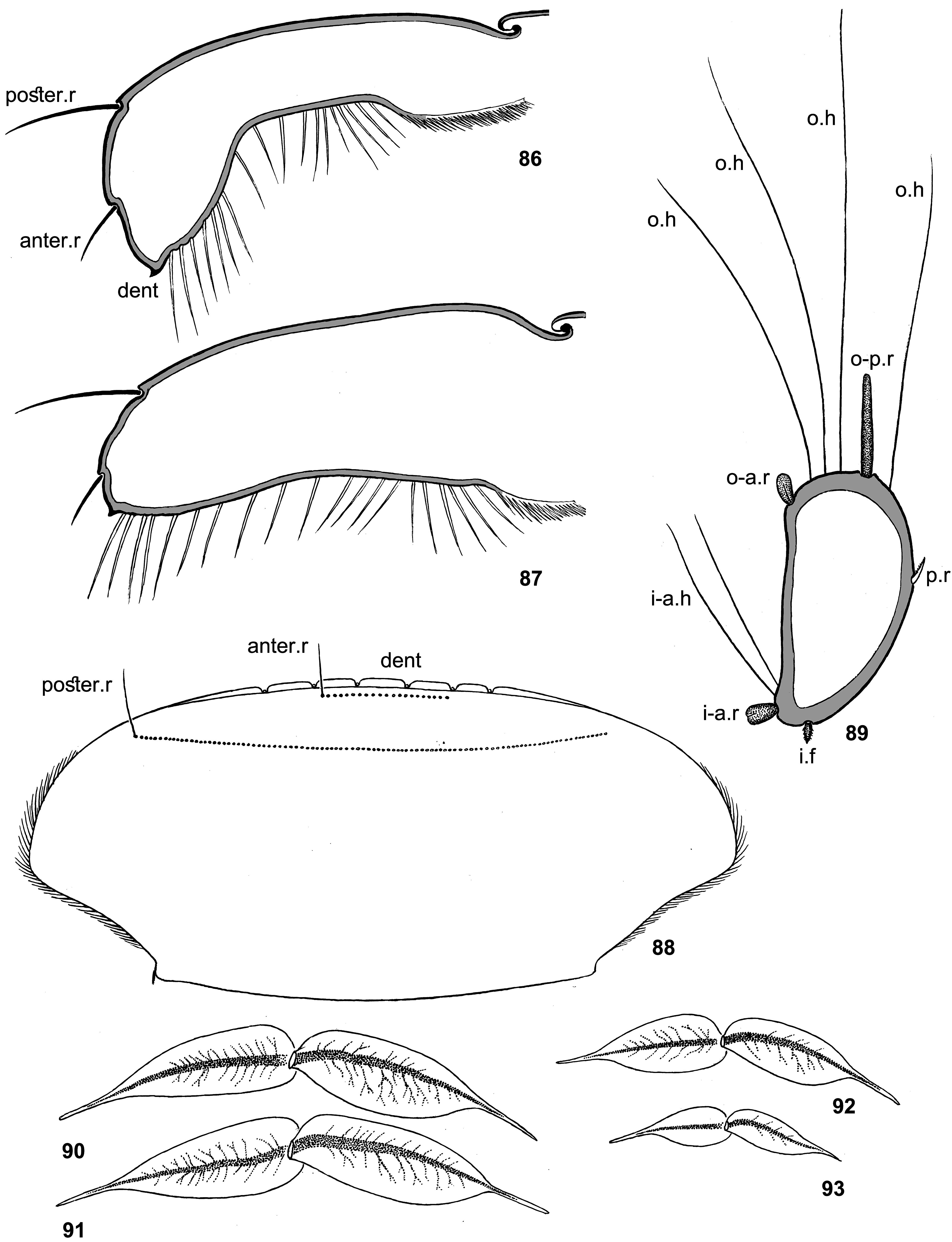
SHAPE AND SETATION. Clypeus widened distally; labrum 1.3–1.6 times wider than clypeus ( Fig. 276 View FIGURES 276–296 ). Labrum widest at 0.4 length from base; initial fore margin (turned ventrally) without median emargination, with all 5 denticles wide; anterior transverse setal row regular (as in Fig. 88 View FIGURES 86–93 ), as wide as all 5 denticles. Maxilla with 17 pectinate setae in apical-ventral row.
Femora: Stout setae on anterior surface widened distally, apically rounded ( Fig. 293 View FIGURES 276–296 ). Irregular row of hairs near inner margin absent on fore femur, present on middle and hind femora.
Fore tibia ( Fig. 294 View FIGURES 276–296 ): outer hairs form two irregular rows; inner-anterior row of recurved hairs absent; inneranterior row of stout setae represented by few (1–3) blunt stout setae near tibia base; inner field of stout pointed setae dense (i.e. setae longer than distances between them), consists of pointed bipectinate and non-pectinate setae, situated irregularly (about 3 setae in cross section).
Hind tibia ( Figs 295–296 View FIGURES 276–296 ): both outer-anterior and outer-posterior rows of stout setae consist of various stout setae: short rounded, long spoon-like and intermediate ones; posterior hairs located between these rows, numerous and form more than one row (besides row of hairs posteriad of outer-posterior row of stout setae); stout setae of inner-anterior row short, widened distally and truncated.
Claws with 6–8 denticles on rigid portion, with several minute denticles on articulatory portion.
Tergalii ( Figs 284–291 View FIGURES 276–296 ): of moderate width; on dorsal lamella main trachea without branches; on ventral lamella main trachea either with one short basal branch directed to costal margin ( Fig. 291 View FIGURES 276–296 ), or without branches; dorsal lamella with costal margin most convex in proximal part and anal margin most convex in distal part, gradually narrowed toward apex, with slender apical filament; ventral lamella widest near base, gradually narrowed toward apex, with slender apical filament.
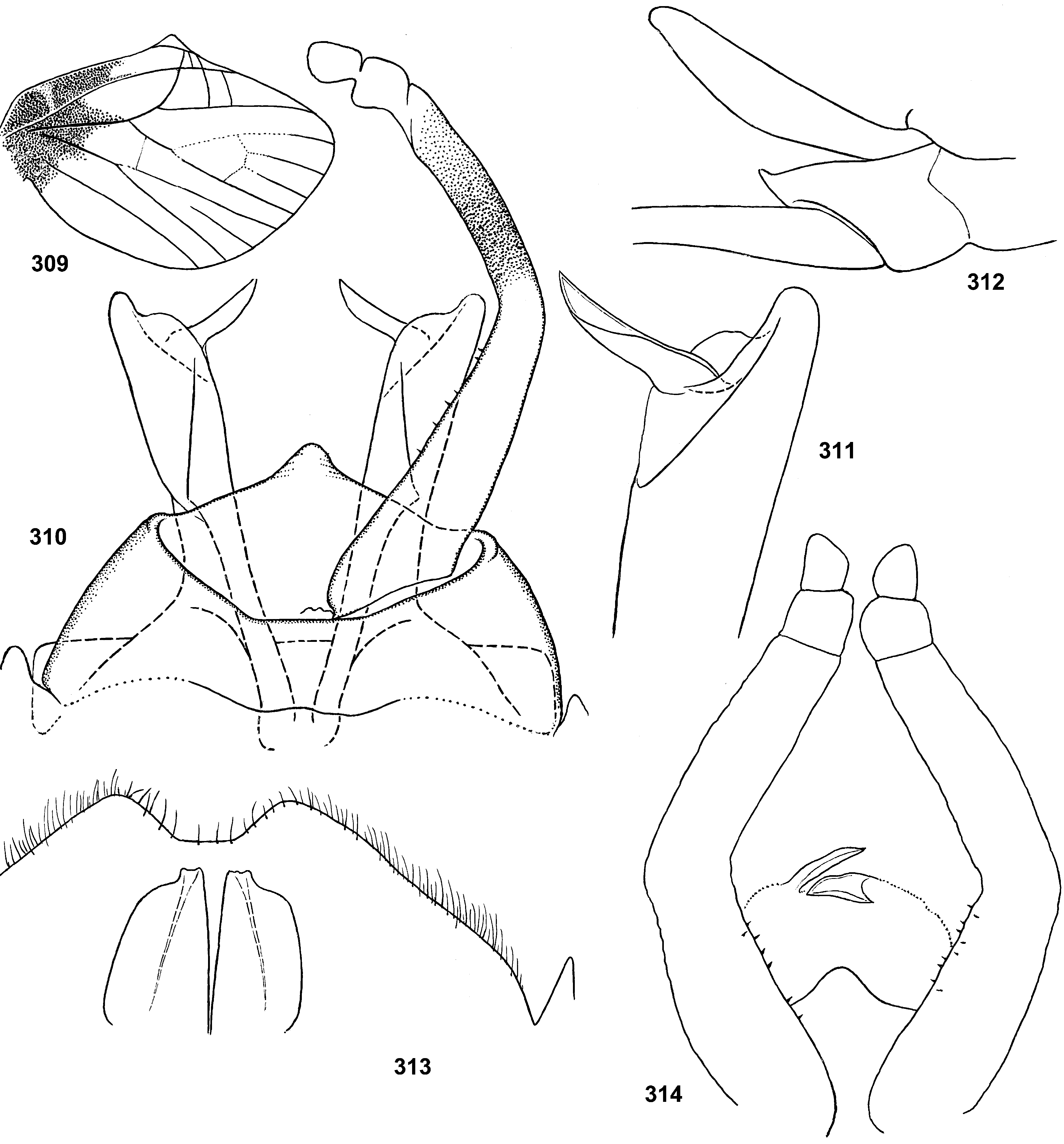
Male genitalia in last larval instar ( Fig. 313 View FIGURES 209–314 ): protogonostyli very short and separated one from another by small and shallow emargination. Each protopenis lobe apically wide, with gonopore-bearing process short, sharply projected caudally and located near median margin; gonopores opened caudally.
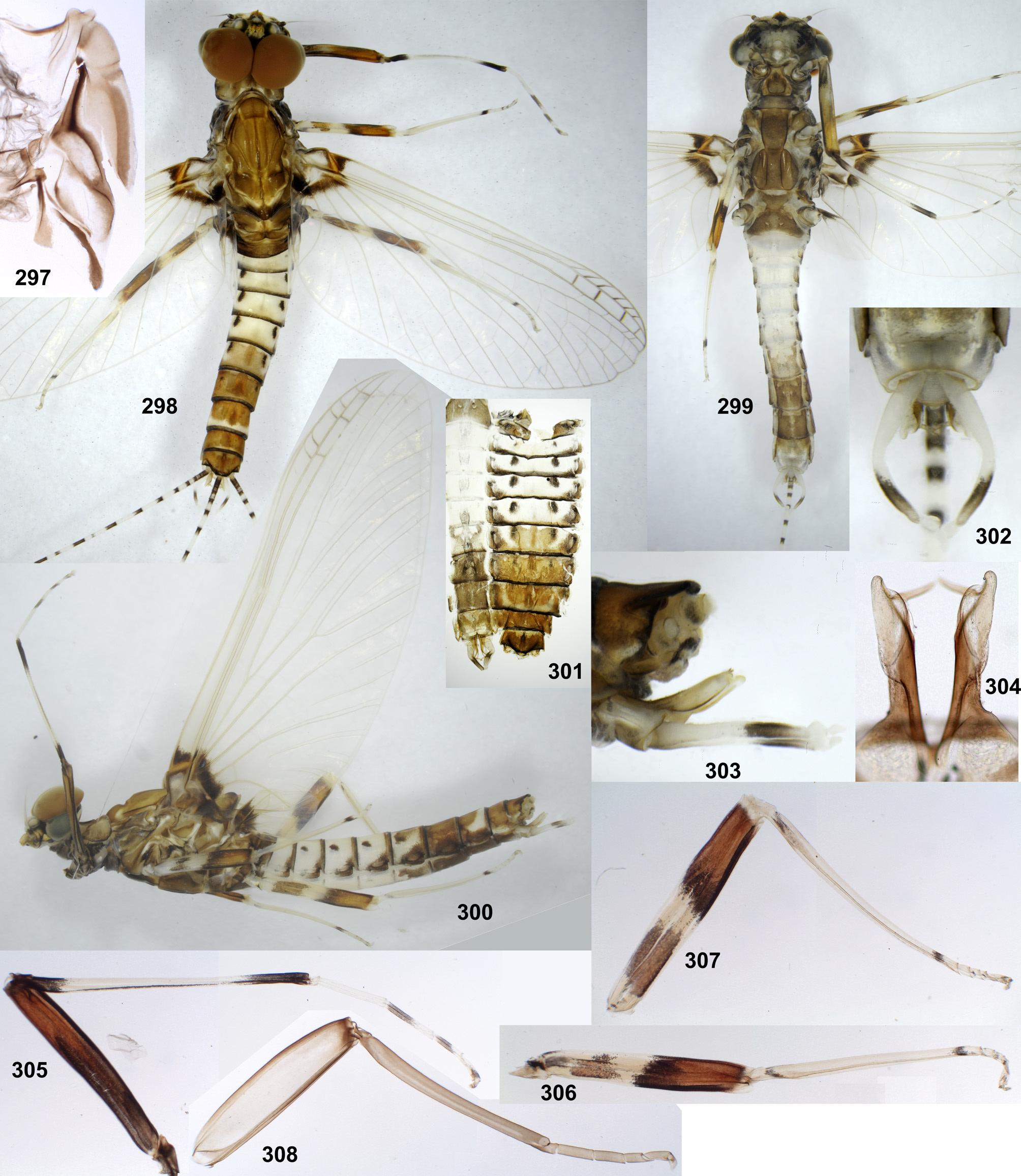
Subimago ( Figs 297, 308 View FIGURES 297–308 , 314 View FIGURES 209–314 ). CUTICULAR COLORATION. Cuticle with colorless and light brownish areas. Flagellum of antenna brownish. Pronotum light brownish. Mesonotum with lighter and darker brown areas; chromozone of medioscutum brown, chromozone of submedioscutum contrastingly lighter brownish ( Fig. 297 View FIGURES 297–308 ). On all legs femur mostly light, bordered by brown on outer and inner margins, with apex brown; tibia brownish; tarsus lighter brownish ( Fig. 308 View FIGURES 297–308 ). Cuticle of wings colorless, microtrichia light brownish. Abdominal terga light brownish, sterna and styliger colorless. Gonostyli and caudalii brownish.
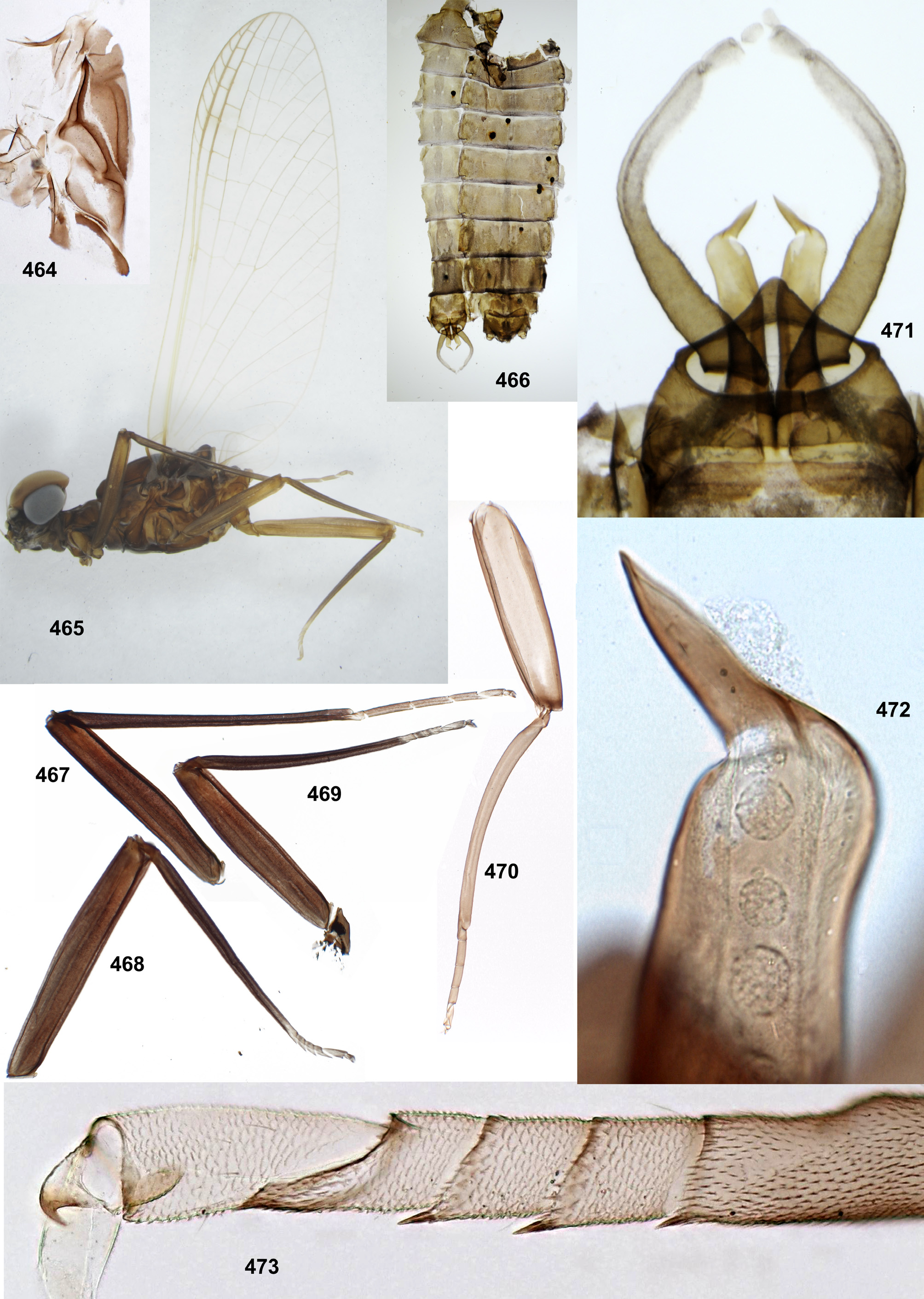
TEXTURE. On tarsi of all legs, 1st tarsomere with microtrichia (as tibia), 2nd–5th tarsomeres coved by blunt microlepides; pointed microlepides present near apical margins of 2nd–4th tarsomeres of all legs (as in Fig. 473 View FIGURES 464–473 ).
Male imago ( Figs 298–307 View FIGURES 297–308 , 309–311 View FIGURES 209–314 ). Head and antennae brown. Dorsal eyes contiguous medially, dull orange. Pronotum light grayish with brown maculae. Mesonotum and metanotum brown or ocher. All thoracic sterna and sclerites of thoracic pleura brown; thoracic pleura with whitish and dark brown hypodermal markings.
Cuticle of legs mostly colorless; on fore leg apex of femur and base of tibia with cuticle light brown. Legs with following hypodermal pigmentation on whitish background: Fore femur entirely pigmented, with proximal part blackish-brown and distal part reddish-brown (on anterior side proximal 2/3 blackish, on posterior side proximal 1/4 blackish), with blackish stripe along inner margin. Fore tibia with narrow longitudinal brown stripe on inner side, basally and apically dark brown. Fore tarsus with 3rd and 4th tarsomeres dark brown proximally, 5th tarsomere dark brown at middle. Middle femur with gray-brown spot on posterior side close to base, larger brown spot on anterior side more distally and multicolored pre-apical band occupying distal half of femur both on anterior and posterior sides; pre-apical band mainly reddish-brown, proximally bordered by blackish brown, on inner side with blackishbrown longitudinal stripe. Hind femur with large pre-basal brown spot on anterior side, beginning at short distance from femur base and occupying most part of proximal half of femur, and with multicolored pre-apical band as on middle femur. On middle and hind legs tibia with brown spots near base (close to patella-tibial suture) and on apex; tarsus with small brown spots on 3rd and 4th tarsomeres. In one of paratypes, relation of fore femur to fore wing length 65:280; proportions femur/tibia/tarsomeres on fore leg 65:78:3:20:16:9:9; on middle leg 63:62:3:4:4:2:7; on hind leg 76:70:3:4:4:2:7.
Fore wing with dark brown band just distad of costal brace; costal brace lighter orange; very base of fore wing non-colored. In area of pterostigma cross veins of costal and subcostal fields brown and narrowly bordered with brown; longitudinal veins C, Sc and RA in pterostigma area either brown, or ocher. In other areas of fore wing longitudinal and cross veins ocher; costal cross veins proximad of bulla colorless and very thin, visible only under high magnification (as in Fig. 142 View FIGURES 133–142 ). Pterostigmatic cross veins not dense, oblique, non-branched. Hind wing short, with costal projection prominent; proximal part of hind wing brown, with costal brace lighter ( Fig. 309 View FIGURES 209–314 ). Proximal coloration of fore and hind wings appears during larval development and present both in subimago and imago; by contrast, coloration of cross veins in pterostigma area exists only in fully molted imagines, being absent in imagines whose apices of fore wings have non-shed subimaginal cuticle.
Abdominal hypodermal coloration: Posterior margins of abdominal terga I–X narrowly bordered by dark brown. Tergum I brown. Terga II–VI partly whitish and translucent, with postero-lateral corners brown, stigmatic spots darker brown; terga III–VI with dark brown midway spots; on tergum VI area between these spots either lighter brown ( Fig. 301 View FIGURES 297–308 ), or whitish with brown spot medially. Terga VII–X mostly brown. Abdominal sterna I and VI–IX brown; sterna II–V whitish, translucent. Caudalii with segments alternated as following: black; colorless; colorless proximally and black distally; colorless.
Genitalia ( Figs 302–304 View FIGURES 297–308 , 310–312, 314 View FIGURES 209–314 ): Styliger light ocher; gonostyli proximally light ocher, distally brown; penis with proximal-median areas brown, distal-lateral areas light ocher (Fig.). Dorsal extension of styliger small and conic. Penis lobes elongate and slightly divergent; each lobe nearly parallel-sided, not widened apically and without lateral pouch. Apico-lateral area forming ear directed caudally; ventro-apical plate moderately developed, hiding base of spear from ventral view. Telopenes in form of spear-like rolls, directed medially and slightly arched caudally, invisible from lateral view ( Fig. 312 View FIGURES 209–314 ), with groove opened dorsally-laterally ( Fig. 311 View FIGURES 209–314 ).
Female imago. Coloration of head, thorax, legs, wings and caudalii as in male (single examined female in subimaginal cuticle has colorless veins of pterostigma, as well as males, whose wings are in the same condition). Abdomen brown with poorly expressed darker brown midway spots on terga III–VI.
Eggs ( Figs 315–316 View FIGURES 315–316 ). Mostly barrel-shaped. Each KTC surrounded by wide, smooth, ring-like cover. Other chorion with large protuberances forming rows equidistantly from each two neighboring KTCs.
Dimension. Fore wing length (and approximate body length) 6.5 mm.
Comparison. Th. fascipennis sp. n. is closely related to Th. zonalis : imago has the similar coloration of legs and wings and similar structure of male genitalia ( Traver & Edmunds 1967: figs 25, 41). The male imago of Th. fascipennis sp. n. differs from Th. zonalis by coloration of abdomen and longer penis lobes. Subimago of Th. fascipennis sp. n. differs from Th. zonalis by lighter cuticle of wings. Last instar male larva of Th. fascipennis sp. n. differs from Th. zonalis by very short protogonostyli with shallow emargination between them ( Fig. 313 View FIGURES 209–314 ). The egg of Th. fascipennis sp. n. differs by arrangement of protuberances, which are located at maximum distance from KTCs ( Figs 315–316 View FIGURES 315–316 ) (in contrast to all other species, whose protuberances are either evenly dispersed, or located mainly near KTCs, or absent). Both male and female imagines and subimagines of Th. fascipennis sp. n. can be distinguished from Th. zonalis by coloration of the hind femora: in Th. fascipennis sp. n. the proximal brown spot on the anterior side does not reach the base of the femur ( Fig. 307 View FIGURES 297–308 ), while in Th. zonalis it reaches the base of the femur ( Fig. 341 View FIGURES 337–345 ).


No known copyright restrictions apply. See Agosti, D., Egloff, W., 2009. Taxonomic information exchange and copyright: the Plazi approach. BMC Research Notes 2009, 2:53 for further explanation.
|
Kingdom |
|
|
Phylum |
|
|
Class |
|
|
Order |
|
|
Family |
|
|
Genus |