
Podranea ricasoliana
|
publication ID |
https://doi.org/10.5252/adansonia2023v45a12 |
|
DOI |
https://doi.org/10.5281/zenodo.8015296 |
|
persistent identifier |
https://treatment.plazi.org/id/039687DC-FFE5-911F-8D31-C0F7FAA2AC40 |
|
treatment provided by |
Felipe |
|
scientific name |
Podranea ricasoliana |
| status |
|
Podranea ricasoliana View in CoL View at ENA
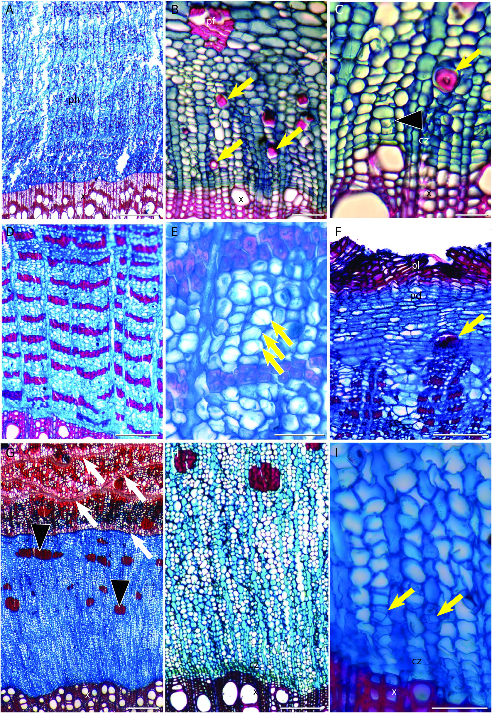
Non-stratified phloem ( Fig. 19G, H View FIG ). Sieve tubes solitary or in multiples of 2-3 ( Fig. 19I View FIG ). Most sieve plates simple, on a transverse to slightly inclined wall. One or two companion cell per sieve-tube element ( Fig. 19I View FIG ), lying on opposite sides of the sieve tube. Companion cells in strands of two cells. Axial parenchyma constitutes the ground tissue ( Fig. 19H, I View FIG ), two cells per parenchyma strand, sometimes up to four. Course of rays straight ( Fig. 19G View FIG ). Ray dilatation seemingly absent ( Fig.19G View FIG ). Rays bi-triseriate, heterocellular mixed,with procumbent, square and upright cells intermixed across the entire ray. Rays smaller than 1 mm, except when two or more rays merge. Rays do not sclerify, not even when touching the fibersclereid clusters ( Fig. 19G, H View FIG ). Sclerenchyma composed of fibersclereids only, differentiated in the nonconducting phloem in discrete, evenly distributed clusters. Non-storied. In young stems, pericyclic fibers forming a ring of discrete strands. Multiple periderms (rhytidome), reticulate ( Fig. 19G View FIG ). The phellem and phelloderm are thin walled, non-stratified ( Fig. 19G View FIG ). The phelloderm is thin, with less than three cell layers ( Fig. 19G View FIG ).
No known copyright restrictions apply. See Agosti, D., Egloff, W., 2009. Taxonomic information exchange and copyright: the Plazi approach. BMC Research Notes 2009, 2:53 for further explanation.
|
Kingdom |
|
|
Phylum |
|
|
Class |
|
|
Order |
|
|
Family |
|
|
Genus |