
Perianthomega
|
publication ID |
https://doi.org/10.5252/adansonia2023v45a12 |
|
DOI |
https://doi.org/10.5281/zenodo.8015255 |
|
persistent identifier |
https://treatment.plazi.org/id/039687DC-FFC9-9133-8F52-C117FAF5ABFE |
|
treatment provided by |
Felipe |
|
scientific name |
Perianthomega |
| status |
|
I. Perianthomega View in CoL View at ENA clade
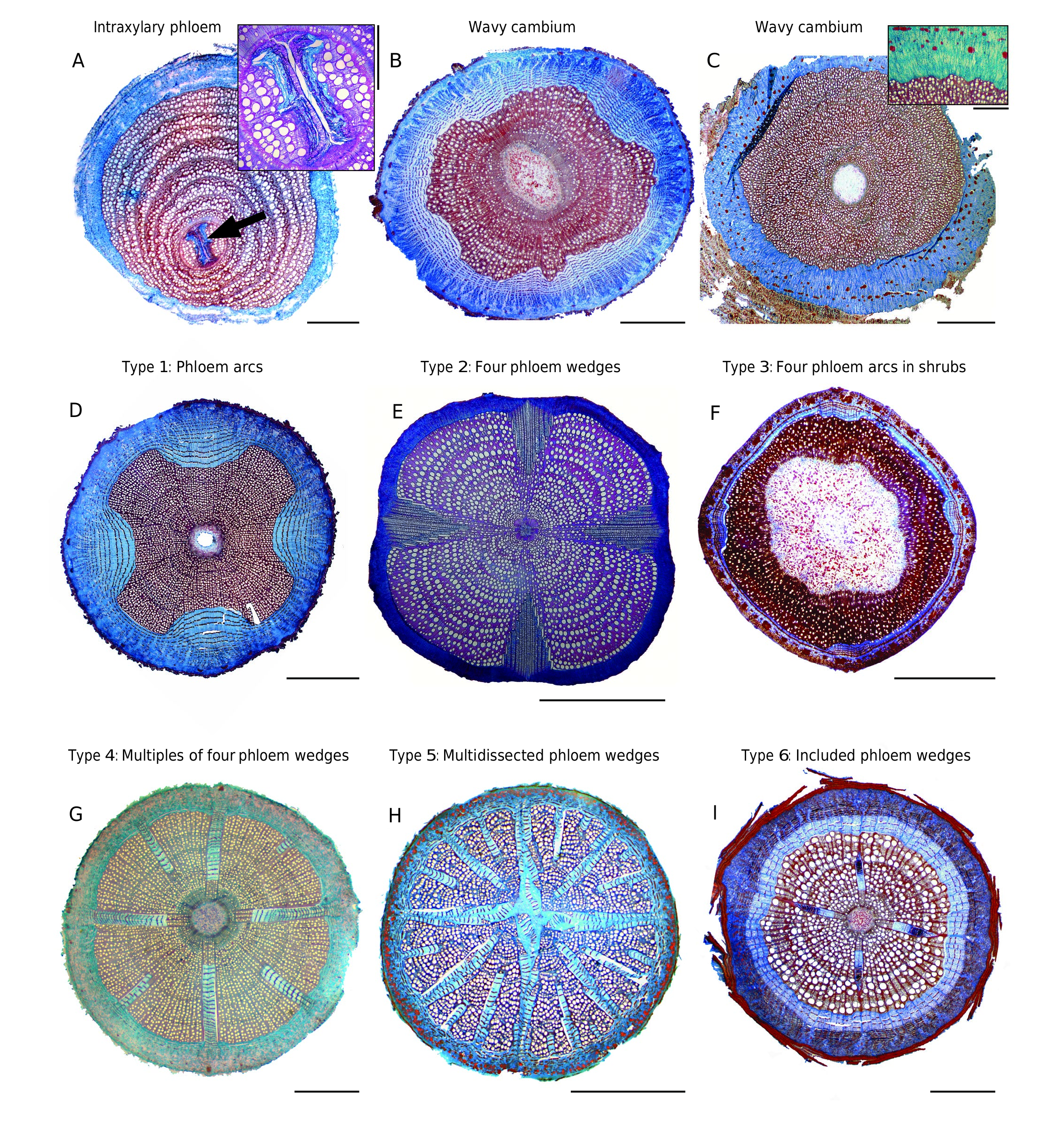
TAXONOMIC INFORMATION. — This clade contains a single genus, Perianthomega , with four phloem arcs in transversal section ( Fig. 3D View FIG ). The genus was placed previously in Tecomeae s.l. ( Gentry 1992; Fischer et al. 2004; Table 1 View TABLE ), but subsequently transferred into Bignonieae ( Lohmann & Taylor 2014).
TOTAL NUMBER OF SPECIES IN THIS CLADE. — One species ( Lohmann & Taylor 2014).
STUDIED SPECIES. — One species, Perianthomega vellozoi Bureau.
Regular phloem
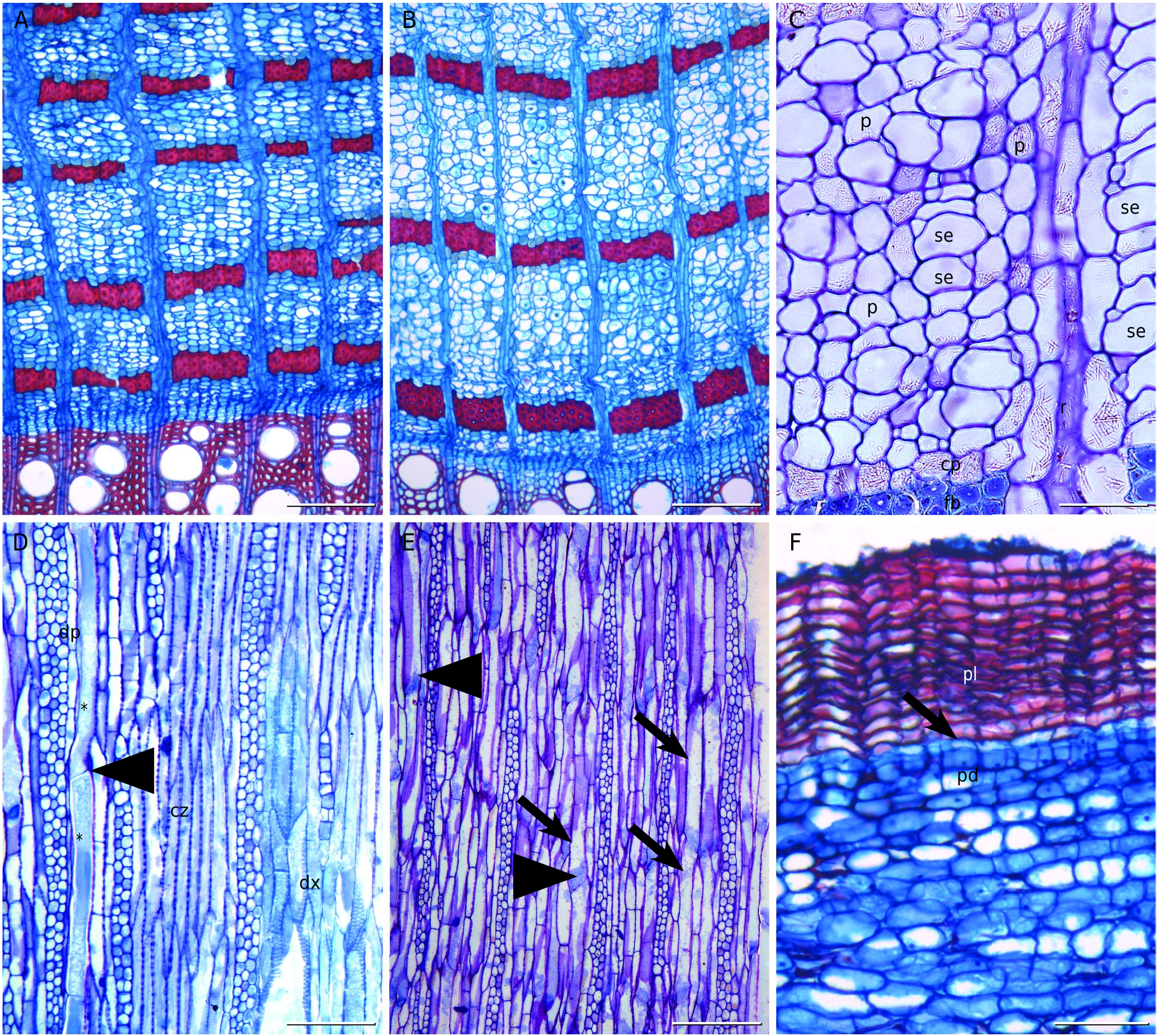
Thin to thick fiber bands ( Fig. 7A View FIG ), without assemblages.
Variant phloem
General configuration. Non-fibrous, stratified, with approximately 12-18 rows of sieve tubes and phloem parenchyma cells between fiber bands ( Fig. 7B View FIG ). A tendency to a storied structure is evident in the cambial zone ( Fig. 7D View FIG ) and secondary xylem, less so in the secondary phloem ( Fig. 7E View FIG ).
Sieve tube elements. As seen in transverse section, each sieve element is associated with one companion cell ( Fig. 7C View FIG ), sometimes two, one at each opposite corner of the sieve element. Sieve tubes are solitary or radial and composed of 2-3 cells ( Fig. 7B, C View FIG ). As seen in longitudinal section, the sieve elements are short (<500 µm) ( Fig. 7E View FIG ) and their end walls are transverse to slightly inclined ( Fig. 7D, E View FIG ), bearing simple sieve plates ( Fig. 7E View FIG ).
Axial parenchyma. The phloem parenchyma forms a matrix where sieve elements and fiber bands are embedded ( Fig. 7B, C View FIG ). Long radial rows of phloem parenchyma, sometimes with more than 10 cells, are present. Furthermore, a crystalliferous parenchyma is found surrounding the fiber bands ( Fig. 7C View FIG ).
Fibers. The fiber bands are composed of 4-6 rows of cells ( Fig. 7B View FIG ).
Rays. Limiting rays are not present in Perianthomega . The rays are non-lignified, not even when crossing the fiber bands ( Fig. 7B View FIG ).
Crystals. Acicular crystals are present and more abundant in the crystalliferous parenchyma, although also found in all other cells of the phloem and ray parenchyma ( Fig. 7C View FIG ).
Periderm
Only one periderm is formed ( Fig. 7F View FIG ). The phellem is composed of evenly thin-walled cells. The phelloderm is thin, with less than three layers of cells ( Fig. 7F View FIG ).
No known copyright restrictions apply. See Agosti, D., Egloff, W., 2009. Taxonomic information exchange and copyright: the Plazi approach. BMC Research Notes 2009, 2:53 for further explanation.
|
Kingdom |
|
|
Phylum |
|
|
Class |
|
|
Order |
|
|
Family |