
Cylapoides Carvalho
|
publication ID |
https://doi.org/10.11646/zootaxa.5074.1.1 |
|
publication LSID |
lsid:zoobank.org:pub:7B3C6765-F0D2-4846-BB95-200258ECC0E1 |
|
DOI |
https://doi.org/10.5281/zenodo.5784480 |
|
persistent identifier |
https://treatment.plazi.org/id/039587FB-AE58-FFB7-FF51-1157432DF9FC |
|
treatment provided by |
Plazi (2021-12-06 08:45:40, last updated 2024-11-28 20:55:07) |
|
scientific name |
Cylapoides Carvalho |
| status |
|
Cylapoides Carvalho View in CoL
Cylapoides Carvalho 1952a: 269 View in CoL (new genus). Type species: Cylapoides bicolor Carvalho, 1952 View in CoL (original designation). Cylapoides: Carvalho 1955: 21 View in CoL (key to genera), 1957: 28 (catalog); Schuh 1995: 22 (catalog); Carvalho & Froeschner 1987: 128 (list); Gorczyca 2000: 48 (list), 2006a: 14 (catalog).
Diagnosis. Recognized by the following set of features: dorsum impunctate ( Figs 3c, d View FIGURE 3 , 11e View FIGURE 11 ); head broad, ca 3.6–5.0 x as wide as long; eyes not pedunculate, covering anterior magin of pronotum ( Figs 3c,d View FIGURE 3 , 11e View FIGURE 11 ); clypeal base weakly removed from ventral margin of eyes ( Fig. 8b View FIGURE 8 ); antennal insertion weakly removed from suture between maxillary and mandibular plates ( Fig. 8b View FIGURE 8 ); antenna relatively short, total length shorter than body length ( Fig. 3c, d View FIGURE 3 ); pronotum short, ca 5 x as wide as long ( Figs 3c, d View FIGURE 3 , 11e View FIGURE 11 ); endosoma composed of inflated, sclerotized lobes ( Fig. 12g View FIGURE 12 ); left paramere apical process with outgrowth basally ( Fig. 12h View FIGURE 12 ; Carvalho 1952: figs 7, 9); right paramere sickle-shaped ( Fig. 12i View FIGURE 12 ; Carvalho 1952: figs 7, 8).
Redescription. Male. COLORATION ( Figs 3c, d View FIGURE 3 ). Body dark brown with yellow and silvery areas. SURFACE AND VESTITURE. Dorsum impunctate ( Fig. 11e View FIGURE 11 ). Head. Covered with long, dense setae; antenna covered with short, semi-recumbent setae; setae on segment III and IV denser than those on segment II. Thorax. Pronotum, mesoscutum, scutellum and hemelytron. Covered with long, irregularly distributed, semi-recumbent setae. Thoracic pleura. Impunctate; covered with sparse, short setae. Abdomen. Covered with dense, reclining setae. STRUCTURE ( Figs 3c, d View FIGURE 3 , 8b View FIGURE 8 , 11e View FIGURE 11 ). Macropterous, elongate-oval. Head. Broad, ca 3.6 –5.0 x as wide as long; eyes not pedunculate, covering anterior margin of pronotum; vertex strongly carinate posteriorly; clypeal base weakly removed from ventral margin of eye; antennal insertion weakly removed from suture between maxillary and mandibular plates antenna relatively short, shorter than body length antenna long as long as body length; antennal segment I short, ca 0.3–0.4 x as long as interocular distance; antennal segment I and II weakly broadened toward apex, segment I slightly ticker than segment II; segments III and IV thinner than segment II, filiform; labium relatively thick, apex reaching II abdominal segment, its segments not subdivided. Thorax. Pronotum. Collar thin, delimited by shallow depression; calli flattened, separated by shallow depression; lateral margin rounded; pronotum short, ca 5 x as wide as long. Mesoscutum and scutellum. Weakly convex. Thoracic pleura. Scent gland evaporative areas moderately developed; peritreme weakly raised above evaporative areas, oval. Hemelytron. Embolium narrow; cuneus longer than wide. Legs. Short; tarsus with tarsomere I about two times shorter than tarsomere II and III combined; pretarsal claw toothed subapically. Genitalia. Endosoma composed of inflated, sclerotized lobes ( Fig. 12g View FIGURE 12 ). Left paramere with apical process with outgrowth basally ( Fig. 12h View FIGURE 12 ; Carvalho 1952: figs 7, 9). Right paramere sickle-shaped ( Fig. 12i View FIGURE 12 ; Carvalho 1952: figs 7, 8).
Female. Like male in coloration, structure, texture, and vestiture. For female genitalia see description of C.unicolor .
Carvalho, J. C. M. (1952 a) Neotropical Miridae, 56: Description of three new genera and five new species from Brazil and British Honduras (Hemiptera). Revista Brasileira de Biologia, 12, 265 - 273.
Carvalho, J. C. M. (1955) Keys to the genera of Miridae of the world (Hemiptera). Boletim do Museu Paraense Emilio Goeldi, 11, 1 - 151.
Carvalho, J. C. M. & Froeschner, R. C. (1987) Taxonomic names proposed in the insect order Heteroptera by Jose Candido de Melo Carvalho from 1943 to January 1985, with type depositories. Journal of the New York Entomological Society, 95, 121 - 224.
Gorczyca, J. (2000) A systematic study on Cylapinae with a revision of the Afrotropical Region (Heteroptera, Miridae). Wydawnictwo Uniwersytetu Slaskiego, Katowice, 174 pp.
Schuh, R. T. (1995) Plant bugs of the World (Insecta: Heteroptera: Miridae). New York Entomological Society, New York, New York, 1329 pp.
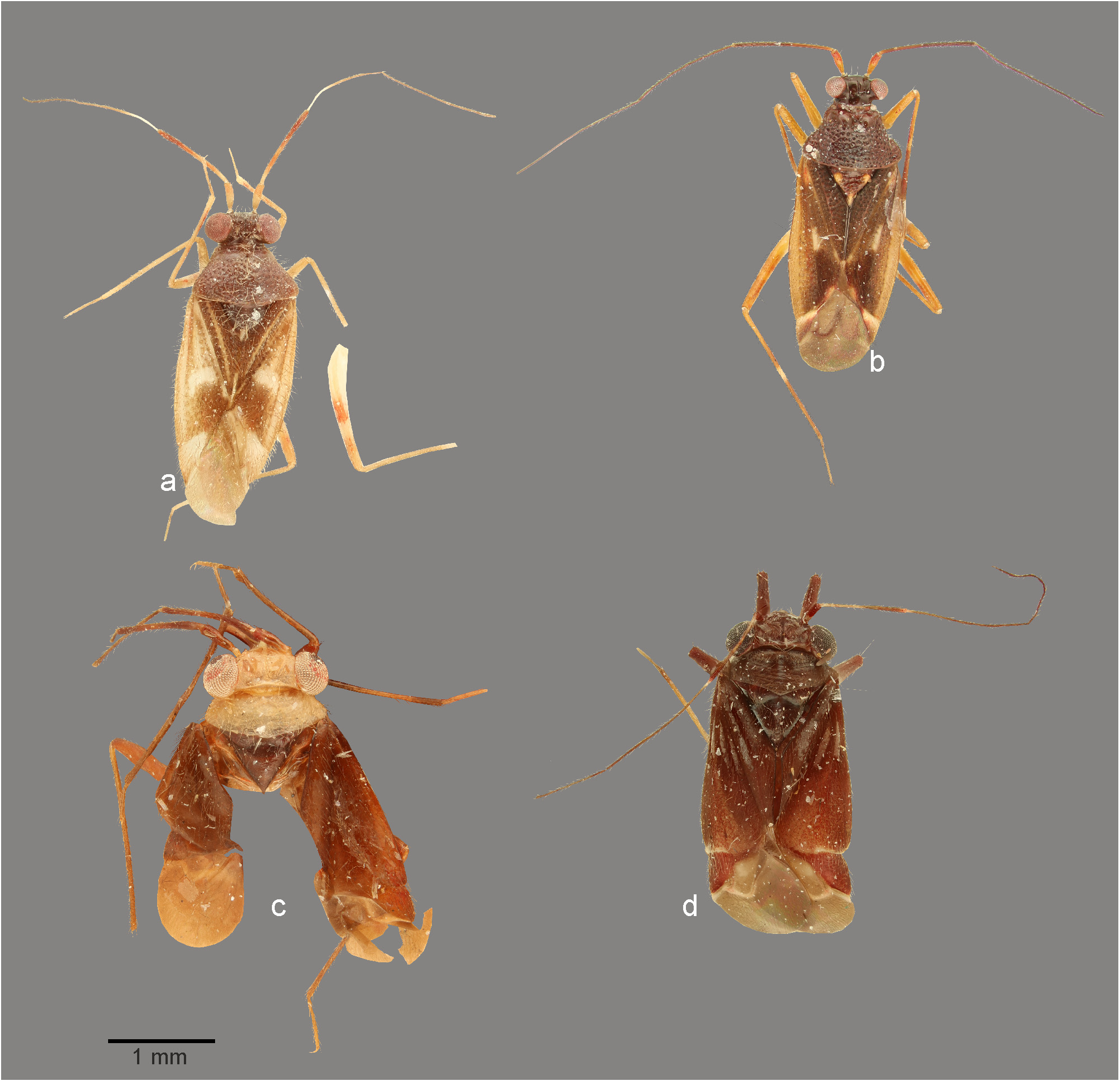
FIGURE 3. Dorsal habitus photographs. a. Cylapinus minusculus (♂, Ecuador); b. Cylapinus yasunagai (holotype); c. Cylapoides bicolor (holotype); d. Cylapoides unicolor (♂, Ecuador).

FIGURE 8. Head and pronotum, anterior (a–j, l, m) and lateral (k) views. a. Cylapinus yasunagai (paratype); b. Cylapoides unicolor (♀); c. Cylapus ruficeps Bergroth (♂); d. Cylapus tenuicornis (♂); e. Peltidocylapus calyciformis (paratype); f. Peltidocylapus caudatus (paratype); g. Peltidocylapus rugosus (holotype); h. Peltidocylapus simplex (paratype); i. Cylapomorpha sp. (♂); j, k. Vanniusoides clypeatus (paratype); l. Fulvius pallens (♂); m. Rhinocylapus vittatus (♀).

FIGURE 11. Scanning electron micrographs. a, e, g, m. Dorsal habitus; b, n. Lateral habitus; h, l. Scutellum; c, f, i. Thoracic pleura; d, j, o. Tarsus. k. Pretarsal claw. a–d. Cylapinus minusculus; e, f. Cylapoides unicolor; g–j. Peltidocylapus caudatus; k. Peltidocylapus calyciformis; l. Peltidocylapus ecuadorensis; m–o. Peltidocylapus scutellaris.

FIGURE 12. Male genitalia. a, d, g, j, p. Endosoma (dorsal view); b, e, h, m, s. Left paramere (right lateral view). c, f, i, o, u. Right paramere (left lateral view); n, t. Left paramere (dorsal view). k, q. Transparent portion of distal sclerotized portion of ductus seminis inside endosoma; l, r. Endosoma (ventral view). a–c. Cylapinus minusculus; d–f. Cylapinus yasunagai; g–i. Cylapoides unicolor; j–o. Peltidocylapus calyciformis; p–u. Peltidocylapus caudatus. ap = apical process; bp = basal process; dss = distal part of ductus seminis inside endosoma; pb = paramere body; sl = sensory lobe. Scale bars 0.2 mm.
No known copyright restrictions apply. See Agosti, D., Egloff, W., 2009. Taxonomic information exchange and copyright: the Plazi approach. BMC Research Notes 2009, 2:53 for further explanation.
|
Kingdom |
|
|
Phylum |
|
|
Class |
|
|
Order |
|
|
Family |
|
|
Tribe |
Cylapini |
Cylapoides Carvalho
| Wolski, Andrzej 2021 |
Cylapoides
| Schuh, R. T. 1995: 22 |
| Carvalho, J. C. M. & Froeschner, R. C. 1987: 128 |
| Carvalho, J. C. M. 1955: 21 |
| Carvalho, J. C. M. 1952: 269 |