
Nitella sonderi A.Braun
|
publication ID |
https://doi.org/ 10.5252/cryptogamie-algologie2022v43a13 |
|
DOI |
https://doi.org/10.5281/zenodo.7819473 |
|
persistent identifier |
https://treatment.plazi.org/id/039587A1-1C4D-FF88-FEC9-603492A59F61 |
|
treatment provided by |
Felipe |
|
scientific name |
Nitella sonderi A.Braun |
| status |
|
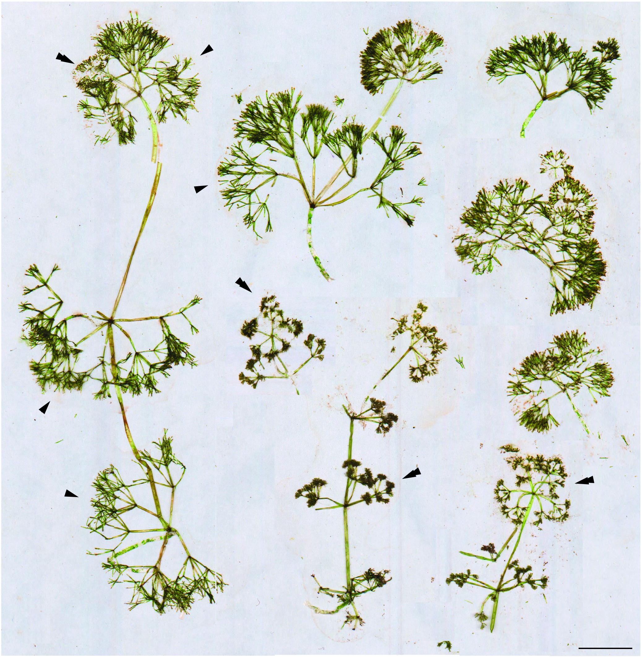
SPECIMENS EXAMINED. — Argentina. Neuquén Province, Alicurá Reservoir, 40°35’19”S, 70°52’34”W, 690 m a.s.l., 07.IV.2019, P. Quiroga & R. Vidal-Russell (LE[A0000321, A0000322], in spirit) ( Figs 1-3 View FIG View FIG View FIG ).
Australia. Queensland, 3.5 mi [5.6 km] S of Stanthorpe post office, W of road, SW shore of metal (gravel) quarry, in clear water, 20.XI.1960, R.D. Wood 60-11-20-22 ( LE [ A 0001490], GenBank accession: rbc L [ OM 311638 View Materials ]); 50.9 mi [81.9 km] S of Miriam Vale on road to Gin Gin, abundant in c. eight inches of water, mud bottom, creek in deep valley (Black Creek?), 30.XI.1960, R.D. Wood 60-11-30-10 ( LE [ A 0001491], GenBank accessions: rbc L [ OM 311639 View Materials ], ITS[ OM 338646 View Materials ]); 3.5 mi [6.5 km] S of Stanthorpe, post office, W of road, selected slender specimens common along S shore of metal (gravel) quarry, 20.XI.1960, R.D. Wood 60- 11-20-20 ( LE [ A 0001488]); Victoria, Benalla, N arm of Broken River, c. 300 yd. [c. 274 m] N of bridge (behind swimming pool), N shore of river, common at water’s edge, in c. 3 inches of fairly clear water, sandy gray mud bottom, 26.III.1963, R.D. Wood 61- 3-26-2 ( LE [ A 0001489]).
PHENOLOGY. — Male plants of N. sonderi were observed in the Patagonian locality during summer and autumn. A single plant producing antheridia has been successfully growing in an indoor aquarium since March 2020.
DESCRIPTION OF PATAGONIAN PLANTS
Plants dioecious (only male plants were found), green, not encrusted with lime, unbranched, longer than 13 cm, with isolated homeoclemous whorls, neither condensed nor clumped at the apex ( Fig. 1 View FIG ), with obvious and wide, somewhat diffluent mucilage envelope of upper part of plants covering fertile whorls and internodes with a layer ( Fig. 2D, F, G View FIG ), approximately as thick as the diameter of the internodes and uniting whole branchlets within common envelope, sometimes resulting in lax apical heads ( Figs 1 View FIG ; 2C, D View FIG ). Stem diameter is (196-)337-509(-540) µm, increasing towards the basal part.
Romanov R. E. et al.
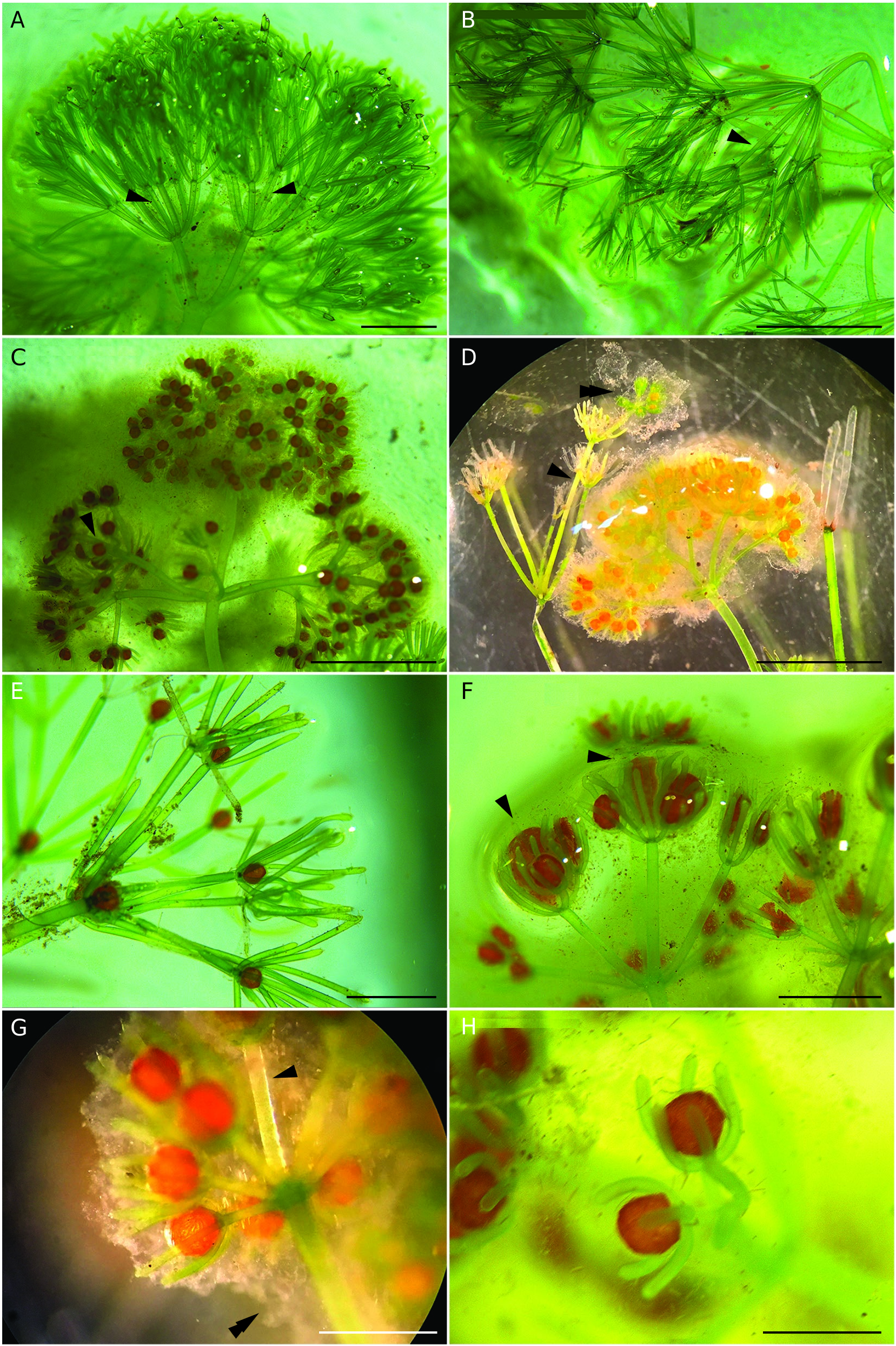
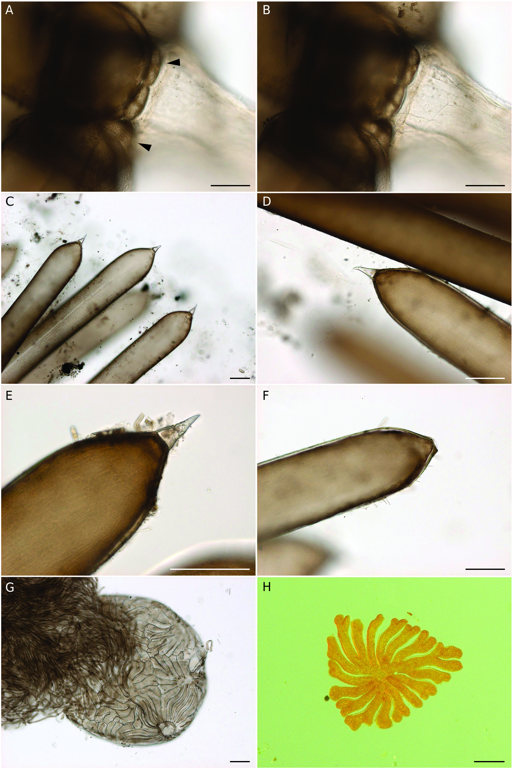
The transversely elliptical cells extending from the base of the branchlet and forming a ring surrounding it are easily recognisable at appropriate magnification ( Fig. 3A, B View FIG ). The pair of these cells is commonly present below the branchlet base, one of the cells as an exception can be missing. These cells contain chloroplasts, not starch, and are not discoloured, 42-109 µm in length, 71-164 µm in width. Their growth seems to be able to culminate in nodal bulbil formation, recognisable as overgrowth of stem nodes inside and outside of the whorl before destruction of branchlets.
The sterile and fertile branchlets are macrodactylous, with similar principal arrangement and quite uniform appearance within the same whorl. However, fertile ones have shorter penultimate rays, resulting in a tassel-like appearance at the ends of the central rays because of aggregated dactyls (cf. Figs 1 View FIG ; 2 View FIG A-D).
The sterile branchlets are 2-2.8-times shorter than the internodes; the length of internodes and the proportion between internode and branchlet length decrease towards the apex. The whorls of sterile branchlets are 32-44 mm in span, more lax and diffuse, spreading more in contrast to fertile ones, which are shorter and compact at their ends, with a diameter of (10-) 16-29 mm. The length of sterile branchlets is (10.9-)12.5-21.5(-23)mm,withprimaryrays(5-)7.7-9.5(-10)mm long, i.e., half of the total branchlet length or slightly shorter.The lateral secondary rays of sterile branchlets are 3.5-6(-6.5) mm in length, whereas the central secondary ones are approximately 4.5-5 mm.
The fertile branchlets form at apical parts of the plants ( Fig. 1 View FIG ). The length of fertile branchlets is 9-15 mm, with primary rays having a length of 5-8.5 mm, i.e., mostly somewhat exceeding half of the total branchlet length. The primary ray at the basal part is 155-382 µm in diameter, and in the apical part, in fertile whorls, it is 110-235 µm in diameter. The lateral and central secondary rays of fertile branchlets are shorter in absolute values (approximately two times less in case of the latter ones) in comparison with sterile branchlets.
Each branchlet is 2-3-times forked, with a central secondary ray, surrounded by 6-8 more or less equal or somewhat shorter lateral secondary rays (sometimes c. 0.4-0.7 the length of the central secondary ray), mostly not differing in width from the central one, but sometimes obviously more robust ( Fig. 2A, C, F, G View FIG ). As an exception, the branchlet can produce 3-furcate “prolification”, looking like a fertile branchlet without a central secondary ray. It forms from the lateral tertiary ray at the central secondary ray furcation ( Fig. 2D View FIG ). The lateral secondary rays are 1- or 1- and 2-times forked; the latter is less frequent at sterile branchlets in contrast with the common pattern of fertile ones. The lateral secondary rays are longer than the dactyls in sterile whorls and longer, nearly equal to mostly slightly or obviously shorter than the dactyls in fertile whorls (to approximately 0.8 of dactyl length). The dactyls of 1- and 2-times forked lateral secondary rays in sterile whorls are 1.8-2-times shorter than those in tertiary rays.
The central tertiary ray is neither formed at the furcation of lateral rays nor at the central secondary ray; the tertiary rays are 1-3 (or more?) in furcation and, as a rule, shorter than dactyls. The central secondary rays are 2-times furcated.
The dactyls are 2-4(-5) in sterile branchlets, and 5-6 in fertile branchlets, strictly bicellulate, 0.8-2.8(-3) mm in length in sterile branchlets, 0.8-1.3 mm long in fertile branchlets, at second and third furcations, and at the first furcation of sterile branchlets, straight at sterile and fertile nodes in case of dactyls significantly longer of the antheridium, usually more or less arcuate in their basal parts at nodes with the antheridium in case of short dactyls, 1.2-2-5-times longer than the antheridium ( Fig. 2C View FIG , E-H). The penultimate cell is long cylindrical with a shortly narrowing end ( Fig. 3 View FIG C-E). The base of the end cell is confluent with the tip of penultimate cells. The end cell is small, conical, pointed, straight or somewhat curved through the whole length or only at the tip, mostly discoloured in living, pressed and fixed states. The dactyls appear single-celled at lower magnification because of tiny and mostly discoloured confluent end cells. The width of dactyls at their base (above the antheridium) is (112-)130-250 µm, at the apical part (below the shortly narrowing end) it is 123-187 µm. The end cells are 46-68 µm in length and 15-31 µm in width. The tip of the end cell has an obviously thickened cell wall ( Fig. 2 View FIG C-D). The end cells can be lost over time ( Fig. 3F View FIG ).
The antheridia are solitary, strictly terminal at the second and third furcations of the branchlet, never at the first one, sessile, round with a slightly attenuated base, octoscutate (i.e., with triangular shields; Fig. 3G, H View FIG ), frequently looking robust in contrast with the length and width of dactyls surrounding them ( Fig. 2 View FIG E-H), (370-)399-655(-762)µm in diameter; their diameter can be different within the same branchlet ( Fig. 2F View FIG ).
| S |
Department of Botany, Swedish Museum of Natural History |
| W |
Naturhistorisches Museum Wien |
| R |
Departamento de Geologia, Universidad de Chile |
| LE |
Servico de Microbiologia e Imunologia |
| A |
Harvard University - Arnold Arboretum |
| L |
Nationaal Herbarium Nederland, Leiden University branch |
| OM |
Otago Museum |
| N |
Nanjing University |
No known copyright restrictions apply. See Agosti, D., Egloff, W., 2009. Taxonomic information exchange and copyright: the Plazi approach. BMC Research Notes 2009, 2:53 for further explanation.
|
Kingdom |
|
|
Phylum |
|
|
Class |
|
|
Order |
|
|
Family |
|
|
Genus |