
Russula indosenecis A.Ghosh, D.Chakr., K.Das & Buyck, 2022
|
publication ID |
https://doi.org/ 10.5852/ejt.2022.847.1985 |
|
DOI |
https://doi.org/10.5281/zenodo.7376910 |
|
persistent identifier |
https://treatment.plazi.org/id/03947A67-FF80-FFF9-9E83-FDF516D7BE33 |
|
treatment provided by |
Felipe |
|
scientific name |
Russula indosenecis A.Ghosh, D.Chakr., K.Das & Buyck |
| status |
sp. nov. |
Russula indosenecis A.Ghosh, D.Chakr., K.Das & Buyck View in CoL sp. nov.
MycoBank: MB842307
Figs 2–4A View Fig View Fig View Fig
Diagnosis
Russula indosenecis sp. nov. resembles Japanese R. senecis Imai but differs from it mainly by the strongly amyloid suprahilar spot on the basidiospores, genetic distance of the nrITS sequences (97.25%–97.79% similarity) and its occurrence under Abies densa Giff. in subalpine forests.
Etymology
Referred to its occurrence in Indian Himalaya and morphological resemblance to R. senecis .
Material examined
Holotype INDIA • East Himalayan Region , Tawang district, on the way to Panga Teng Tso Lake; 27°38′15.5″ N, 91°51′12.1″ E; alt. 3935 m a.s.l.; in subalpine forest under Abies densa ; 30 Aug. 2021; A. Ghosh AG-21- 06A; GenBank: OL701269 View Materials (ITS); CAL[1856]. GoogleMaps
Paratype INDIA • East Himalayan Region , Tawang district, on the way to Panga Teng Tso Lake; 27°38′15.2″ N, 91°51′11.6″ E; alt. 3919 m a.s.l.; in subalpine forest under Abies densa ; 29 Aug. 2021; A. Ghosh AG-21- 04A; GenBank: OL701254 View Materials (ITS); CAL[1857] GoogleMaps .
Description
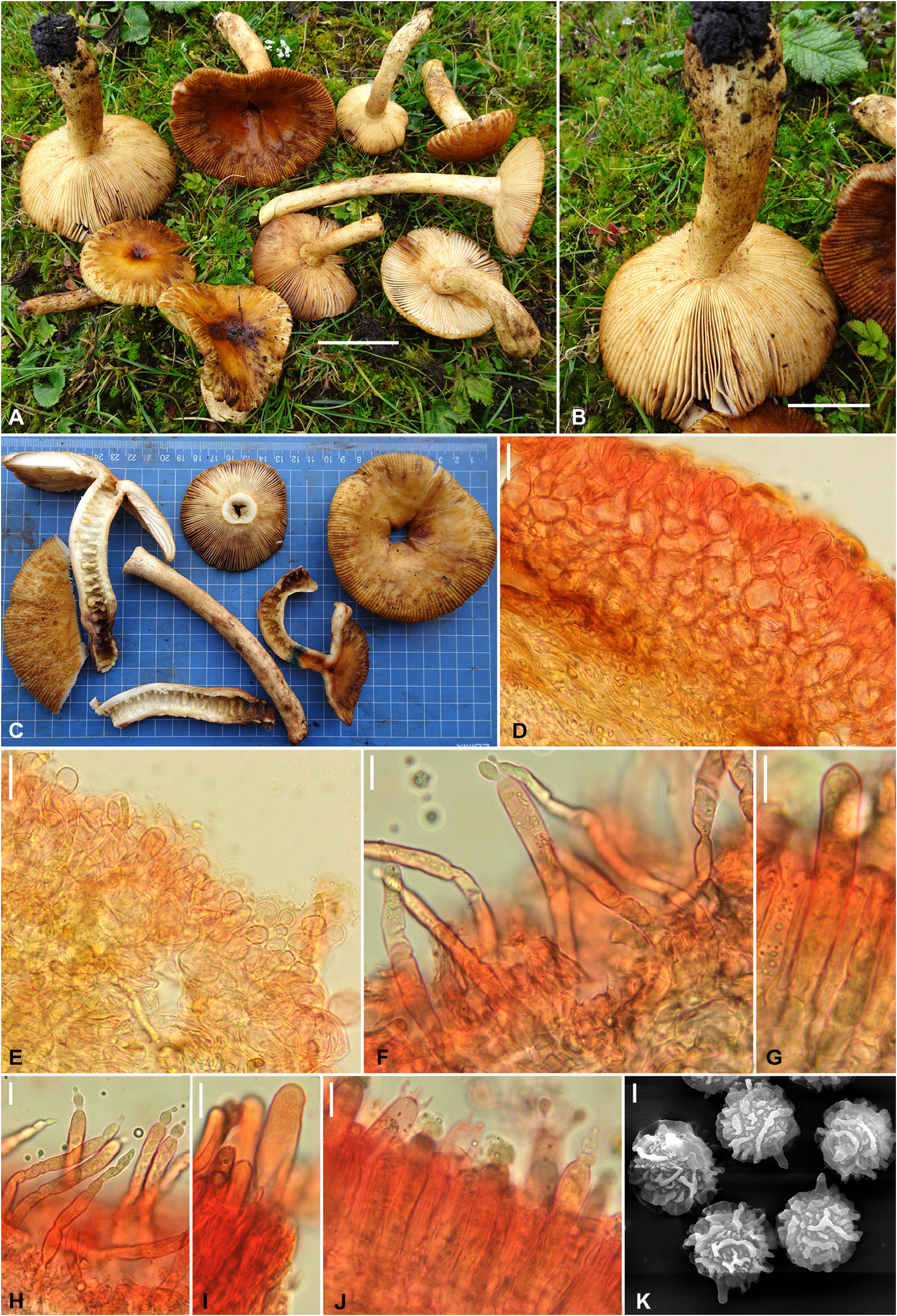
Pileus medium to large sized, 65–140 mm in diameter, convex, planoconvex to applanate with broadly depressed center, becoming infundibuliform with maturity; margin decurved to plane or uplifted with maturity, entire, strongly tuberculate-striate; surface viscid and glutinous when moist, dull with drying, quickly cracked, easily peeled off ⅓ rd to ¾ th toward center, light orange or melon yellow or apricot yellow or golden yellow (5A–B5–7), centrally turning dark brown (6–7E6–8) with maturity or age, turning orange (6A8) with KOH. Pileus context up to 6 mm thick at the disc, compact, brittle, firm, chalky white (1–2A1), unchanging after bruising or on exposure. Lamellae shortly adnate to subfree, equal or with rare lamellulae, subdistant (7–10/cm at pileus margin), rarely forked, chalky white (1A1) to pale cream (3A2) when young, becoming concolorous to pileus colour with age or maturity, unchanging after bruising or on exposure; edges punctuated with brownish orange (6C5–7) or light brown (6D5–7), entire. Stipe long and slender, 90–160 × 13–30 mm, firm, brittle, cylindrical to subclavate, centrally attached; surface dry, smooth, longitudinally striate, light yellow to maize yellow (4A4–6) with light brown (6D5–7) to brown (6D6–7) tinges. Stipe context light orange or apricot yellow or golden yellow (5A–B5–7), multichambered, soon hollowing, unchanging on exposure; turning deep to dark turquoise (24E–F7–8) with guaiacol, insensitive to FeSO 4. Odor indistinctive. Taste acrid and very strong to hurting. Spore print pale cream (IIb).
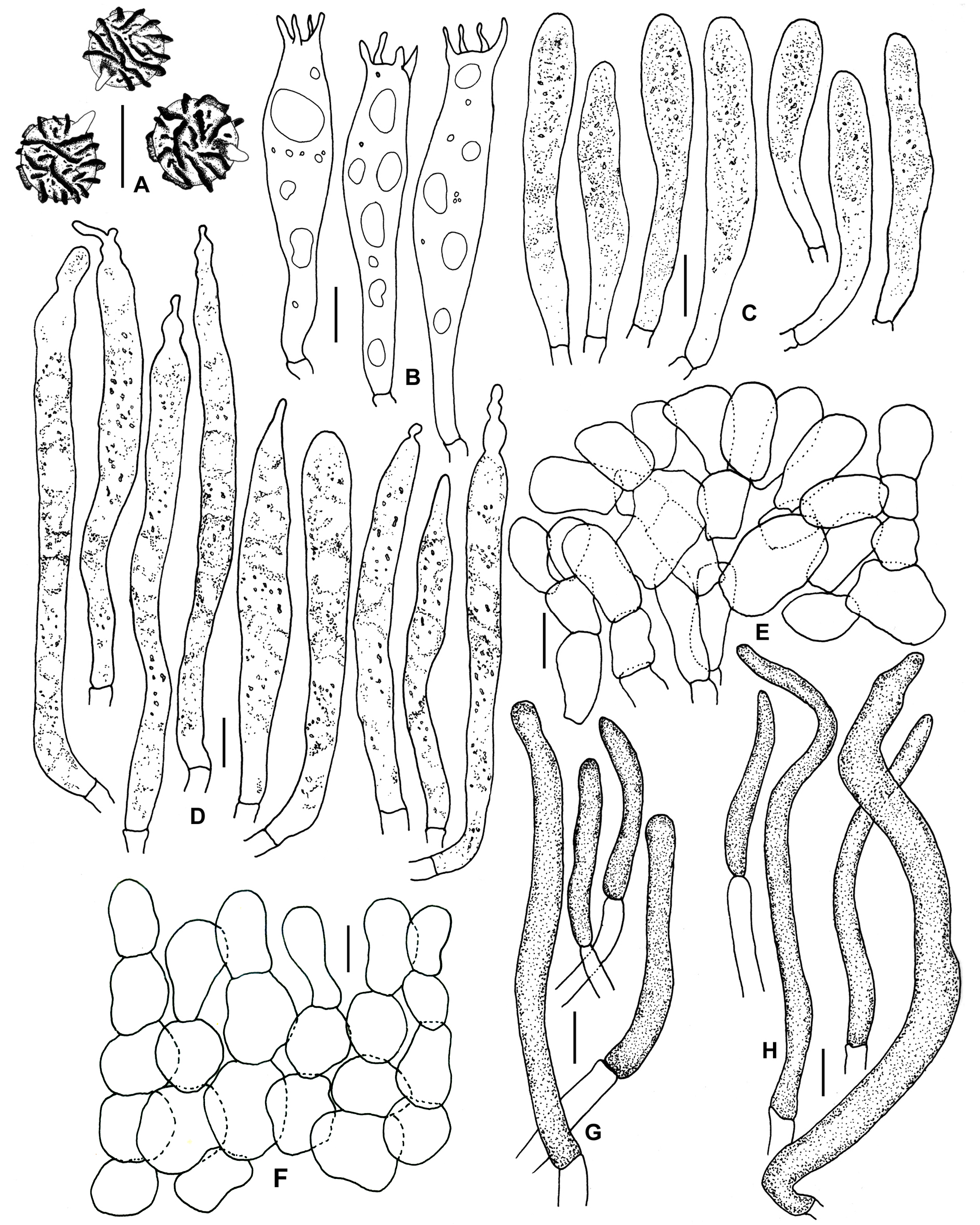
Basidiospores globose to subglobose, (8.4–)8.8–9.3–9.8(–10.5) × (8.2–)8.6–9.0–9.5(–10.4) μm, Q = (1–)1.01–1.03–1.06(–1.10); ornamentation amyloid, composed of up to 1.8 μm high wings running over more or less long distances on the spore surface or even nearly encircling the spores, mixed with dense, low network of short, laterally flattened, blunt ridges and warts forming an incomplete network, intermixed with crowded, isolated warts and large spines (up to 1.5 μm high), some spines partly connected; suprahilar spot indistinct, warted, sometimes partially amyloid; apiculi up to 2.7 μm long. Basidia (52–)58–64–71(–75) × 11–13–14(–15) μm, 4-spored, subclavate to clavate, tapered at the base; sterigmata up to 6 μm long. Hymenial cystidia on lamellae sides (68–)73.9–85.7–97.5(–115) × 5.5–8–10.5(–16) μm, abundant, cylindrical to lanceolate with obtuse-rounded, mucronate to capitate or subcapitate, appendiculate to lageniform or moniliform apex, emergent up to 50 μm beyond the basidiole tips, few deeply embedded; content dense, finely crystalline with refractive granular bodies, turning grayblack with sulfovanillin. Lamellae edges fertile with basidia and cystidia. Hymenial cystidia on lamellae edges (37–)44.7–51.5–58 × (6–)6.8–7.5–8 μm, cylindrical to lanceolate with obtuse-rounded apex; content dense, finely crystalline with refractive granular bodies, turning gray-black with sulfovanillin. Marginal cells not differentiated. Subhymenium layer up to 35 μm thick, pseudoparenchymatous. Hymenophoral trama composed mainly of large nests of sphaerocytes and few hyphal elements. Pileipellis orthochromatic in Cresyl Blue, sharply delimited from the underlying sphaerocytes of the context, 140–150 μm thick, twolayered; subpellis 65–70 μm deep, composed of more or less densely intermixed, horizontally oriented hyphae and dispersed pileocystidia; suprapellis pseudoparenchymatous, an ixo-palisade, 75–80 μm thick, mainly composed of ascending to erect, densely septate hyphal extremities forming chains of mostly strongly inflated cells. Acidoresistant incrustations absent. Hyphal terminations near the pileus margin thin-walled, composed of chains of 3–5 cells, sometimes branched at the terminal cells; terminal cells (12–)13.9–20.6–27.3(–42) × 7–10.1–12.9(–19) μm, mainly clavate to subglobose or cylindrical with rounded apex; subterminal cells inflated or cylindrical. Hyphal terminations in the pileus center also thinwalled, rarely branched at the subterminal cells; terminal cells measuring (11–)14.3–19.9–25.5(–36) × 6–9.1–12.2(–18) μm, mainly cylindrical or clavate; subterminal cells mainly cylindrical or inflated. Pileocystidia near the pileus margin single celled, long, flexuous, thin-walled, (40–)35.1–57.3–79.4(– 104) × 5–6.1–7.2(–8) μm, mainly cylindrical, apically mainly obtuse-rounded; contents finely crystalline with refractive granular bodies, turning gray-black in sulfovanillin. Pileocystidia near the pileus center similar, but comparatively longer and broader, (42–)60–92.3–124(–140) × (5–)4.6–6.6–8.7(–10) μm, and sometimes with lateral projections. Oleiferous hyphae present in pileus context. Clamp connections absent from all tissues.

No known copyright restrictions apply. See Agosti, D., Egloff, W., 2009. Taxonomic information exchange and copyright: the Plazi approach. BMC Research Notes 2009, 2:53 for further explanation.
|
Kingdom |
|
|
Phylum |
|
|
Class |
|
|
Order |
|
|
Family |
|
|
Genus |