
Uteriporus pacificus? Sluys, 1989
|
publication ID |
https://doi.org/ 10.1080/00222930410001671309 |
|
persistent identifier |
https://treatment.plazi.org/id/039387D4-E50E-927F-4289-A186DE8C3710 |
|
treatment provided by |
Felipe |
|
scientific name |
Uteriporus pacificus? Sluys, 1989 |
| status |
|
Uteriporus pacificus? Sluys, 1989 View in CoL
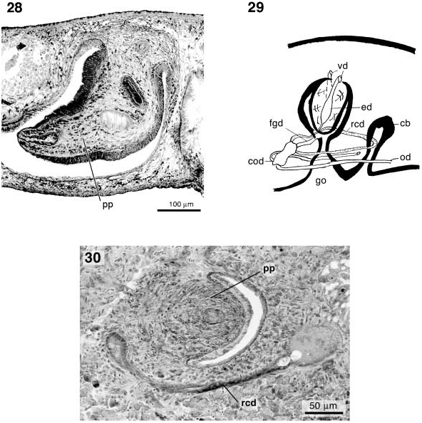
( Figures 29, 30 View Figures 28–30 )
Comparative discussion
Poljakova (1991) mentioned a presumably new species of Uteriporus from the Avachinska Bay and the Gulf of Kronotski, on the east coast of Kamchatka, and from the Komandorskyi Archipelago, namely Uteriporus sp. n. The preserved specimens measured 5–9× 4 mm, with rounded front and hind end. Dorsal surface red-brown, ventral surface pale. The posterior gut trunks fuse in the hind end of the body. Testes numerous, distributed between the gut diverticula and occurring throughout the body length. According to Poljakova (1991), the oviducts fuse posterior to the male copulatory apparatus to form a common oviduct, which subsequently gives rise to three ducts ( Figure 29 View Figures 28–30 ): two receptacular ducts, opening into the anterior copulatory bursa, and the female genital duct that communicates with the atrium. According to Poljakova (1991), common oviduct and female genital duct are penetrated by shell glands.
In our opinion, Poljakova’s reconstruction of the female copulatory apparatus most likely is not fully correct. In the two species of the genus Uteriporus Bergendal, 1890 that have been described up to the present, the posterior, expanded portions of the receptacular ducts do not communicate directly with the female genital duct. The swollen portions of the receptacular ducts only receive a short, narrow branch of the oviduct before the latter communicates with the female genital duct (cf. Bergendal 1896: pl. 1, figure 4; Sluys 1989: figures 101–104). In her reconstruction, Poljakova (1991) may have been misled by Tomkiewicz and Ball’s (1973) description of the copulatory apparatus of U. vulgaris Bergendal, 1890 , since she bases her description of this species on the account of these workers and reproduces their reconstruction figure of the copulatory complex (incorrectly, Poljakova refers to Ball, 1973). However, Tomkiewicz and Ball’s reconstruction is incorrect in that it describes a direct connection between the expanded portions of the receptacular ducts and the female genital duct, as already noted by Sluys and Ball (1983).
On the basis of Poljakova’s (1991) description it is difficult to decide whether her specimens from the Far East represent U. vulgaris , U. pacificus or a third, new species. For U. pacificus a light brown or reddish brown dorsal surface was reported, which is in agreement with the Kamchatkan specimens, while the dorsal body surface of U. vulgaris varies from milky white to pale brown. In U. vulgaris the posterior sections of the receptacular ducts may be expanded to greater or lesser extent and thus may approach the generally smaller expansions in U. pacificus as well as those reported by Poljakova (1991) for the Russian animals. According to Sluys (1989), diagnostic features for U. pacificus are the testes occupying the entire dorso-ventral space (contrasting with the ventral follicles in U. vulgaris ), and the absence of an anterior copulatory bursa (contrasting with the welldeveloped bursa in U. vulgaris ). Unfortunately, Poljakova (1991) does not describe the vertical dimension of the testes, but a photomicrograph of a histological section (her figure 16A) seems to suggest that a testis follicle extends considerably towards the dorsal body surface. In U. pacificus the receptacular ducts fuse to form a common duct that opens to the exterior, whereas in U. vulgaris they open into an anterior bursa that subsequently opens to the outside. It must be noted that re-examination of the type material of U. pacificus revealed that the receptacular ducts do not merely fuse to form a common duct but expand before uniting and/or open into a small expansion ( Figure 30 View Figures 28–30 ). In the Russian specimens there seems to be an anterior bursa, which is suggested in Poljakova’s reconstruction drawing ( Figure 29 View Figures 28–30 ) but also in a series of photomicrographs of serial sections (her figure 17). However, it must be noted that her reconstruction is not drawn to scale and that the photomicrographs show a distinct duct leading to the second, anterior gonopore. Such a duct is present in U. pacificus , whereas in U. vulgaris there is only a short and shallow opening of the anterior bursa to the exterior (cf. Bergendal 1896: pl. 4, figure 32; Sluys 1989: pl. 4, figure F).
Although we do not consider it to be impossible that an Atlantic species, like U. vulgaris , also occurs on the coasts of the Bering Sea, we do here attribute Poljakova’s animals to the species U. pacificus , albeit with some reservations in view of the absence of more detailed anatomical information. This record considerably extends the range of U. pacificus since the species was originally recorded for the coast of British Columbia, Canada ( Sluys 1989).
No known copyright restrictions apply. See Agosti, D., Egloff, W., 2009. Taxonomic information exchange and copyright: the Plazi approach. BMC Research Notes 2009, 2:53 for further explanation.