
Mallinella wiputrai, Dankittipakul, Pakawin, Jocqué, Rudy & Singtripop, Tippawan, 2010
|
publication ID |
https://doi.org/10.5281/zenodo.276153 |
|
DOI |
https://doi.org/10.5281/zenodo.6204687 |
|
persistent identifier |
https://treatment.plazi.org/id/03938749-FF8A-FFD6-05BA-BB19FCC3FF12 |
|
treatment provided by |
Plazi |
|
scientific name |
Mallinella wiputrai |
| status |
sp. nov. |
Mallinella wiputrai View in CoL sp. nov.
Figs 9–13 View FIGURES 9 – 13 , 25–31
Types: Holotype: 3, INDONESIA, Palau Belitung, Gunung Tajam, between Gurok Beraye Waterfalls ( 2º47'01''S, 107º51'47''E) and summit ( 2º46'40''S, 107º51'37''E), 150-450 m, primary forest, 21.-23., 26.ix.2008, leg. P.J. Schwendinger [MHNG, IND-08/03]. Paratypes: 13, 5♀, data as holotype [MHNG]; 2♀, Palau Belitung, Gunung Tajam, near Gurok Beraye Waterfalls ( 2º47'01''S, 107º51'47''E) and summit ( 2º46'40''S, 107º51'37''E), 150 m, primary forest, 20.ix.2008, leg. P.J. Schwendinger [MHNG, IND-08/02].
Etymology: The species name is an eponymous noun in the genitive case in honour of Welly Antahmandro Wiputra-Jayasugianto (The University of Auckland, New Zealand).
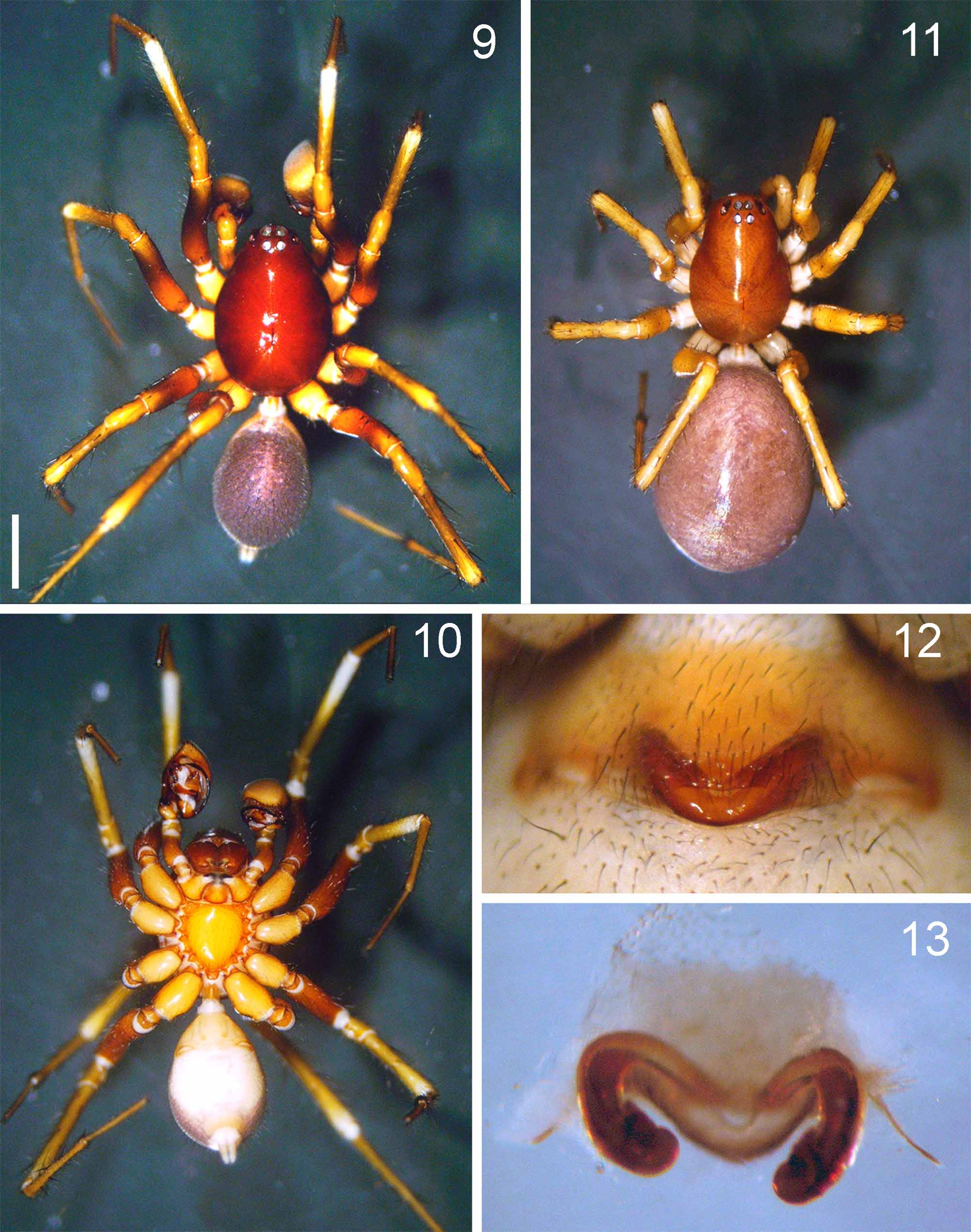
Diagnosis: Mallinella wiputrai sp. nov. can be easily separated from all other Mallinella species in having a shiny reddish brown carapace and dark purplish opisthosoma without any color pattern which is covered with numerous erect spines ( Figs 9, 11 View FIGURES 9 – 13 ); the triangular sternum with four pairs of fused pits situated along the lateral margin ( Fig. 10 View FIGURES 9 – 13 ); the bicoloured tibia, the proximal half being yellowish, the distal half whitish. Males are recognized by the flat, flange-like, subterminally twisted embolus (Figs 25, 28), TA with triangular retrolateral fold and sharply pointed apex directed anteriad, the gradually narrowing apex of the apico-prolateral process pointing mesad (Figs 25–26, 28). Females are recognized by the V-shaped median plate of the epigyne ( Fig. 12 View FIGURES 9 – 13 ) and by the reniform spermathecae situated terminally on elongated tubular IDs ( Figs 13 View FIGURES 9 – 13 , 29–31).
Description: Male ( holotype). Total length 4.88; prosoma 2.72 long, 2.16 wide; opisthosoma 2.58 long, 1.60 wide. Eye sizes and interdistances: AME>PME>ALE=PLE; ratio: AME 1.0, ALE 0.45, PME 0.90, PLE 0.45, AME-AME 0.38, AME-ALE 0.96, PME-PME 0.42, PME-PLE 1.58; MOQ: 1.00 anterior width, 1.10 posterior width, 0.95 long. Leg formula: 4123. Leg measurements: I 11.9 (3.0, 3.5, 2.9, 2.5), II 11.3 (2.7, 3.4, 2.8, 2.4), III 10.1 (2.5, 3.2, 2.4, 2.0), IV 13.0 (3.2, 3.7, 3.2, 2.9).
Pattern and coloration ( Figs 9–10 View FIGURES 9 – 13 ): Carapace pear-shaped, in profile highest between PME and longitudinal fovea; tegument smooth and shiny, reddish brown. Chelicerae dark reddish brown. Labium triangular, yellowish orange, basal and lateral margins slightly darker. Endites yellowish orange, apices pale, with anteromesal brush of black hairs. Sternum yellowish orange, triangular, with bluntly pointed extensions fitting coxal and intercoxal concavities; anterior margin straight, protruding posteriorly, its margin straight; lateral margins with pairs of fused pits. Legs with femur distinctly brown, distal half dark chestnut brown; coxa and trochanter yellowish; other leg segments yellowish brown, except for anterior tibia distally whitish.
Opisthosoma elongated ovoid, covered with numerous erect spines. Dorsum purplish, without pattern. Dorsal scutum indistinct, represented by slightly sclerotized cardiac region, margin poorly delimited. Venter pale. Posterior ventral spines thin and elongate, apices sharply pointed, arranged in a single row.
Palp (Figs 25–28): RTA digitiform in ventral view, broad at base, gradually tapering towards its bluntly pointed apex. Cymbial fold fairly broad, approximately half the length of cymbium. TA sickle-shaped, apicoprolaterally with thin, sharply pointed process directed mesad, retrolaterally with broad, triangular subterminal process, its apex sharply pointed, directed anteriad in lateral view. Conductor beak-shaped, apex sharp, pointing downwards; its dorsal lobe blunt. Embolic base triangular, distinctly broad anteriorly, narrowing posteriorly, retrolateral margin slightly excavated; anterior membranous portion triangular, relatively large. Embolus: originating at 180° flat, relatively broad, constricted and twisted subterminally, its apex rounded.
Female ( paratype). Total length 5.78; prosoma 2.68 long, 1.98 wide; opisthosoma 3.10 long, 2.02 wide. Eye sizes and interdistances: AME<PME>ALE=PLE; ratio: AME 1.0, ALE 0.78, PME 1.20, PLE 0.80, AME-AME 0.25, AME-ALE 1.24, PME-PME 1.62, PME-PLE 2.15; MOQ: 1.00 anterior width, 1.22 posterior width, 1.12 long. Leg formula: 4123. Leg measurements: I 11.1 (2.8, 3.2, 2.8, 2.3), II 10.6 (2.5, 3.3, 2.5, 2.3), III 9.6 (2.5, 3.0, 2.2, 1.9), IV 12.1 (3.1, 3.5, 3.0, 2.5).
Pattern and coloration ( Fig. 11 View FIGURES 9 – 13 ): Carapace smooth and shiny, brown in color. Chelicerae dark brown. Sternum yellowish. Legs bicoloured: coxa and trochanter whitish, other leg segments yellowish brown. Dorsum of opisthosoma purplish brown, without pattern. Dorsal scutum absent. Venter pale. Posterior ventral spines cylindrical, apices bluntly pointed, arranged in a single row.
Genitalia ( Figs 12–13 View FIGURES 9 – 13 , 29–31): Median plate of epigyne a narrowed V-shaped band. Lateral lobes retracted. IDs extremely elongate, curved, descending downwards; reniform spermathecae very compact; FDs situated posteriorly between border of IDs and spermathecae.
Natural history: The type specimens were collected in primary lowland forests of a small island. Distribution: Belitung Island, Indonesia ( Fig. 51 View FIGURE 51 ).

No known copyright restrictions apply. See Agosti, D., Egloff, W., 2009. Taxonomic information exchange and copyright: the Plazi approach. BMC Research Notes 2009, 2:53 for further explanation.
|
Kingdom |
|
|
Phylum |
|
|
Class |
|
|
Order |
|
|
Family |
|
|
Genus |