
Cyclocephala marqueti Dechambre, 1997
|
publication ID |
https://doi.org/10.11646/zootaxa.5026.1.1 |
|
publication LSID |
lsid:zoobank.org:pub:07E0C922-6B0F-4916-85C2-AD95146A8F1E |
|
persistent identifier |
https://treatment.plazi.org/id/03932D70-B626-C370-FF7B-0C91A1B3DB63 |
|
treatment provided by |
Plazi |
|
scientific name |
Cyclocephala marqueti Dechambre, 1997 |
| status |
|
Cyclocephala marqueti Dechambre, 1997
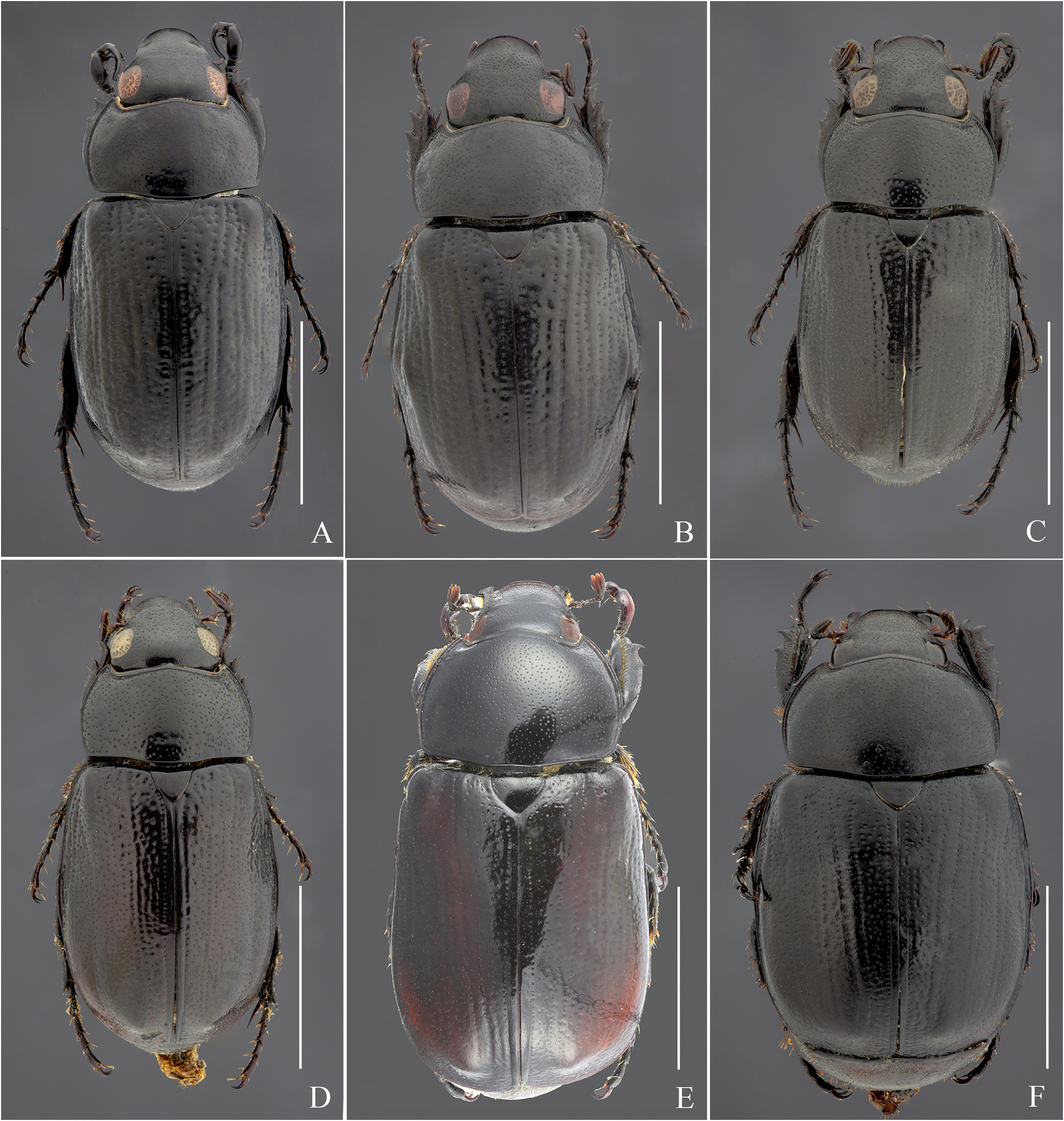
( Figs. 1A–B View FIGURE 1 ; 7A–B View FIGURE 7 ; 8A–B View FIGURE 8 ; 12A View FIGURE 12 ; 13A–B View FIGURE 13 ; 15A View FIGURE 15 ; 16A View FIGURE 16 ; 17A View FIGURE 17 ; 18A View FIGURE 18 ; 19A View FIGURE 19 ; 20A View FIGURE 20 ; 21A View FIGURE 21 ; 24A View FIGURE 24 , 26A–B View FIGURE 26 ; 31A View FIGURE 31 ; 32C; 33A–D; 34A–D; 35A–G; 36A–C; 37A–C; 38)
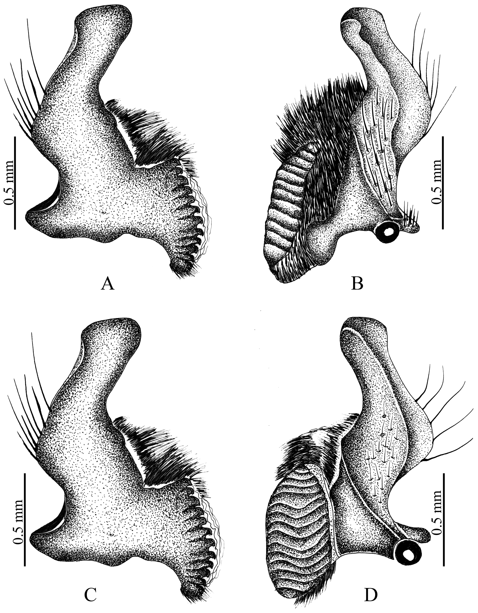
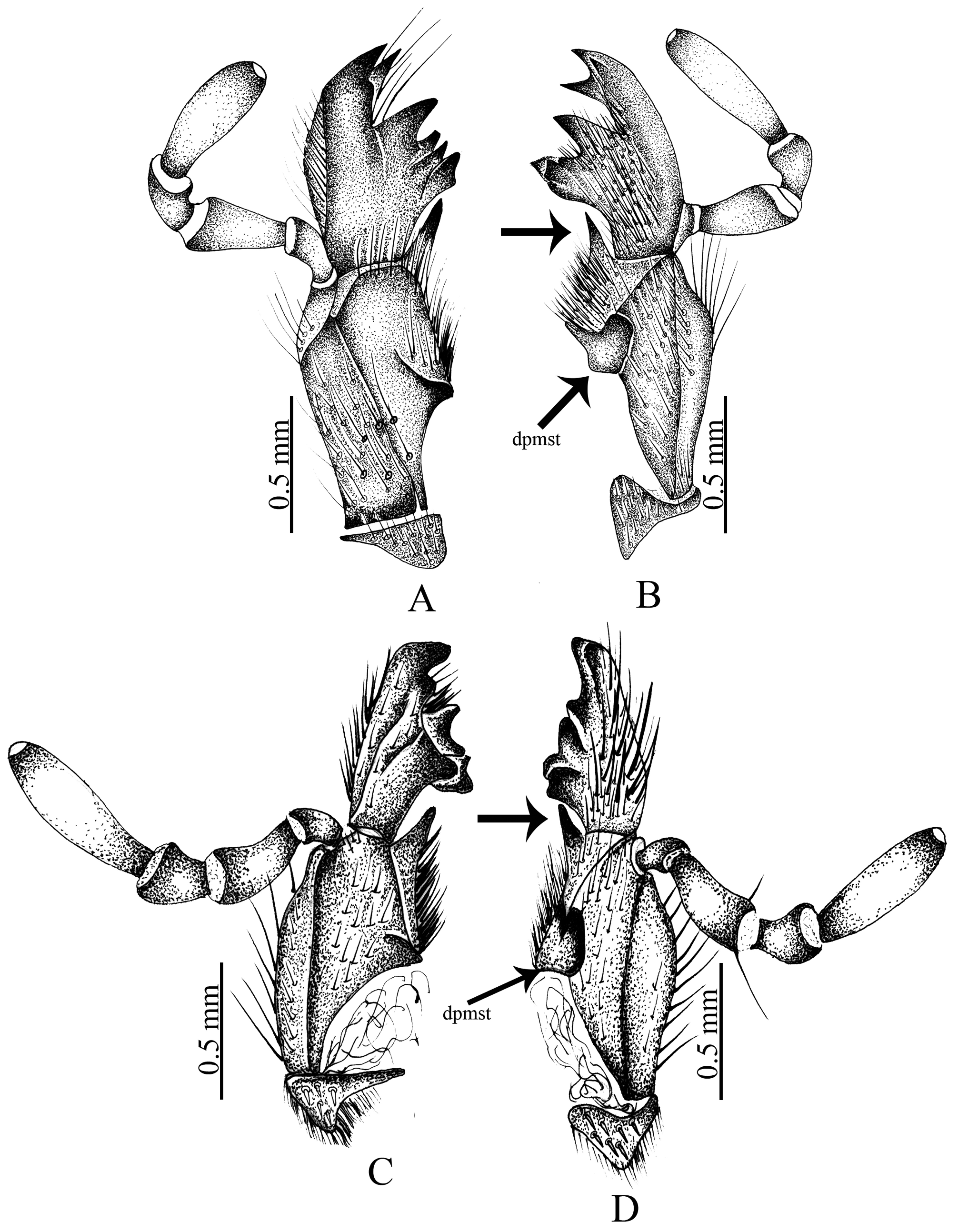
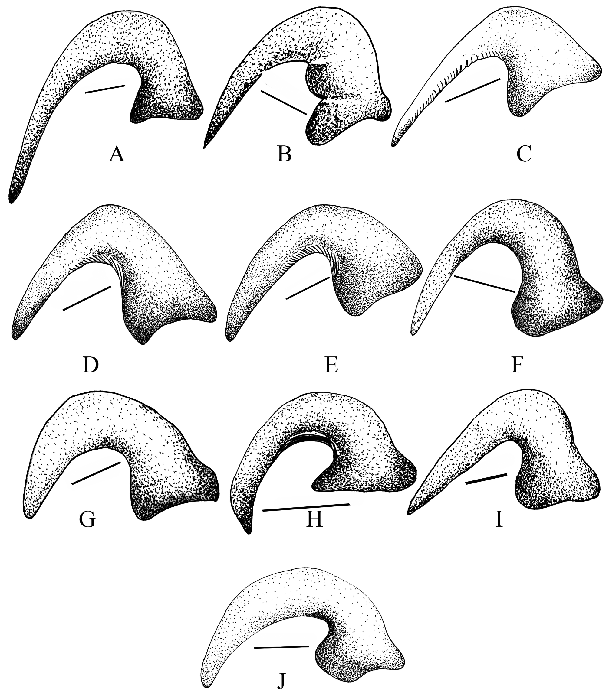
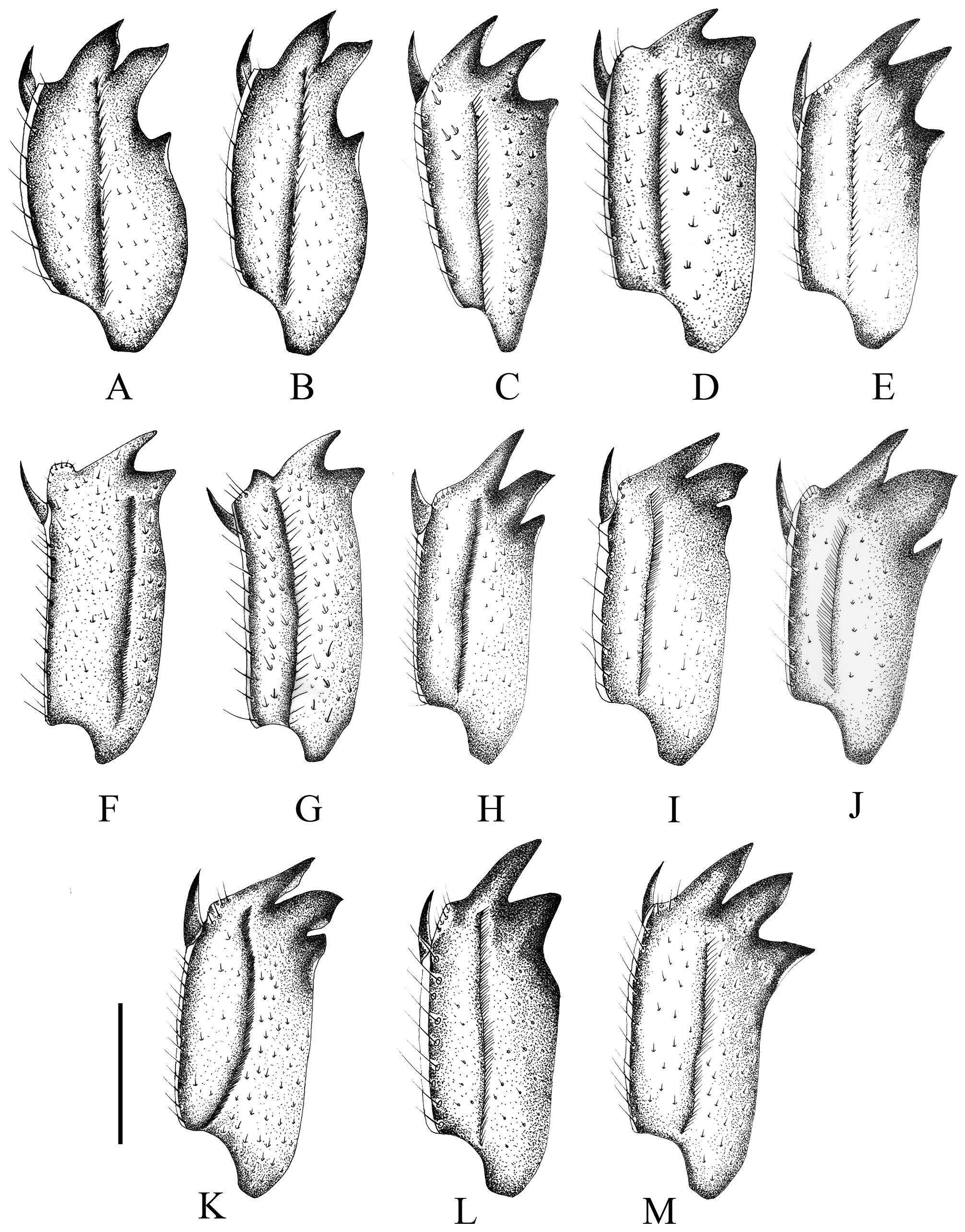
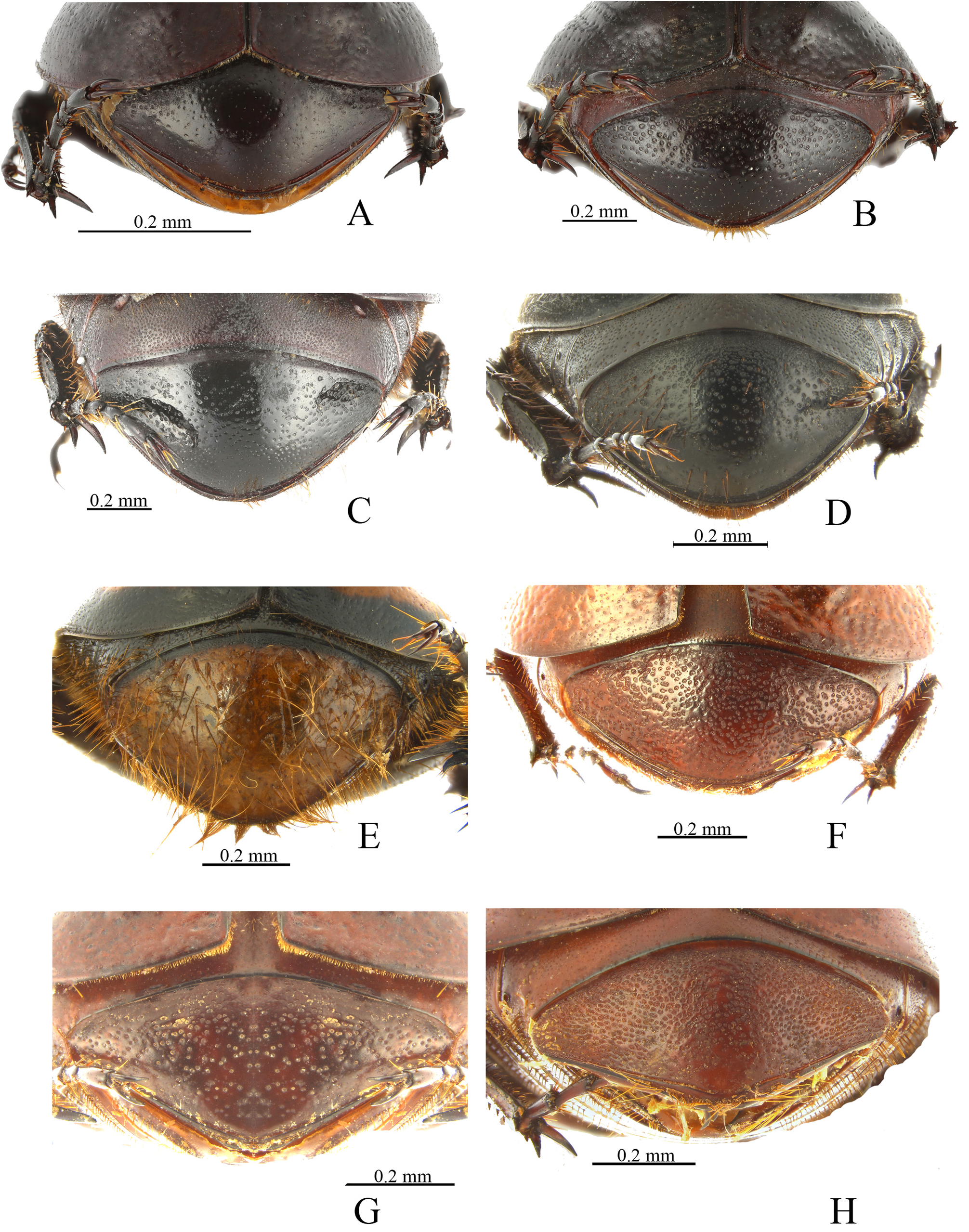
Diagnosis. Cyclocephala marqueti has a longitudinal keel on the inner edge of the metatibia ( Fig. 19A, C View FIGURE 19 ), whereas C. proxima does not ( Fig. 16B View FIGURE 16 ), and C. marqueti has sparse punctures on the pygidium, and the punctures are dense in C. proxima . In addition, the male protibial large claw lacks a basal tooth ( Fig. 19B View FIGURE 19 ), but the tooth is present in C. proxima ( Fig. 19B View FIGURE 19 ).
Cyclocephala marqueti is similar to C. rogerpauli because the inner edge of the metatibia has a longitudinal keel ( Fig. 19A View FIGURE 19 ), and the surface of the pygidium has sparse punctures ( Fig. 21A View FIGURE 21 ), but C. marqueti can be distinguished by sparse punctures on the metasternite. The female epileuron is weakly enlarged at the level of the second abdominal sternite ( Fig. 20A View FIGURE 20 ). The internal sac ( Fig. 28A View FIGURE 28 ) has 6 copulatory lamellae.
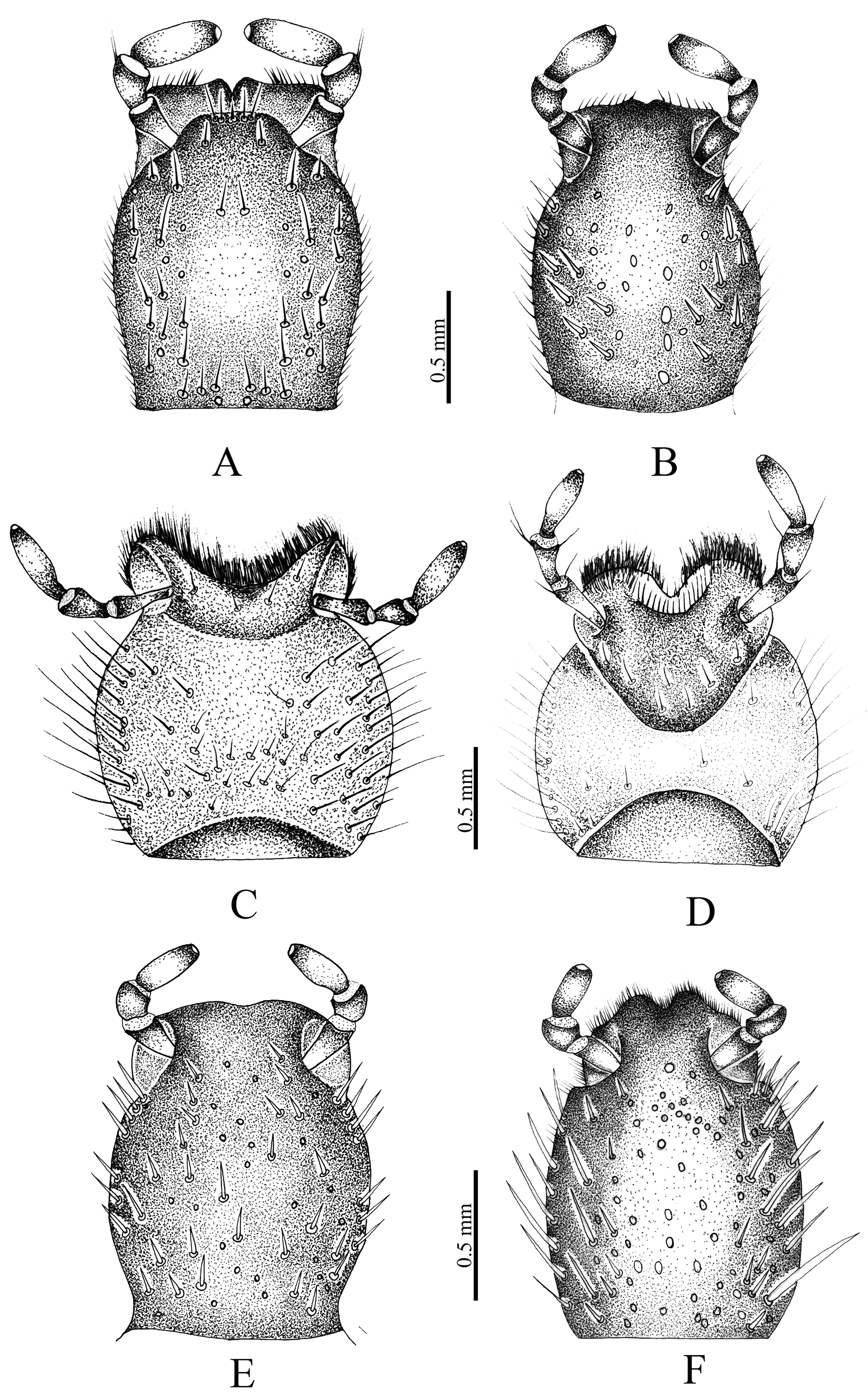
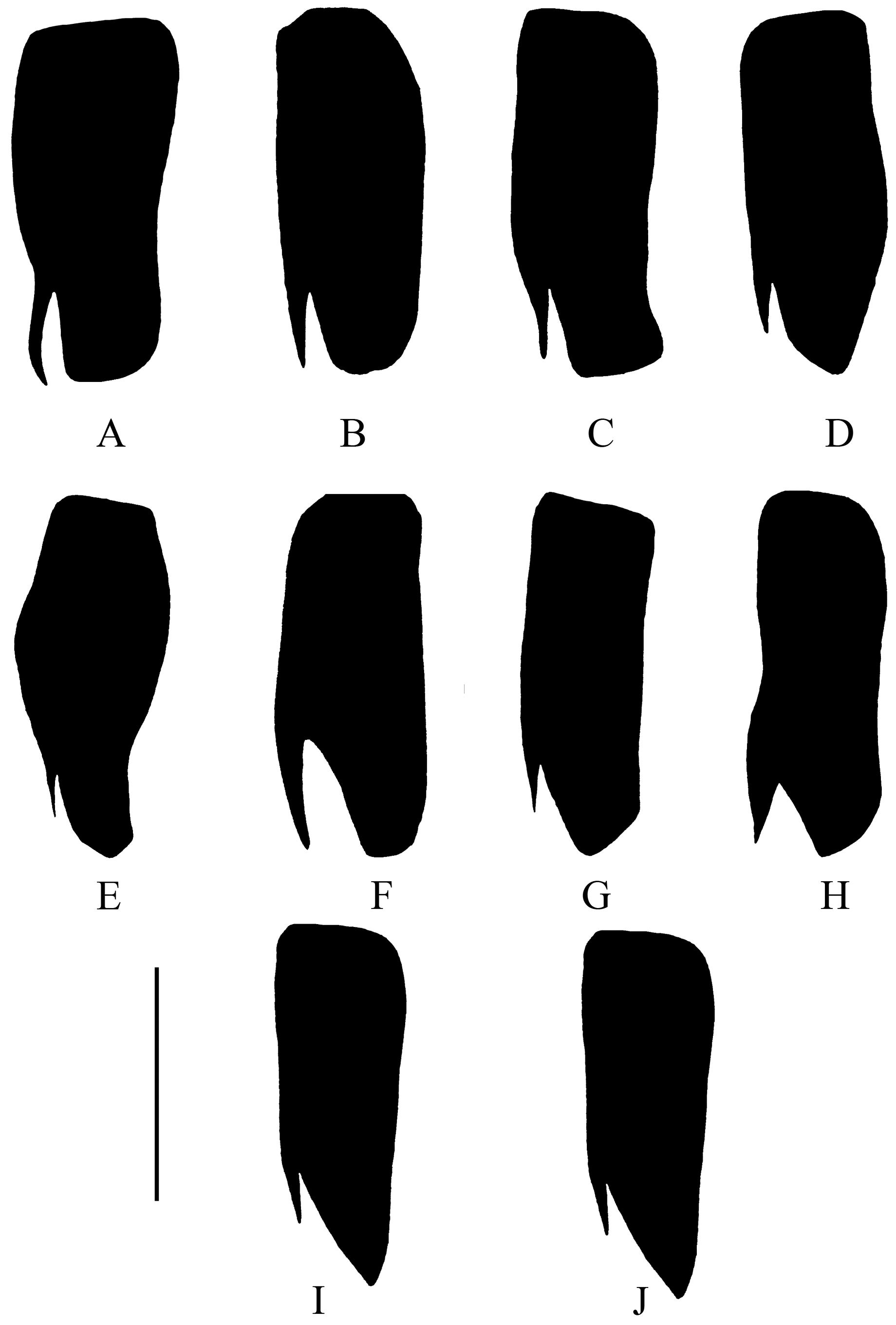
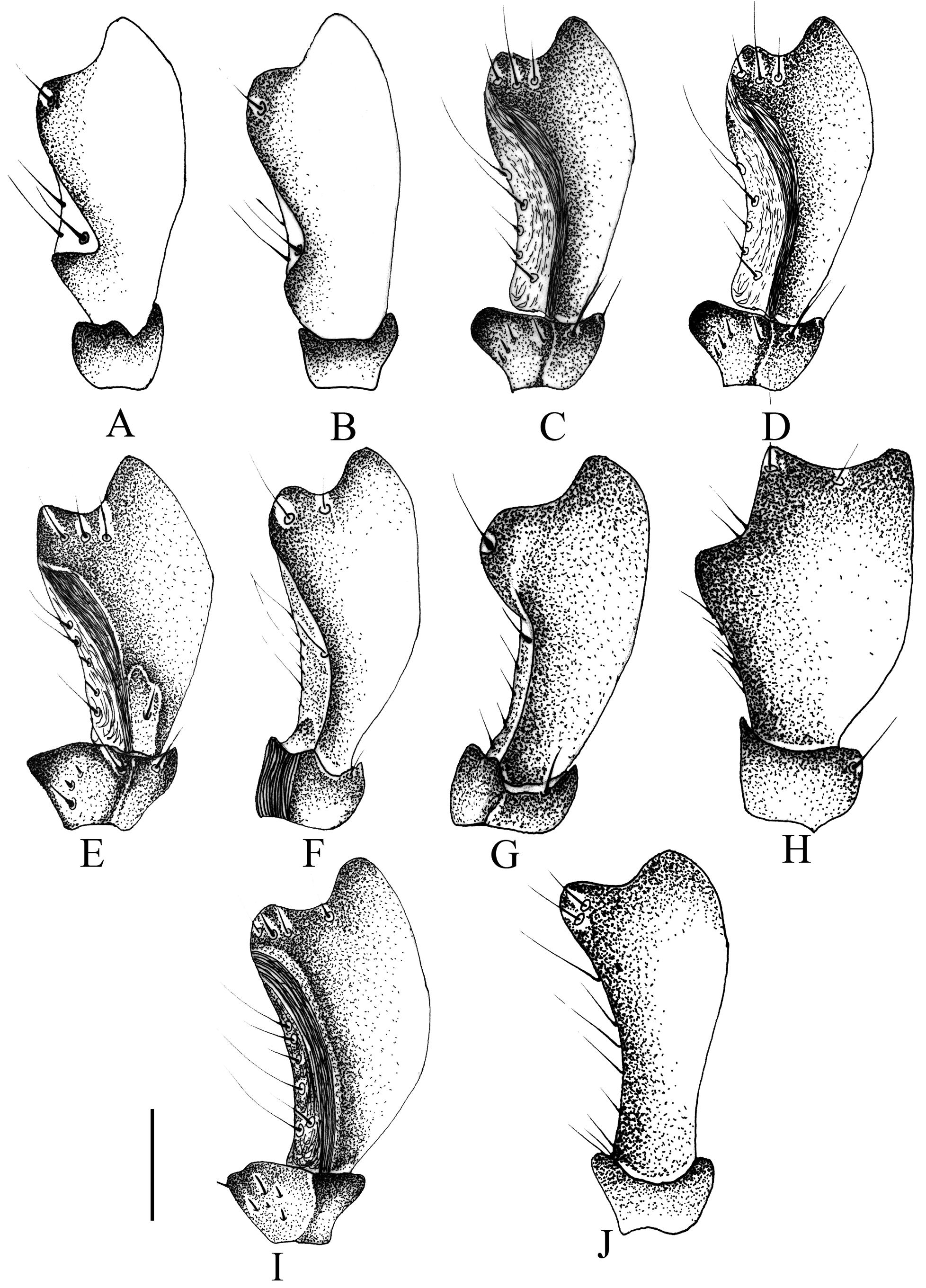
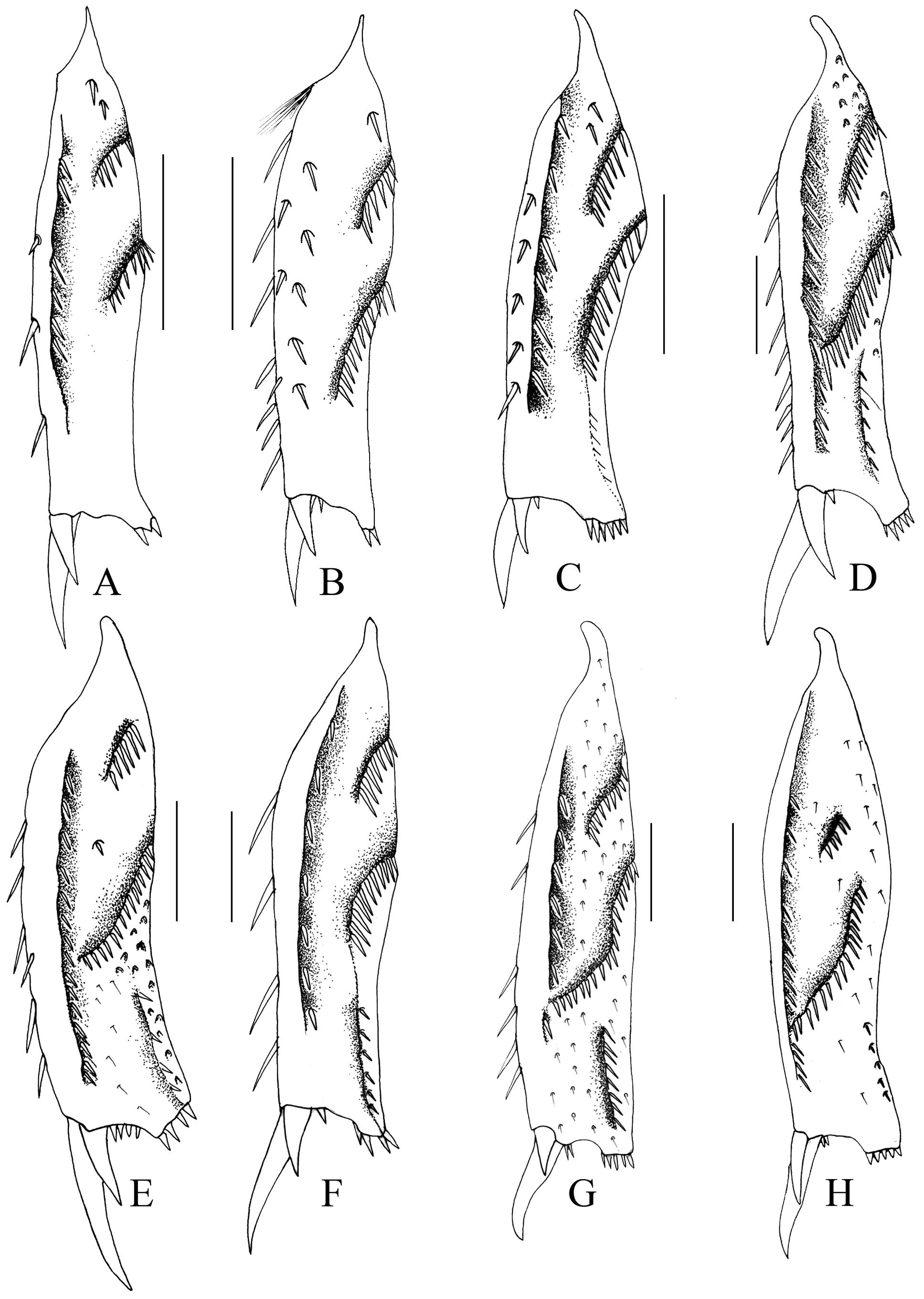
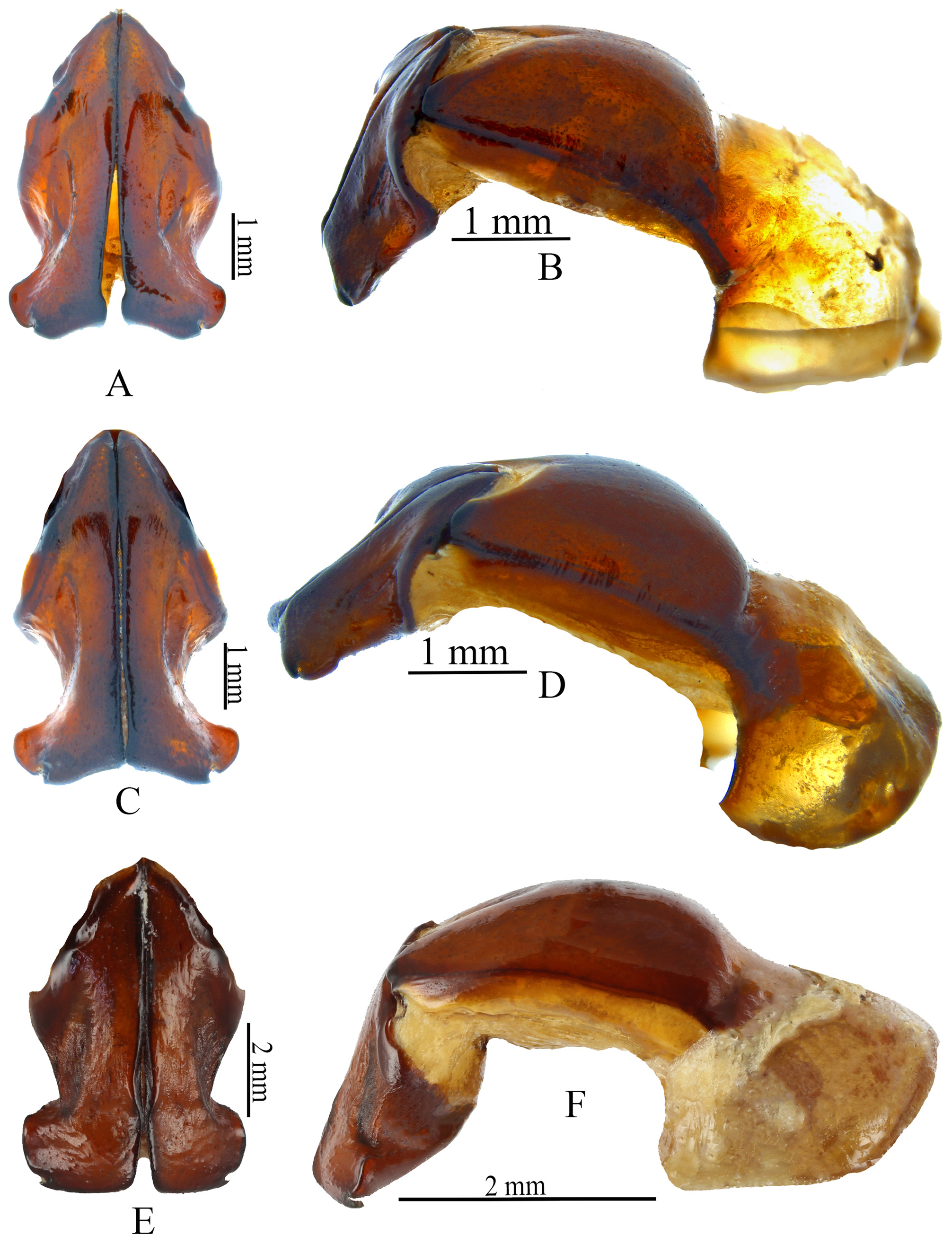
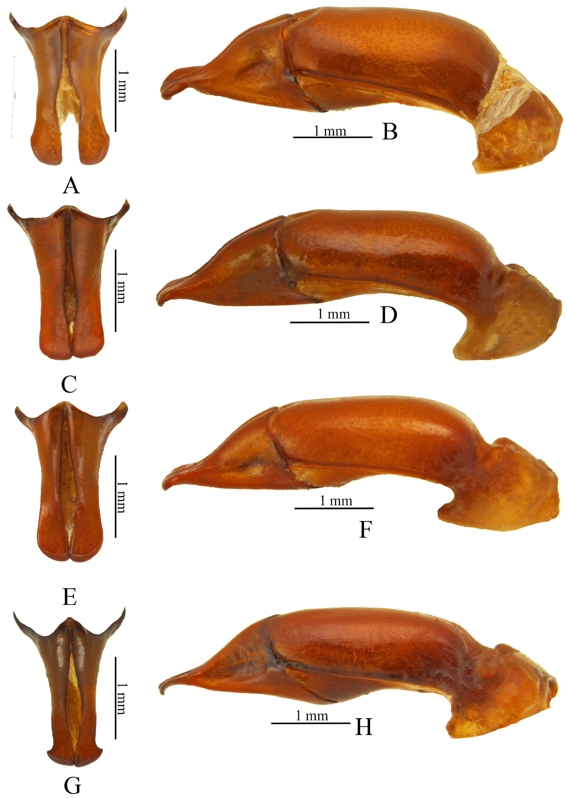
Redescription. Male ( Fig. 1A View FIGURE 1 ). Length 25.4– 22.4 mm; width 11.9– 10.7 mm. Color black. Head: Frons moderately densely punctate, punctures mostly moderately large (rarely small). Clypeal surface similar to frons, apical half often with only a few small punctures: apex convexly rounded, not margined, usually with weak angle at center, and weakly reflexed. Mandibles apically rounded, internal face with a groove ( Fig. 7A–B View FIGURE 7 ); labium densely setose, paraglossa undeveloped, apex slightly notched ( Fig. 12A View FIGURE 12 ); maxilla with galea developed, with 7 teeth, lacinia sclerotized ( Fig. 8A–B View FIGURE 8 ). Epipharynx ( Figs. 13A–B View FIGURE 13 ) ventrally with stout, moderately long setae; dorsum on lateral edge with slender, long setae, surface with small setae. Interocular width equals 4.0 transverse eye diameters. Antenna with 10 antennomeres, club slightly longer than antennomeres 2–7 and with a few long setae. Pronotum: Surface with punctures moderately dense, moderately large to large (especially on sides), ocellate, deep. Base lacking marginal bead. Elytra: Surface with punctate rows, punctures of striae and intervals similar to those of pronotum; entire surface often coarsely and transversely wrinkled obscuring rows of punctures. Pygidium: Surface with sparse, ocellate, setigerous punctures; setae short and tawny ( Fig. 24A View FIGURE 24 ). In lateral view, surface evenly convex. Legs: Protibia tridentate, basal tooth slightly removed from others ( Fig. 18A View FIGURE 18 ) Protarsus enlarged ( Fig. 1A View FIGURE 1 ), tarsomeres 2–4 successively gradually larger, fifth large, curved, with small angulation at base on ventral side ( Fig. 17A View FIGURE 17 ); median claw at base with small tooth, claw large, curved ( Fig. 16A View FIGURE 16 ), apex cleft ( Fig. 15A View FIGURE 15 ). Metatibia on inner edge with longitudinal keel ( Fig. 19A View FIGURE 19 ). Metatarsus subequal in length to metatibia. Venter: Prosternal process moderately long, columnar, apex obliquely flattened into a transverse oval with anterior 2/3 to 4/5 raised into convex “button”. Genitalia: Parameres as in Fig. 26A–B View FIGURE 26 . Internal sac ( Fig. 31A View FIGURE 31 ) with 6 copulatory lamellae and speculum (with many short, spine-like setae).
Female ( Fig. 1B View FIGURE 1 ). Length 25.9– 19.9 mm; width 11.9– 10.2 mm. Similar to male, but pronotal disc more densely punctate. Epipleuron weakly enlarged at level of second abdominal sternite. Elytral lateral margin slightly enlarged just before middle ( Fig. 20A View FIGURE 20 ); in dorsal view, ventral margin of epipleuron without tooth or angulation ( Fig. 1B View FIGURE 1 ). Pygidium slightly convex. Protarsus simple ( Fig. 1B View FIGURE 1 ). Gonocoxite larger than gonocoxal sternite, gonocoxite at center with a membranous area (Fig. 32C).
Distribution. Cyclocephala marqueti is known from Ecuador ( Dechambre 1997; Ratcliffe et al. 2020). This species is widespread in the eastern piedmont of the Eastern Cordillera in Colombia (new country record).
Locality records ( Fig. 35 View FIGURE 35 ). Eight specimens examined from IAvH. “ Colombia, Caquetá, Municipio de Solano. Bosque. / Emergido de larva asociada a termitero. 0º33´15.408´´S; 72º15´39.384´´W. / GWS 84, 174 m. Cría, emergido de/ larva asociada a termitero, 2020-03, / C. A. Medina ” [♀ IAvH-E-198645]. “ Colombia, Meta, Cubar- ral, Finca / Rosania, Bosque húmedo tropical, / 3º49´46.56´´N; 73º49´59.21´´W. / WGS 84, 620 m, Luz Establo, 1-30.iv.2018. A. Lopera; W. Chamorro ” [3♂ -IAvH-E-216418, IAvH- E-216420, IAvH- E-216419; 3♀ -IAvH-E- 216421, IAvH-E-216422, IAvH-E-216423] GoogleMaps .
Life history. Adults are active during the night and attracted to lights. The larvae are found in decomposed trees and associated with termites. The larva and pupa are described here for the first time.
This is the first species in the C. cribrata species group to have the immature stages described. The descriptions are based on third instars and one exuvium from a third instar associated with one adult (female) with the following data: Colombia, Caquetá, Municipio de Solano , forest, emerged from larvae associated with a termite mound, 0º33´15.408´´S, 72º15´39.384´´W, 174 m, iii.2020 [♀ IAvH-E-198645] GoogleMaps .
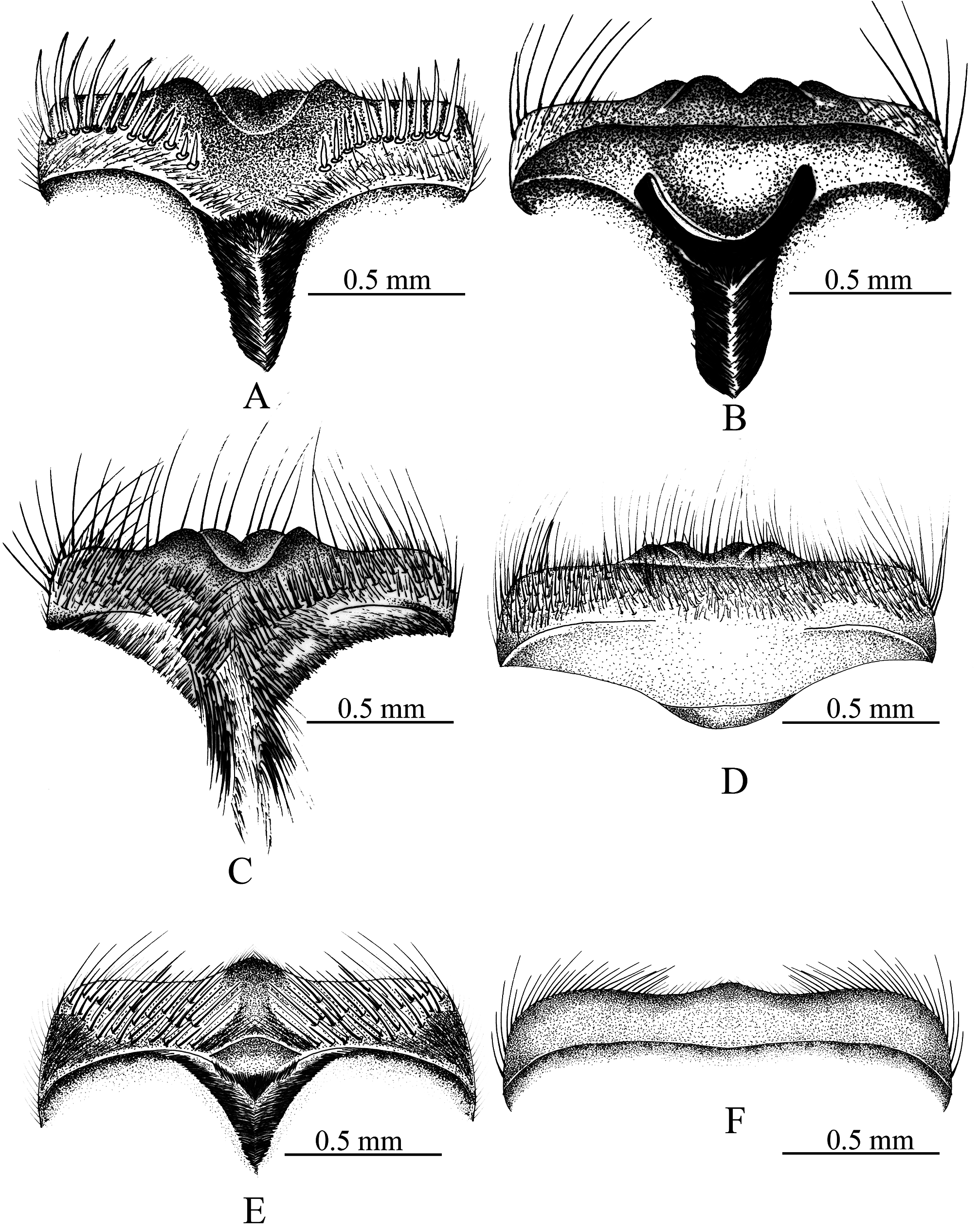
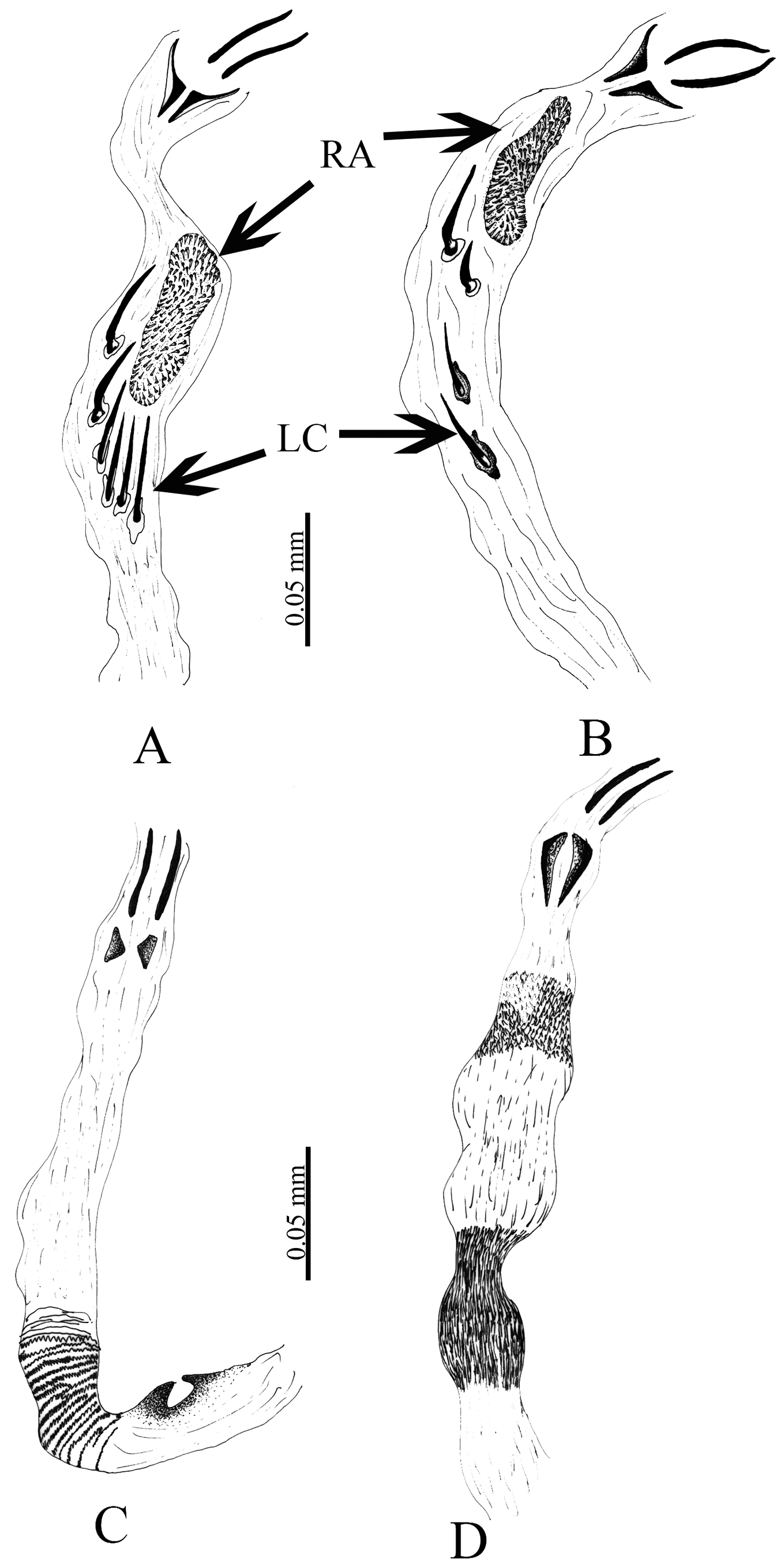
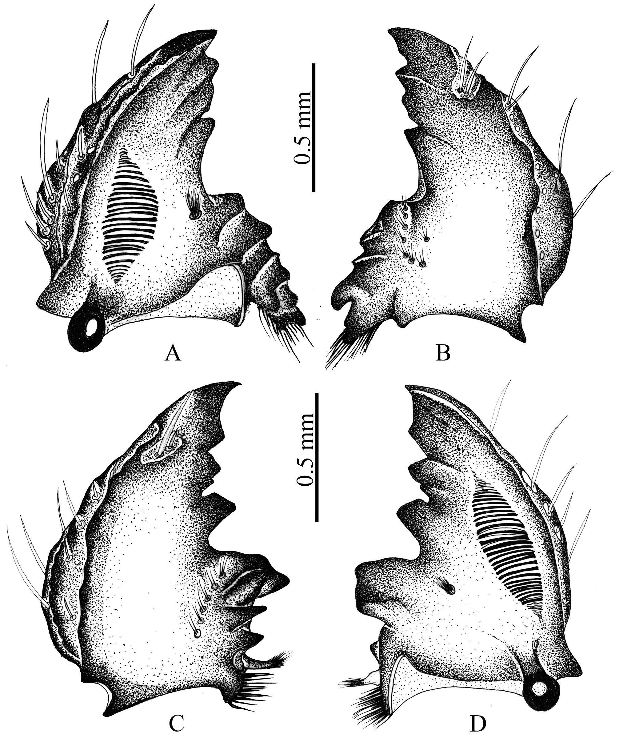
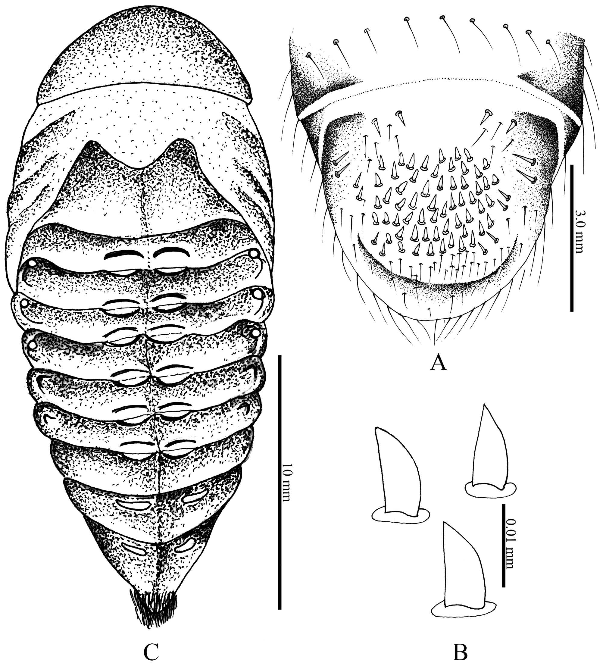
Third instar description. Figs. 33A–D; 34A–D; 35A–G; 36A–C; 37A–C. Head: Cranium (Fig. 33A): Width 6 mm. Surface densely punctate, dark brown. Frontal suture and clypeofrontal suture distinct. Frons (Fig. 33A): 1 anterior frontal seta on each side; exterior frontal seta and posterior frontal setae absent; 1 seta on each anterior angle. Remaining cranial surface with 3 dorsoepicranial setae, 6 epicranial setae distributed irregularly, 6–7 paraocellar setae on each side. Stemmata present. Clypeus: Form trapezoidal. Surface of postclypeus dark brown, well sclerotized, densely punctate; surface of preclypeus light brown, with 2 central setae and 2 lateral setae on each side. Labrum: Rugopunctate, slightly asymmetrical, 7 posterior setae, 3–4 lateral setae on each side, 14 central setae. Epipharynx (Fig. 33D): Lateral margins angulate, asymmetrical. Haptomeral process prominent, entire; right chaetoparia with 58 setae; left chaetoparia with 52 setae and some sensillae; gymnoparia narrow; acroparia each with 14 straight, long, thick setae; corypha with 5 fine, long setae; acanthoparia with 10–11 short, curved, spine-like setae; pedium large, ovate. Dexiotorma elongate, narrow, laeotorma slightly shorter than dexiotorma. Dexiophoba absent; laephoba poorly developed between haptolechus and inner side of laeotorma, with 17 fine setae. Sclerotized plate of right nesium elongate, truncate at apex; sense cone on left nesium represented by longitudinal, well-sclerotized plate, apex with 4 sensilla. Crepis poorly defined. Left mandible ( Fig. 34C–D View FIGURE 34 ): Scissorial region with 4 teeth, apical tooth blade-like, S 1 + S 2 fused; S 1-3 separated from S 4 by scissorial notch; S 3 with a basal tubercle. Scrobis with 7 long, fine setae. Dorsal surface with line of 8 fine, moderately long setae; acia well developed, sharp, setae at apex absent, without basolateral setae. Ventral surface with elongate-oval stridulatory area formed by 34 narrowly separated ridges; ventral process well developed, rounded, with many asperites; dorsomolar area with row of 10 stout, fine, moderately long setae; brustia with 16 stout, long setae arranged in form of a “U”. Molar area with 3 lobes, first molar lobe (M 1) large. Right mandible ( Fig. 34A, B View FIGURE 34 ): Scissorial region with 4, teeth S 1-3 separated from S 4 by scissorial notch; scrobe with 9 fine, long setae. Dorsal surface with line of 14 fine, long setae. Ventral surface with elongate-oval stridulatory area formed by 33 narrowly separated ridges; ventral process well developed, rounded, with many asperites. Brustia with 16 long, stout setae. Calx large, without basolateral setae. Molar area with 3 wide, convex, ridged lobes (M 1-3) and with 6 long, fine setae. Maxillae ( Fig. 35B–C View FIGURE 35 ): Cardo subrectangular. Stipes longer than wide. Galea with many stout setae and 1 well-developed uncus at apex. Lacinia with many stout setae and 2 unci separated at their bases ( Fig. 35C View FIGURE 35 ). Maxillary palpi with 4 palpomeres, all palpomeres of different lengths, palpomere 4 twice as long as palpomere 2. Stridulatory area with 5 truncate ridges and an anterior truncate process ( Fig. 35D View FIGURE 35 ). Hypopharynx ( Fig. 35A View FIGURE 35 ): Glossa with 12 fine, long setae and 26 stout, short setae. Hypopharyngeal sclerome asymmetrical ( Fig. 35A View FIGURE 35 ), concave medially, sharp process produced dorsally; left and right lateral lobes with 11–13 fine, moderately long setae. Left margin with row of 10–12 stout, moderately long setae directed toward center of sclerome and 16 basal setae at sclerome. Antennae: 4 antennomeres, antennomeres 2 and 3 about 1/4 times longer than antennomeres 1 and 4; terminal antennomere slightly longer than antennomere 1; apical antennomere oval in dorsal and ventral view, almond-shaped in lateral view; dorsal surface with 13 sensory spots (Fig. 33B), ventral surface with 12 sensory spots (Fig. 33C).
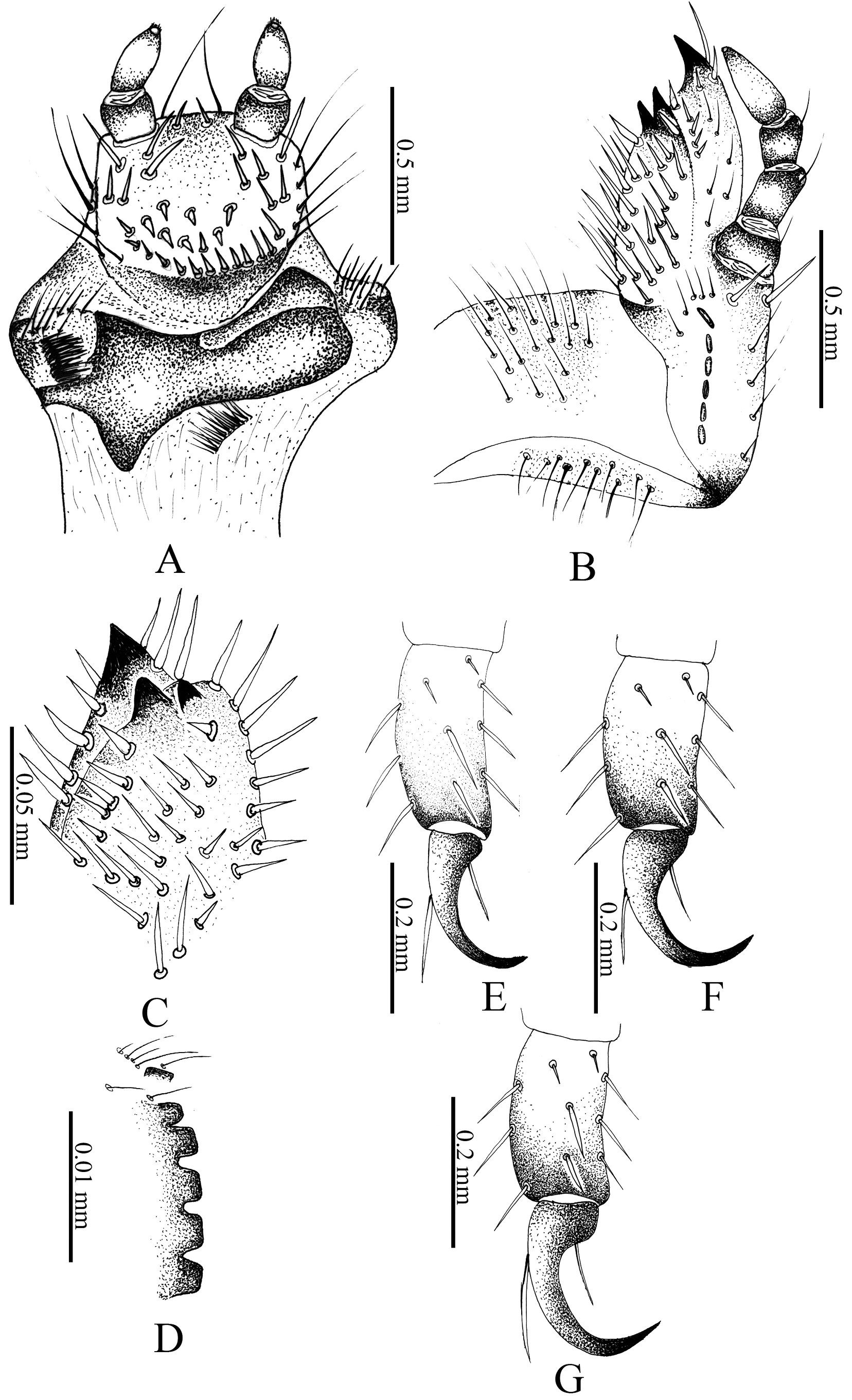
Thorax: Pronotum irregularly and weakly sclerotized, with 8 long, fine setae. Prothoracic spiracles ( Fig. 37C View FIGURE 37 ) 0.62 mm long, 0.34 mm wide; respiratory plate dark brown, bulla not prominent; distance between respiratory lobes less than diameter of bulla; plate with 38 holes across diameter at middle, holes with irregular edges ( Fig. 37B View FIGURE 37 ). Dorsum of prothorax with transverse row of 28 long, fine setae. Mesoprescutum with transverse, irregular row of 24 long, fine setae and 22 short, spine-like setae; metaprescutum with 30 long, fine setae; metascutellum with 6 long, fine setae and 40 short, fine setae; spine-like setae absent. Legs ( Fig. 35E–G View FIGURE 35 ): Tarsal claws with enlarged apical process, 1 basoexternal seta, and 1 internal, preapical seta. Tarsal claw on protarsus larger than claws on mesothoracic and metathoracic legs. Coxa, trochanter, and tibiotarsus of all legs setose, setae fine or stout.
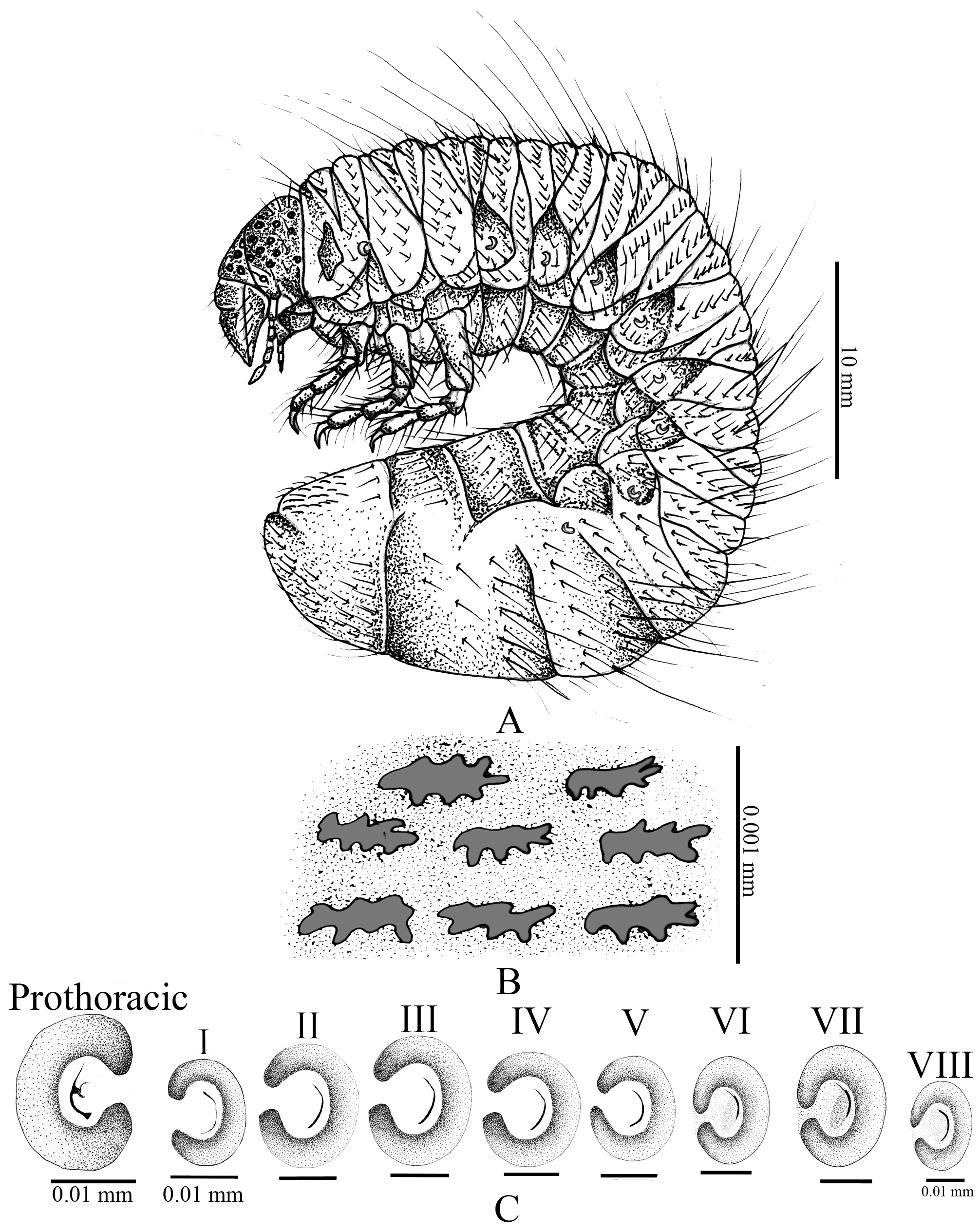
Abdomen: Abdominal spiracle VIII (0.34 mm long, 0.38 mm wide) shorter than spiracles I–VII. Spiracles V (0.32 mm long, 0.48 mm wide) and VI (0.38 mm long, 0.49 mm wide) shorter than spiracles II–IV and VII. Spiracles on segments II (0.26 mm long, 0.53 mm wide), III (0.43 mm long, 0.59 mm wide), IV (0.43 mm long, 0.56 mm wide) and spiracles on segment VII larger than preceding (0.61 mm long, 0.73 mm wide) ( Fig. 34C View FIGURE 34 ). Abdominal segment I with 18 long, fine setae on prescutum and 42 short, fine setae; subscutum with 16 long, fine setae; scutum with 8 long, fine setae, 16 short, fine setae, and 11 short, spine-like setae; scutellum with 8 long, fine setae and 30 short, fine setae. Abdominal segment II on prescutum with 21 long, fine setae and 44 short, fine setae; subscutum with 16 short, fine setae; scutum with 8 long, fine setae, 18 short, fine setae, and 13 short, spine-like setae; scutellum with 8 long, fine setae and 30 short, fine setae. Abdominal segment III on prescutum with 16 long, fine setae, 62 short, fine setae, and 22 short, spine-like setae; subscutum with 51 short, fine setae; scutum with 10 long, fine setae, 102 short, fine setae, and 48 short, spine-like setae; scutellum with 12 long, fine setae, 110 short, fine setae, and 89 short, spine-like setae. Spiracular area with 63 long, fine setae. Abdominal segment IV on prescutum with 18 long, fine setae, 14 short, fine setae, and 118 short, spine-like setae; subscutum with 59 long, fine setae and without short, spine-like setae; scutum with 18 long, fine setae, 167 short, fine setae, and 126 short, spine-like; scutellum with 12 long, fine setae, 186 short, fine setae, and 168 short, spine-like setae. Spiracular area with 61 long, fine setae. Abdominal segment V with 18 long, fine setae, 196 short, fine setae, and 202 short, spine-like setae on prescutum; subscutum with 51 short, fine setae; scutum with 16 long, fine setae, 116 short, fine setae, and 226 short, spine-like setae; scutellum with 12 long, fine setae, 212 short, fine setae, and 109 short, spine-like setae. Spiracular area with 61 long, fine setae. Abdominal segment VI on prescutum with 18 long, fine setae, 120 short, fine setae, and 206 short, spine-like setae; subscutum with 62 short, fine seta and without short, spine-like setae; scutum with about 128 long, fine setae, 199 short, fine setae, and 200 short, spine-like setae; scutellum with about 12 long, fine setae, 120 short, fine setae, and 181 short, spine-like setae. Spiracular area with 58 long, fine setae. Abdominal segment VII with 2 rows of setae (anterior and posterior), each with 8 long, fine setae. Spiracular area with 8 long, fine setae. Abdominal segment VIII with 2 rows of setae, anterior row with 8 long, fine setae; posterior row with 6 long, fine setae. Spiracular area with 32 long, fine setae. Abdominal segment IX with 1 anterior row of 4 fine setae, and a posterior row with 8 long, fine setae. Spiracular area with 32 long, fine setae. Abdominal segment X with approximately 52 moderate to long, fine setae and 102 short, spine-like setae. Spiracular area with 36 long, fine setae. Pleural lobes with 12 long, fine setae, short setae absent. Raster: Surface without palidia; campus with 22 long, fine setae; teges with 67 short and stout setae projecting toward superior anal lobe ( Fig. 36A View FIGURE 36 ), barbula with 56–66 long, fine setae. Anal slit transverse. Approximate dorsal body length 23.5 mm ( Fig. 37A View FIGURE 37 ).
Pupa description. Female ( Figs. 36C View FIGURE 36 ). Length 19.8 mm; width of pronotum 17.2 mm. Body elongate, oval, stout, exarate. Color dark reddish orange. Entire body with fine, velvet-gold vestiture. Head. Surface glabrous, bent sharply beneath thorax; antenna, labrum, mandibles, maxillae, and palps discernible; antennal tecae expanded, stout, with rounded apex. Compound eyes sunken, scarcely visible. Thorax. Surface glabrous. Elytral and posterior wing thecae closely appressed, curved ventrally around body; elytral thecae extending to middle of abdominal segment IV; posterior wing thecae extending to middle of abdominal segment V. Protibia with 3 distinct teeth on external edge. Mesotibiae and metatibiae with inner and external spines well developed at apex. Abdomen. Segments III–X well defined in ventral view. Segment VII slightly longer than preceding, segment VIII 0.25 times longer than segment VII, segments VIII and IX fused, segment X without genital ampulla but with a small ventral plate. Segments I–X with well-defined dioneiform organs in dorsal view, sclerotized between segments I–II, II–III, III–IV, IV–V, V–VI, and VI–VII. Pleural lobes rounded. Spiracle I elongate, with fine peritreme and covered by wing thecae; spiracles II–IV ovate, prominent, with strongly sclerotized peritreme; spiracle V–VIII closed. Abdominal apex rounded, with fine and short setae.
Comments. This is the first described larva of the Cyclocephala cribrata species group. Based on this larva, there are some characters not shared with other Cyclocephala larvae ( Ritcher 1966; Morelli 1991; Morelli and Alzugaray 1994; Morón et al. 2014; Moore et al. 2018; Rodrigues et al. 2018). These characters are: the haptomeral process of the epipharynx is raised in C. marqueti but bilobed in other larvae described (with the exception of C. fasciolata ) ( Morón et al. 2014; Rodrigues et al. 2018). The mandibles of other known larvae have the scissorial area with a sinuate, blade-like apical tooth (S 1 +S 2) and 1 rounded tooth (S 3) after the scissorial notch; in C. marqueti , the scissorial area has 4 teeth with teeth S 1-3 separated from S 4 by a scissorial notch (similar to that in C. fasciolata and C. jalapensis Casey ).
The last antennomere has 13 dorsal sensory spots and 12 ventral sensory spots, but in other described larvae, the last antennomere has only 2 ventral sensory spots and 2 dorsal sensory spots (with the exception of C. barrerai Martínez that also has a third spot). The lateral margins of the epipharynx are rounded in other species, but in C. marqueti , the edges are angled (similar to that of C. fasciolata and C. jalapensis ).
The size of abdominal spiracle VIII is smaller than the preceding spiracles in C. marqueti , but in other Cyclocephala larvae, the spiracles on segments VII and VIII are larger than those on the preceding segments (with the exception of C. fasciolata ) ( Ritcher 1966; Morelli 1991; Morelli & Alzugaray 1994; Morón et al. 2014; Moore et al. 2018; Rodrigues et al. 2018). The raster lacks palidia as in other species of Cyclocephala , but the palidia are present in C. testacea Burmeister , C. modesta Burmeister , and C. tucumana Bréthes ) ( Morelli and Alzugaray 1994; Fuhrmann et al. 2019).
No known copyright restrictions apply. See Agosti, D., Egloff, W., 2009. Taxonomic information exchange and copyright: the Plazi approach. BMC Research Notes 2009, 2:53 for further explanation.
|
Kingdom |
|
|
Phylum |
|
|
Class |
|
|
Order |
|
|
Family |
|
|
Genus |